Archived: Revealing a Piece of Cilia's Puzzle
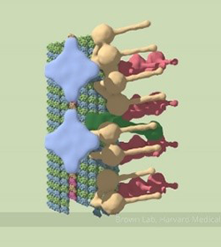 A partial model of a doublet microtubule. Credit: Veronica Falconieri.
Cilia (cilium in singular) are complex organelles found on all of our cells except red blood cells. Their rhythmic beating moves fluid or materials over the cell to help transport food and oxygen or remove debris. For example, cilia in our windpipe prevent bacteria and mucous from traveling to the lungs. Some pick up signals like antennae, such as cilia in our ears that help detect sounds. One component of cilia is the doublet microtubule, a major part of cilia's skeleton that gives it strength and rigidity.
Thanks to advances in cryo-electron microscopy, researchers recently mapped the full structure of the doublet microtubule and its associated proteins. Containing 451 protein chains, it's one of the largest protein structures ever mapped. This achievement may help researchers learn more about how cilia grow and function and better understand their role in disease.
Take a look at the doublet microtubule's intricate structure and learn more with an animated video and news story from Harvard Medical School in Boston, Massachusetts.
This research was supported in part by NIGMS grant R01GM032843.
About the Author