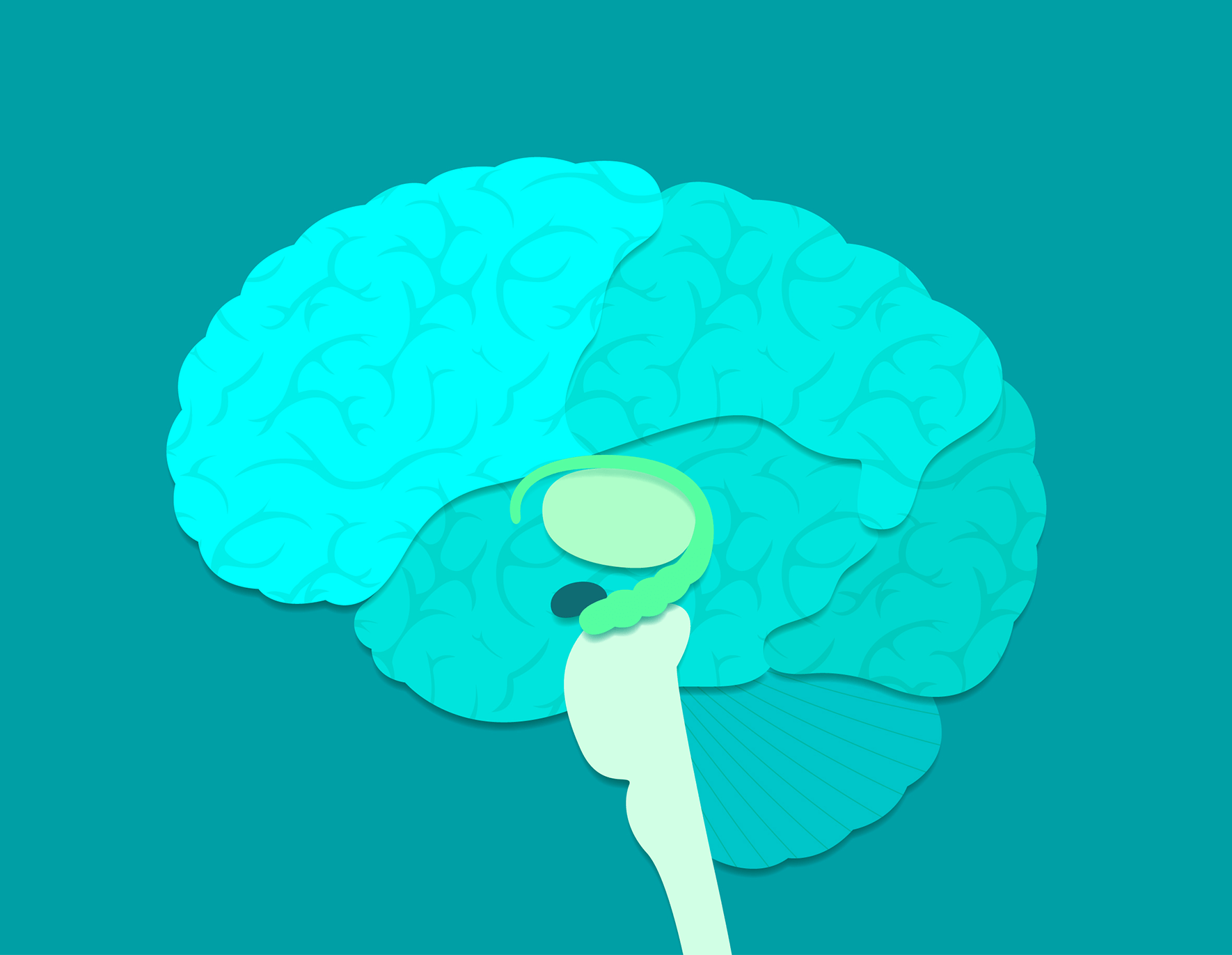
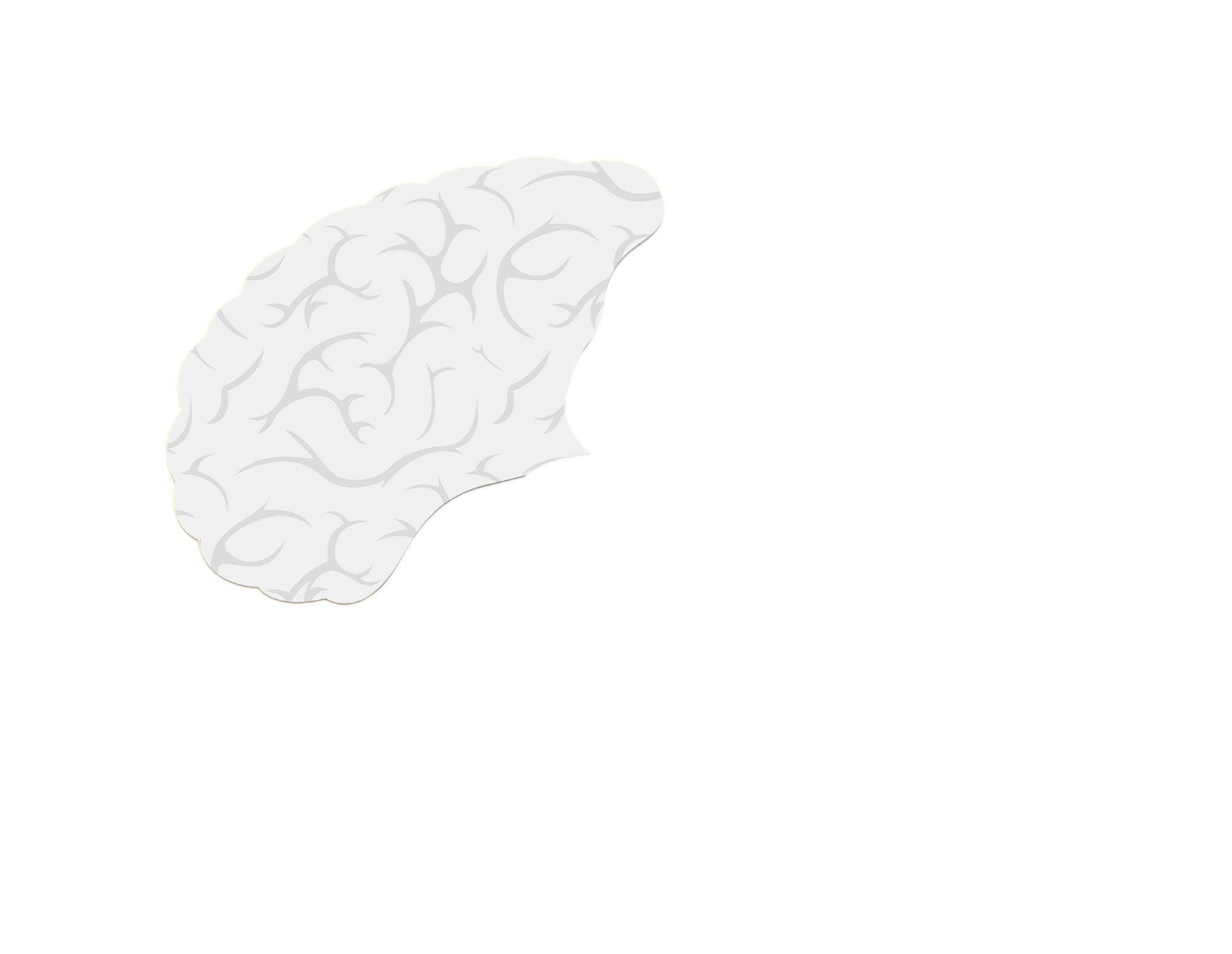


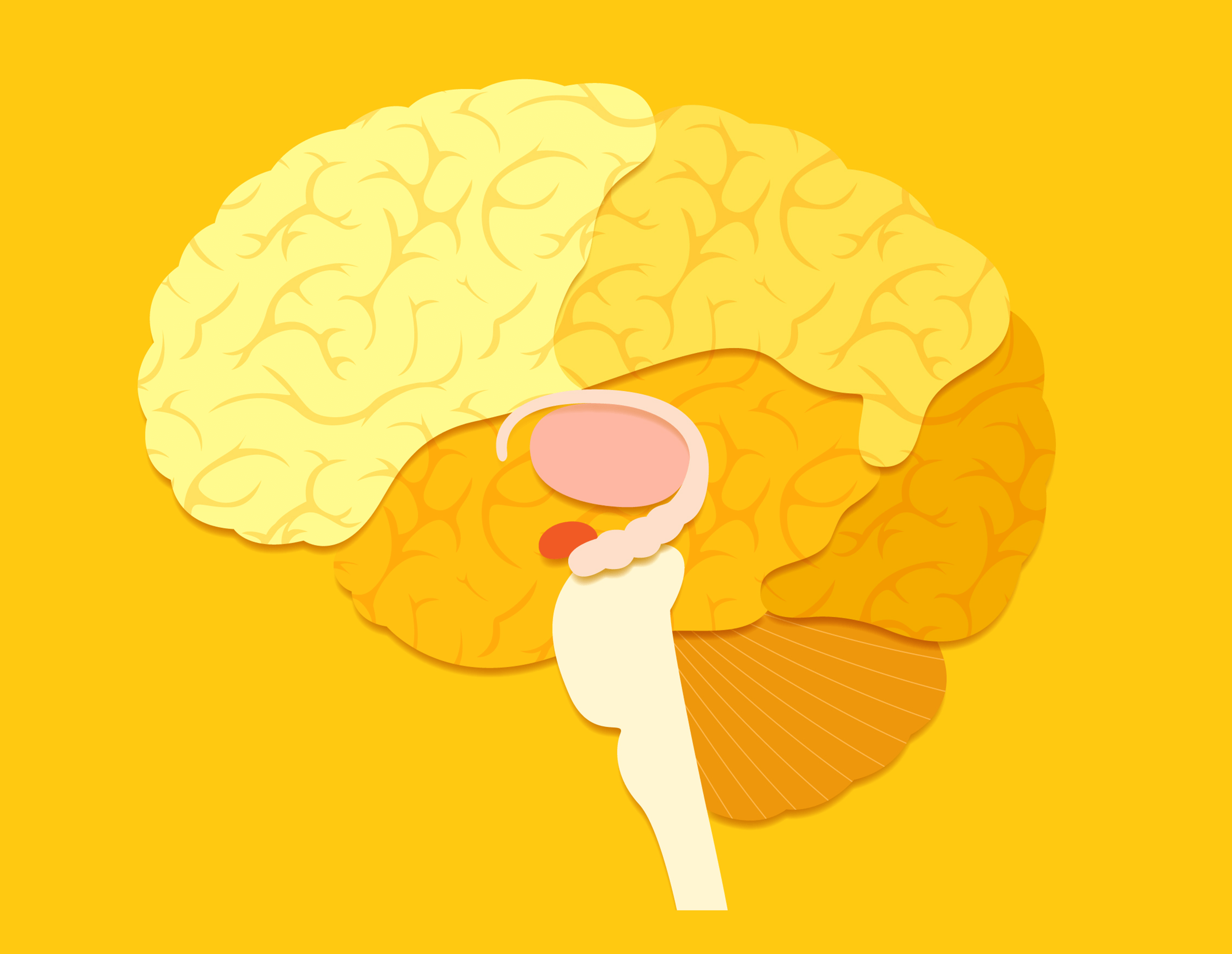
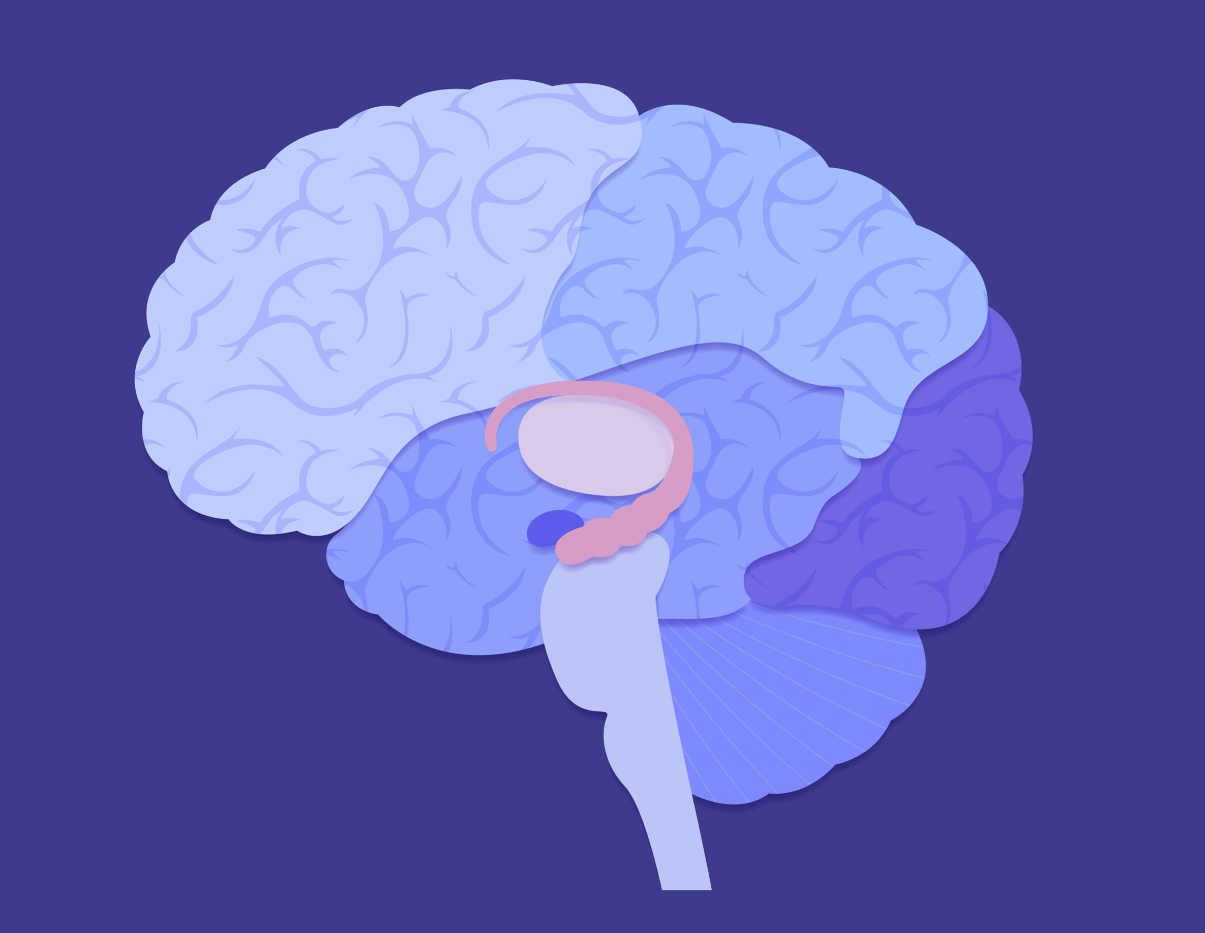
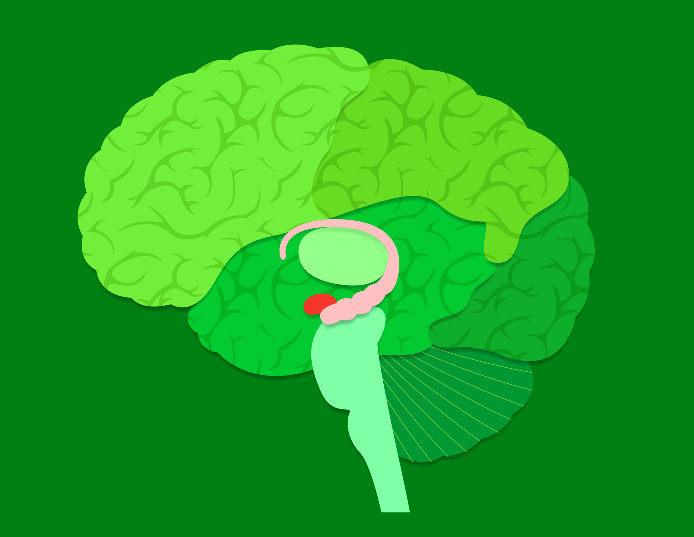
Frontal lobe
Parietal lobe
Occipital lobe
Temporal lobe
Brain stem
Hippocampus
Amygdala
Cerebellum
Thalamus
NextFrontal lobe: Manages planning and organizing and controls your limbs
Temporal lobe: Is where language and recognition memory occurs (contains the hippocampus and amygdala)
Parietal lobe: Manages sensation and perception, spatial awareness, and navigational skills
Occipital lobe: Is the center of visual perception; organizes information to be sent to other brain areas for processing
Keeps you breathing and your heart beating—without you having to think about it (involuntary)
Plays a major role in learning and memory
Is the center for emotions (especially fear) and motivation
Coordinates and regulates muscles
Relays sensory information to the cerebral cortex


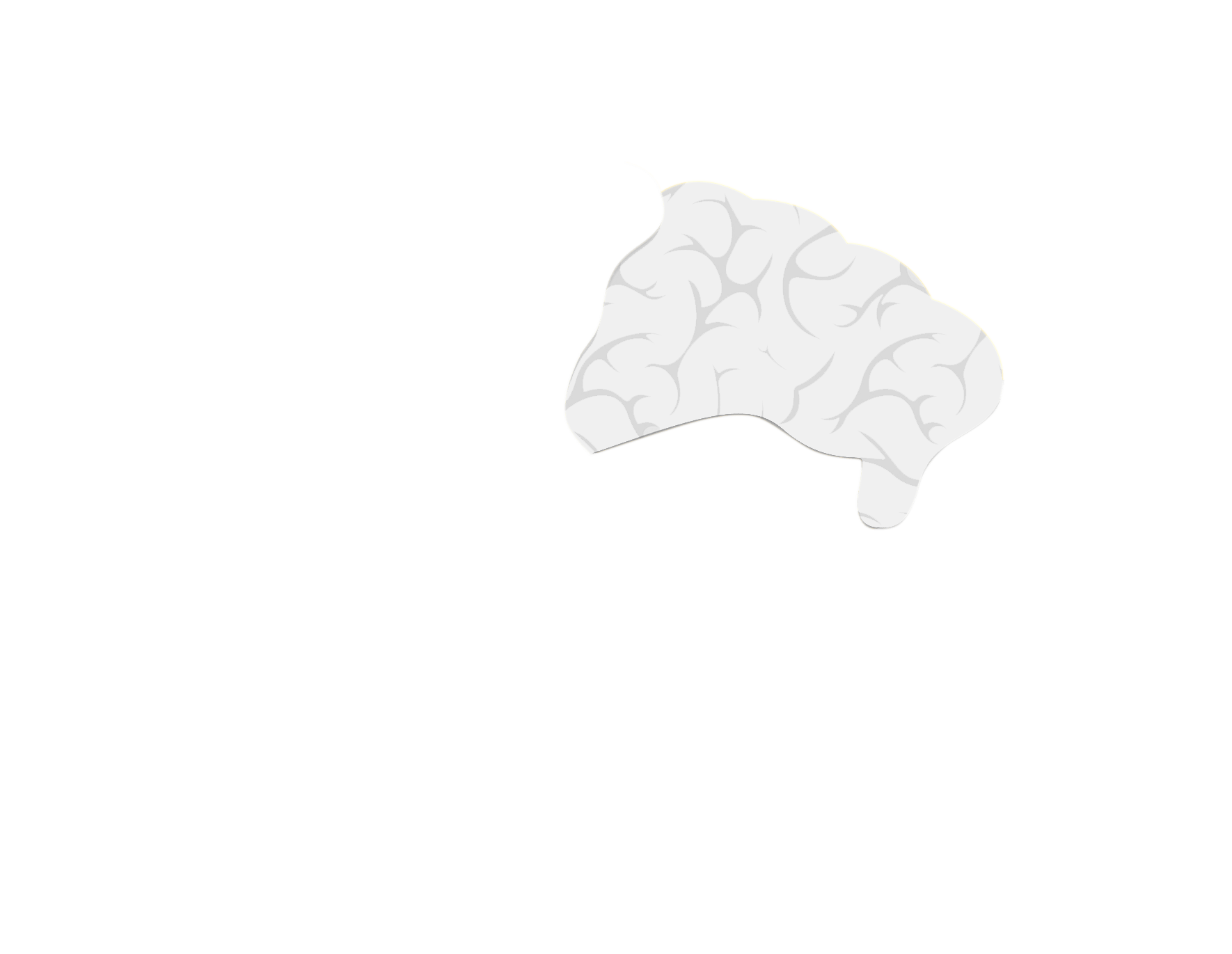
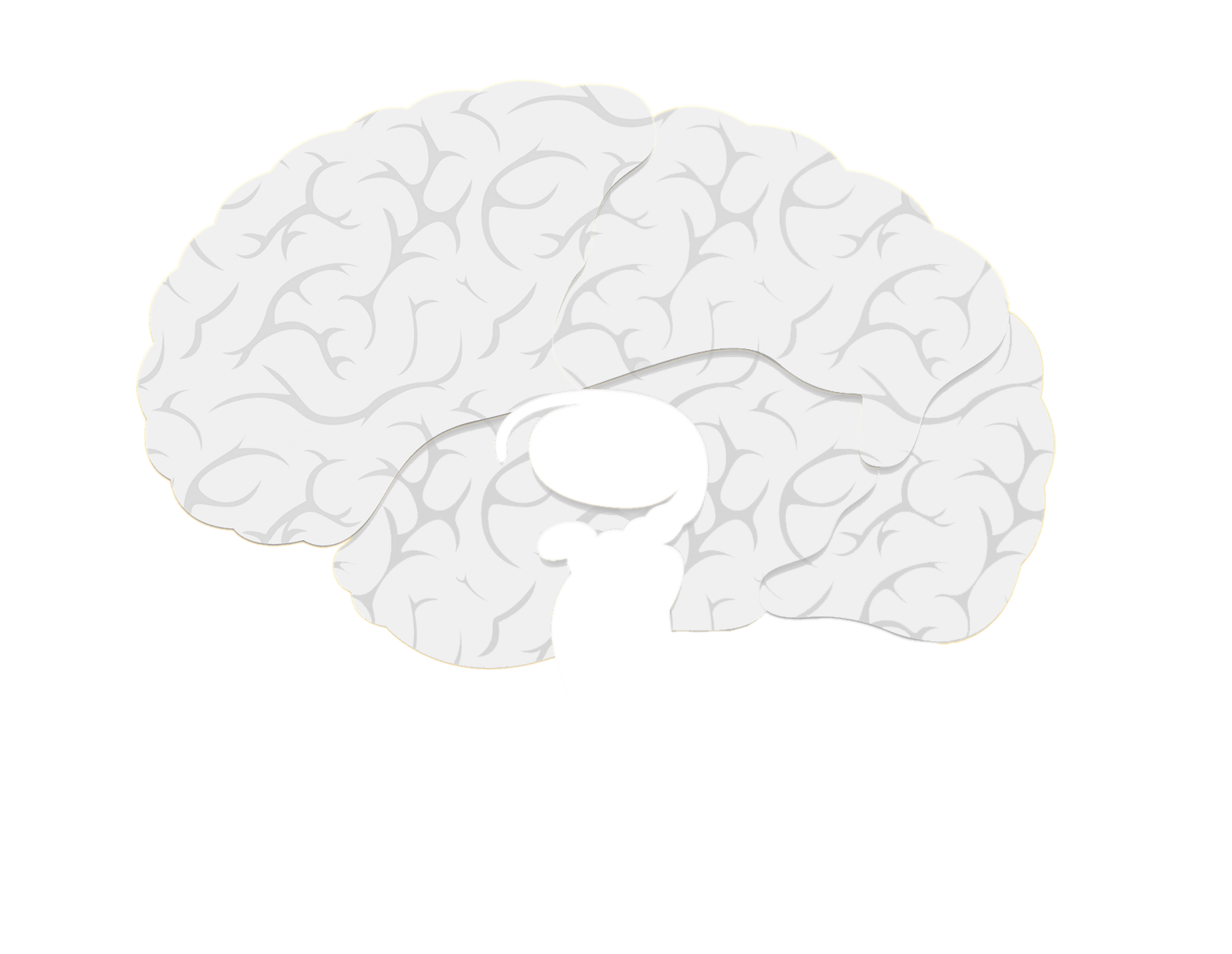
Frontal lobe
Temporal lobe
Hippocampus
Amygdala
Cerebellum


Frontal lobe: The frontal lobe stores working memory (a system for storing multiple bits of knowledge temporarily in the forefront of your mind while performing a task or solving a problem).
Temporal lobe: The temporal lobe regulates recognition memory (recognizing if a person or object is familiar, plus recalling who or what it is).

The hippocampus is involved in memory, especially mental maps of places. It also works on memory consolidation, the slow process by which memories are converted from short-term to long-term memory.
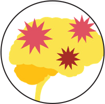
The amygdala encodes emotional learning into memory, such as
fear conditioning (learning to associate something with a
negative event so you can try to avoid it later).
Memory research has shown that the more intense your emotions
are during an event, the greater the chance that you’ll
remember the event.
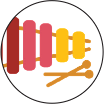
The cerebellum controls balance for walking and standing and other complex motor functions, such as learning to play a musical instrument.


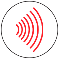
You’re aware of sounds, and you can remember and learn from what you hear.

You can retrieve old memories (names, places, info) to help you navigate life. You can make new memories.

You can feel pain, which may prompt you to take action to ease this negative feeling. You can also remember what you learn about the pain’s cause, so you can try to avoid that pain in the future. During a medical procedure, if an anesthesiologist or dentist gives you a local or regional anesthetic, it will block pain while you remain conscious.

 We know that general anesthetics seem to cause specific
changes in brain rhythms, but there is much that we don’t
know. What is consciousness? What neural activity defines it?
What neural activity defines the lack of consciousness? This is
an important question because we don’t want patients to be
conscious during surgeries. Another important question is: What
is pain? What neural activity can be used to define pain? Is
there a biomarker for pain?
We know that general anesthetics seem to cause specific
changes in brain rhythms, but there is much that we don’t
know. What is consciousness? What neural activity defines it?
What neural activity defines the lack of consciousness? This is
an important question because we don’t want patients to be
conscious during surgeries. Another important question is: What
is pain? What neural activity can be used to define pain? Is
there a biomarker for pain?
I work on understanding
how pain is perceived and regulated by the brain. Specifically,
we are looking at how different regions in the brain are
connected in the presence of a painful stimulus, and
how chronic pain alters the normal processing of a painful
stimulus.
Our lab also works on translating what we learn into treatments
for chronic pain.
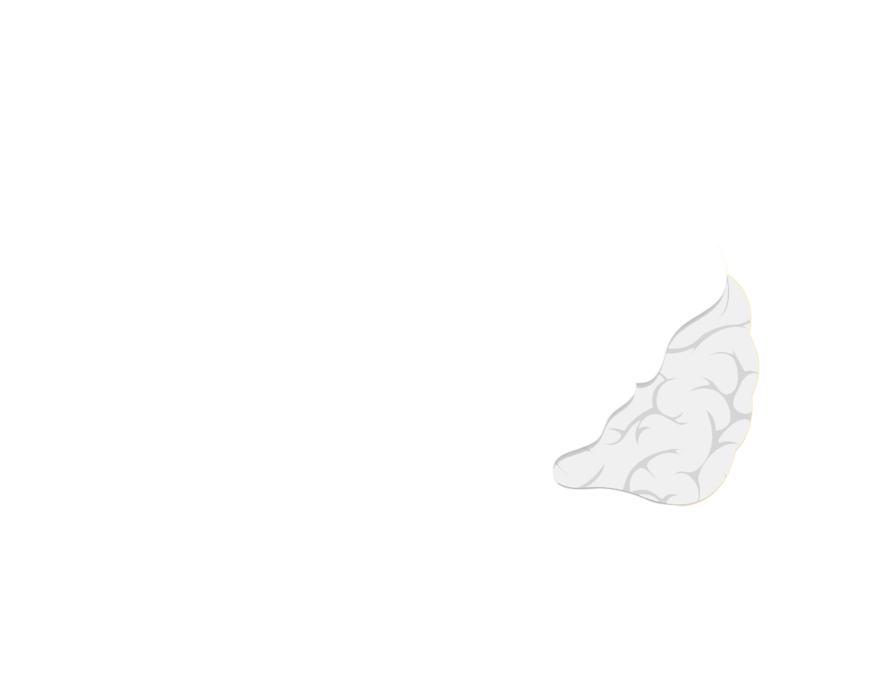
Hippocampus & Cortex
Amygdala
Brain stem
Thalamus

The brain stem helps control the transitions between waking and
sleeping while continuing to maintain involuntary heartbeat and
breathing.
During REM sleep, the brain stem sends signals to relax muscles
needed for limb movements, so that we don’t act out our
dreams.

The hippocampus transfers new memories to the cortex, which replays the memories to consolidate them into long-term memory.
The amygdala becomes increasingly active during REM sleep.
Dreaming’s exact purpose isn’t known, but it may
help you process your emotions.
During most stages of sleep, the thalamus (which relays sensory
information to the cerebral cortex) reduces its activity so you
can tune out your surroundings.
But during REM sleep, the thalamus is active, sending the
cortex images, sounds, and other sensations that fill your
dreams.
As you sleep, you cycle through a few different stages several times per night.
Your brain ignores most sounds, but loud sounds are likely to wake you from sleep, especially sounds that might indicate danger.
Sleep is essential for transferring recent memories into long-term memory. That’s why it’s helpful to get a good night’s sleep after studying for a test.
Pain can disrupt sleep, and poor sleep can make it harder for the body to deal with pain. Like much about the brain, the relationship between pain and sleep isn’t fully understood, and scientists continue to investigate.

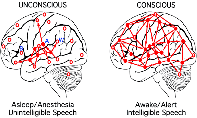
 The brain is a big collection of highly interconnected regions
that are like electrical circuits. These circuits naturally
produce rhythms. The brain uses these rhythms (its brain waves)
to control communications among its many regions. Anesthesia
drugs take over these rhythms and thereby block communication
between brain regions. Loss of communication between brain
regions is one of the ways anesthesia drugs produce
unconsciousness.
The brain is a big collection of highly interconnected regions
that are like electrical circuits. These circuits naturally
produce rhythms. The brain uses these rhythms (its brain waves)
to control communications among its many regions. Anesthesia
drugs take over these rhythms and thereby block communication
between brain regions. Loss of communication between brain
regions is one of the ways anesthesia drugs produce
unconsciousness.
As an anesthesiologist and an
anesthesiology researcher, I want to develop perfectly
controllable anesthesia techniques so that when patients undergo
surgery, they receive only the amount of anesthesia they need,
no more and no less. I want patients to have no side effects
after surgery, such as nausea, uncontrolled pain, or confusion.
The science we uncover through these investigations into how
anesthesia works in the brain may also help people sleep
better and identify new ways to treat depression.
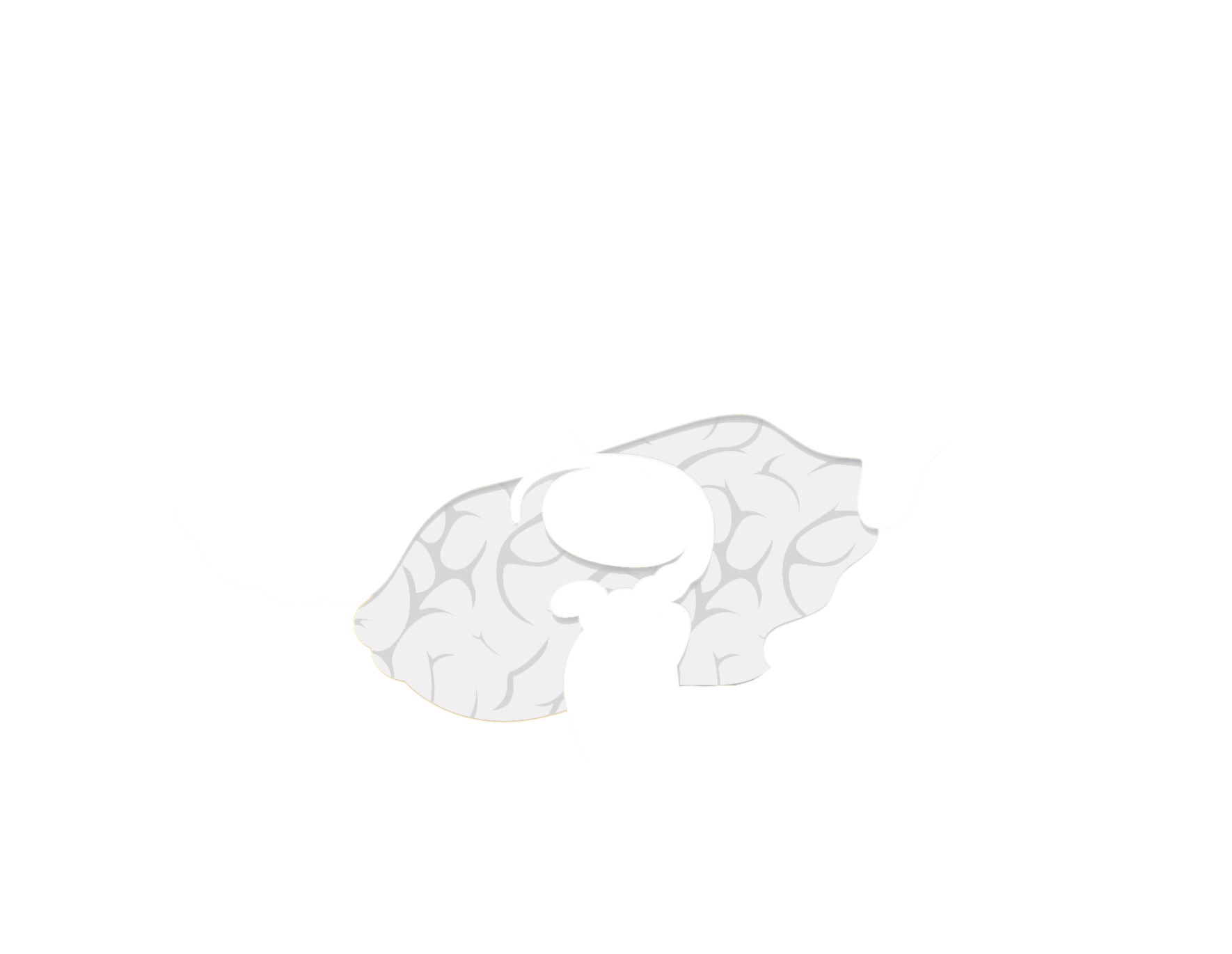
Hippocampus
Amygdala
Brain stem
Thalamus
Cerebral cortex

Under general anesthesia, auditory processing begins in the cortex, but it doesn’t continue, so we lose the meaning and understanding of vocal commands as well as our ability to respond verbally. Learning is suppressed.
Reprinted from British Journal of Anesthesia, Vol 121, S.L. Eagleman, M.B. MacIver, “Can you hear me now? Information processing in primary auditory cortex at loss of consciousness”, Page 526., Copyright Sep 1, 2018, with permission from Elsevier.
General anesthesia causes changes in breathing pattern, heart rate, and body temperature; thus, anesthesiologists carefully monitor surgery patients to keep them safe.
General anesthesia disrupts communication in synaptic networks, disrupting memory formation.

General anesthesia activates neurons in the amygdala that block pain.

General anesthesia interrupts and overpowers the sensory signals that travel back and forth between the thalamus and cortex.
Auditory and sensory processing is interrupted under general anesthesia, so patients can remain unaware (fortunately!) of what’s going on during surgery.
General anesthesia disrupts memory formation, so patients don’t remember what happens while they are unconscious.
General anesthetics block pain and awareness, allowing people to have life-saving surgeries like heart transplants.

 The focus of my research has always been how
volatile anesthetics work. Volatile anesthetics, which
are inhaled in gas form, are a complete general anesthetic, used
every day in operating rooms all over the world. I am trying to
find what these compounds bind to in our cells, on a molecular
level, that can make us completely unaware during surgery.
The focus of my research has always been how
volatile anesthetics work. Volatile anesthetics, which
are inhaled in gas form, are a complete general anesthetic, used
every day in operating rooms all over the world. I am trying to
find what these compounds bind to in our cells, on a molecular
level, that can make us completely unaware during surgery.
To
be able to reversibly produce a state of unconsciousness that is
oblivious to pain is a very amazing feat. How do we have
receptors in us for chemicals that have just recently been
discovered, when our brains are the result of an evolution that
began millions of years ago? And not just us. All across the
animal kingdom, gas anesthetics can work well, even on worms! So
whatever is making this happen, it is an ancient pathway in our
brains that was there hundreds of millions of years ago when
organisms were slowly evolving into different animals.
If we could understand how volatile anesthetics work, it
could really tell us something about how the central nervous
system functions and the nature of consciousness
itself.
I think that would be a contribution to medicine that would have
many, many benefits.