
Mature Synchrotron Resources for Structural Biology (P30) Program
The Mature Synchrotron Resources (MSRs) for Structural Biology (new NOFO forecast PAR-26-029) connect biomedical researchers with state-of-the-art X-ray beamlines, hands-on user support, and expert training to generate and analyze structural and cellular biology data.
MSRs are accessible to all biomedical researchers whose projects are vetted through a peer review process. Explore the Available MSRs below to find available techniques and learn how to request access
What MSRs Provide
Support
MSRs provide assistance with data collection, processing, and analysis. This includes guidance on sample preparation and shipping, as well as support throughout the experimental process.
Access
User access is available on an equal-opportunity, nationwide basis, with supported techniques including fiber diffraction, macromolecular crystallography, small- and wide-angle X-ray scattering, soft X-ray tomography, X-ray spectroscopy and imaging, and X-ray footprinting with mass spectrometry.

Training
Training is available through on-site and remote sessions, workshops, and instructional videos.
Available MSRs
ALS-ENABLE @ ALS/Lawrence Berkeley National Laboratory
Macromolecular Crystallography, Small-Angle X-ray Scattering,
X-ray Footprinting with Mass Spectrometry.
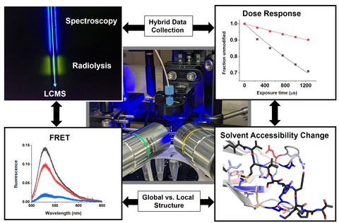
X-Ray Footprinting with Mass Spectrometry
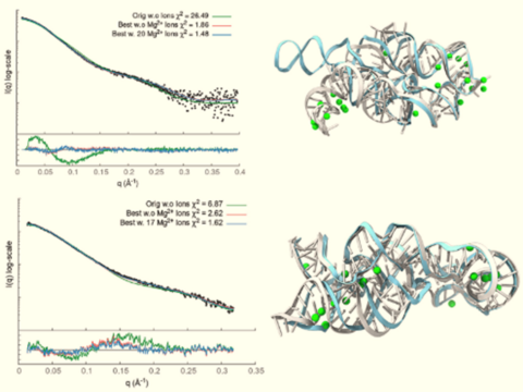
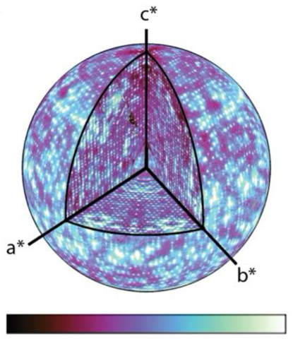
SCOPER program combines deep learning with small angle X-ray scattering (SAXS) experimental data for more accurate structure predictions
Images courtesy of Dr. Paul Adams
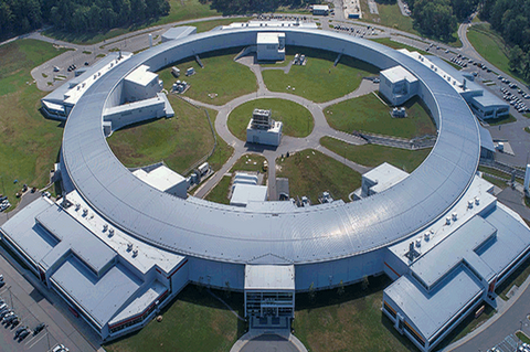
BioCAT @ APS/Argonne National Laboratory
Small-Angle X-ray Scattering, Fiber Diffraction
An ensemble of conformational states modeled based on SAXS data

X-ray diffraction of muscle fiber
Images courtesy of Dr. Thomas Irving
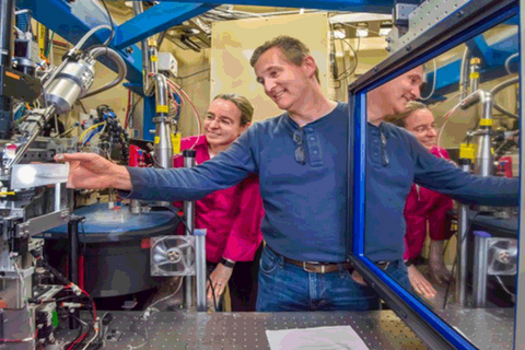
CBMS @ NSLS-II/Brookhaven National Laboratory
Macromolecular Crystallography, Small- and Wide-Angle X-ray
Scattering
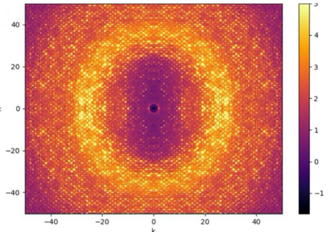
Automated SAXS data collection
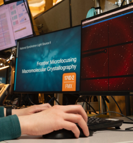
Frontier Microfocusing Macromolecular Crystallography
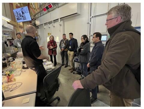
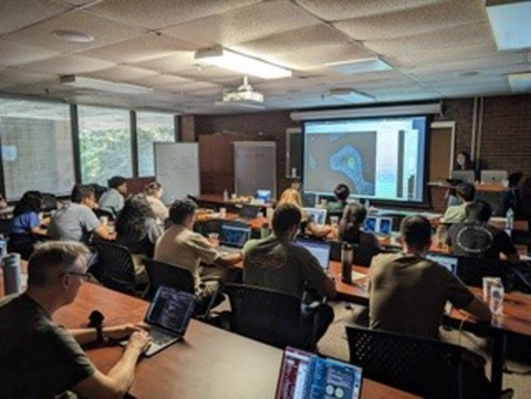
A science workshop.
Images courtesy of Dr. Sean McSweeney
GM/CA @ APS/Argonne National Laboratory
Macromolecular Crystallography
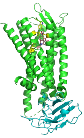
Crystal structure of the κ-opioid receptor bound to nalfurafine (salmon) and stabilized by a nanobody (cyan)
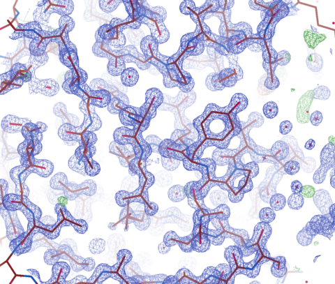
2Fo-Fc Electron density for trypsin (1.0-Å resolution)
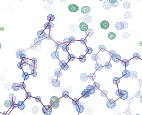
2Fo-Fc Electron density for crambin (0.7-Å resolution)
Images courtesy of Dr. Robert Fischetti and Dr. Janet Smith
MacCHESS @ CHESS/Cornell University
Macromolecular Crystallography, Small-Angle X-ray Scattering
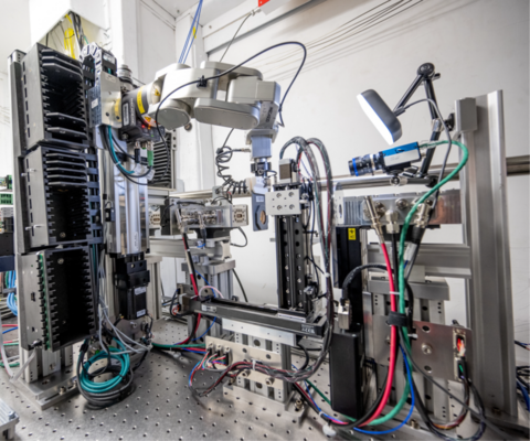
The diffuse scattering setup at FlexX.
A scattering map from the Ando group at Cornell.
A central section of a 3D scattering map from the Thorne group at Cornell.
Images courtesy of Dr. Richard Cerione
NCXT @ ALS/Lawrence Berkeley National Laboratory
Soft X-ray Tomography
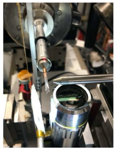
NE-CAT @ APS/Argonne National Laboratory
Macromolecular Crystallography
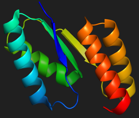
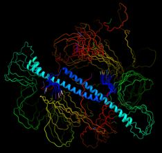
The structural model of a de novo designed protein that neutralizes lethal snake venom toxins
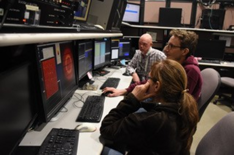
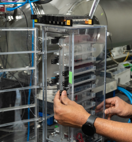
Scientists at the beamline
A NE-CAT workshop
Images courtesy of Dr. Frank Murphy
SMB @ SSRL/SLAC National Accelerator Laboratory
Macromolecular Crystallography, Small-Angle X-ray Scattering,
X-ray Spectroscopy, and Imaging
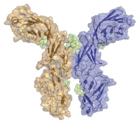
Surface representation of the dimeric LAG-3 protein
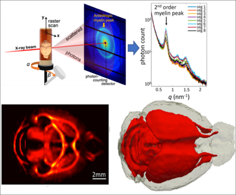
Surface representation of the dimeric LAG-3 protein (left). The X-ray crystal structure was solved using data collected after screening more than 3,000 crystals. The small-angle X-ray scattering (SAXS) tensor tomography image of myelin levels in nervous tissue
Automated and high-throughput X-ray absorption spectroscopy (XAS) for remote access use
Images courtesy of Dr. Keith Hodgson
For Current MSR Awardees
Guidance on preparing and submitting annual Research Performance Progress Reports (RPPRs) is available below.
RPPR Guidance for Funded Resources Word Template [DOCX]
Program Contacts
For more information or assistance contact the program staff: