Image and Video Gallery
This is a searchable collection of scientific photos, illustrations, and videos. The images and videos in this gallery are licensed under Creative Commons Attribution Non-Commercial ShareAlike 3.0. This license lets you remix, tweak, and build upon this work non-commercially, as long as you credit and license your new creations under identical terms.
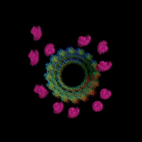
5752: Genetically identical mycobacteria respond differently to antibiotic 2
In this video, genetically identical mycobacteria are growing in a miniature growth chamber called a microfluidic chamber. Using live imaging, the researchers found that individual mycobacteria will respond differently to the antibiotic, depending on the growth stage and other timing factors. The researchers used genetic tagging with green fluorescent protein to distinguish cells that can resist rifampicin and those that cannot. With this gene tag, cells tolerant of the antibiotic light up in green and those that are susceptible in violet, enabling the team to monitor the cells' responses in real time.
To learn more about how the researchers studied antibiotic resistance in mycobacteria, see this news release from Tufts University. Related to image 5751.
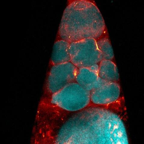
6753: Fruit fly nurse cells during egg development
2636: Computer model of cell membrane
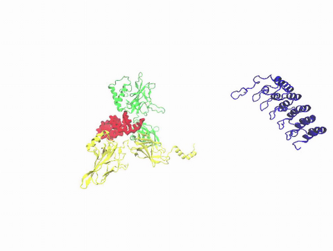
6571: Actin filaments bundled around the dynamin helical polymer
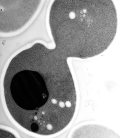
5770: EM of yeast cell division
This image shows an electron microscopy (EM) thin section taken at 10,000x magnification of a dividing yeast cell over-expressing the protein ubiquitin, which is involved in protein degradation and recycling. The picture features mother and daughter endosome accumulations (small organelles with internal vesicles), a darkly stained vacuole and a dividing nucleus in close contact with a cadre of lipid droplets (unstained spherical bodies). Other dynamic events are also visible, such as spindle microtubules in the nucleus and endocytic pits at the plasma membrane.
These extensive details were revealed thanks to a preservation method involving high-pressure freezing, freeze-substitution and Lowicryl HM20 embedding.
3427: Antitoxin GhoS (Illustration 1)
6801: “Two-faced” Janus particle activating a macrophage
Related to video 6800.
3729: A molecular switch strips transcription factor from DNA
3488: Shiga toxin being sorted inside a cell
6539: Pathways: What is Basic Science?
2406: Hen egg lysozyme (2)
2759: Cross section of a Drosophila melanogaster pupa lacking Draper
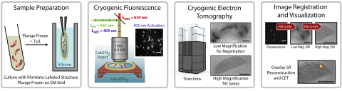
6568: Correlative imaging by annotation with single molecules (CIASM) process
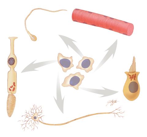
1294: Stem cell differentiation
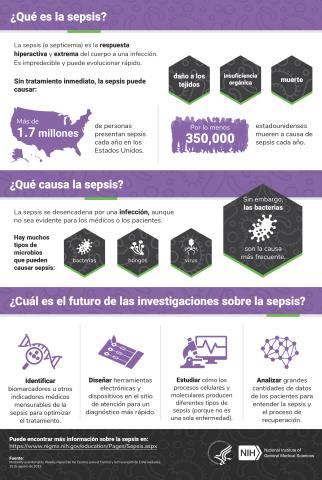
6551: ¿Qué es la sepsis? (Sepsis Infographic)
Vea 6536 para la versión en inglés de esta infografía.
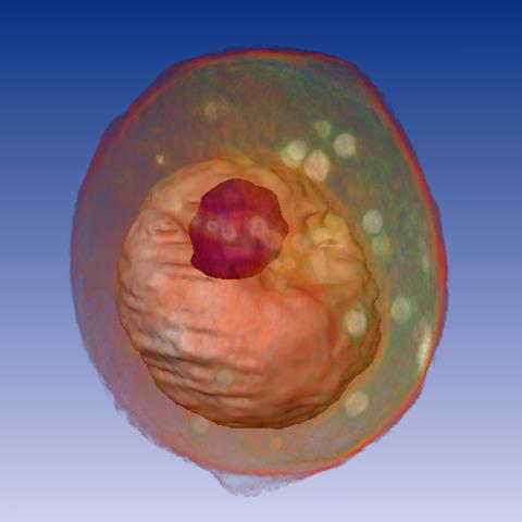
1092: Yeast cell
6777: Human endoplasmic reticulum membrane protein complex
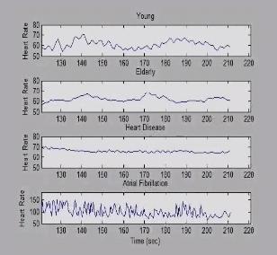
3596: Heart rates time series image
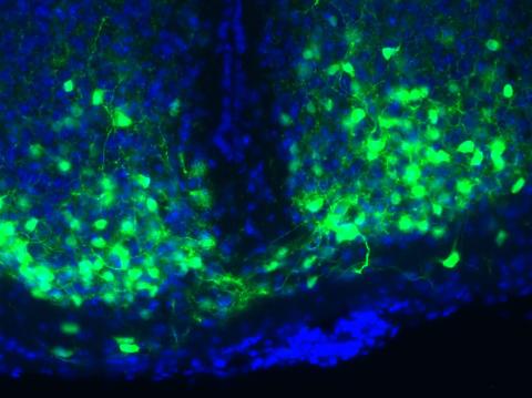
3547: Master clock of the mouse brain
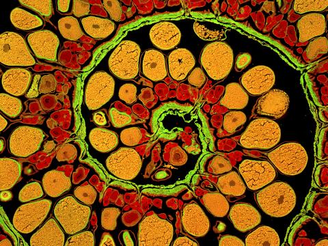
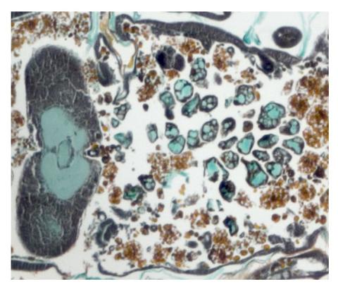
3620: Anglerfish ovary cross-section
This image was part of the Life: Magnified exhibit that ran from June 3, 2014, to January 21, 2015, at Dulles International Airport.
5762: Panorama view of golden mitochondria
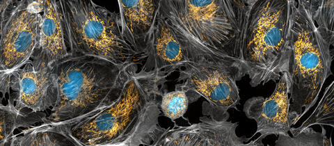
6891: Microtubules in African green monkey cells
Related to images 6889, 6890, and 6892.
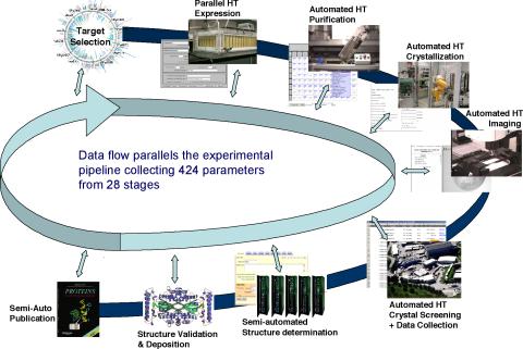
2364: High-throughput protein structure determination pipeline
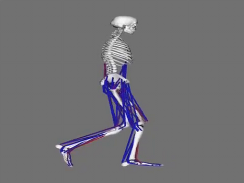
6598: Simulation of leg muscles moving
3764: Movie of the 19S proteasome subunit processing a protein substrate
3306: Planarian stem cell colony
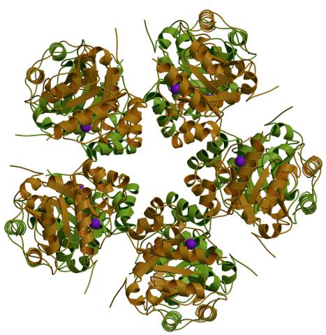
6762: CCP enzyme
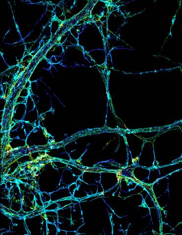
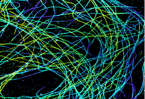
3678: STORM image of axonal cytoskeleton
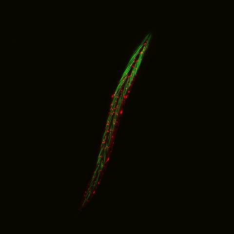
6581: Fluorescent C. elegans showing muscle and ribosomal protein
View group of roundworms here 6582.
View closeup of roundworms here 6583.
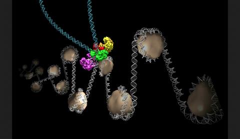
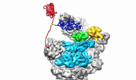
6346: Intasome
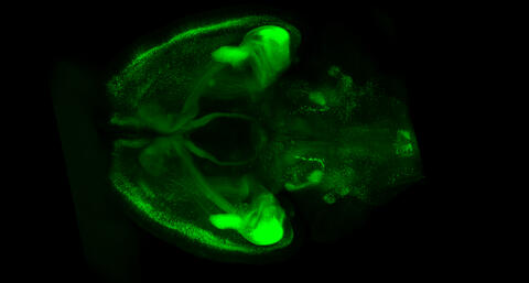
6929: Mouse brain 1
This image was captured using a light sheet microscope.
Related to image 6930 and video 6931.
2435: Developing fruit fly nerve cord
2331: Statistical cartography
2605: Induced stem cells from adult skin 03
6588: Cell-like compartments emerging from scrambled frog eggs 2
For more photos of cell-like compartments from frog eggs view: 6584, 6585, 6586, 6591, 6592, and 6593.
For videos of cell-like compartments from frog eggs view: 6587, 6589, and 6590.
2749: Cytoscape network wiring diagram 2
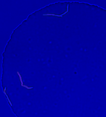
5777: Microsporidia in roundworm 1
2502: Focal adhesions
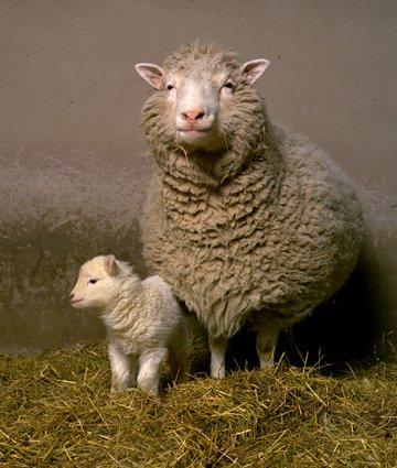
2690: Dolly the sheep
6589: Cell-like compartments emerging from scrambled frog eggs 3
For more photos of cell-like compartments from frog eggs view: 6584, 6585, 6586, 6591, 6592, and 6593.
For videos of cell-like compartments from frog eggs view: 6587, 6588, and 6590.
2490: Cascade reaction promoted by water
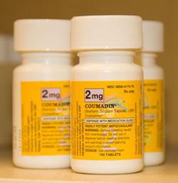
2579: Bottles of warfarin
2399: Bence Jones protein MLE
6587: Cell-like compartments emerging from scrambled frog eggs
For more photos of cell-like compartments from frog eggs view: 6584, 6585, 6586, 6591, 6592, and 6593.
For videos of cell-like compartments from frog eggs view: 6588, 6589, and 6590.
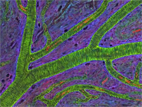
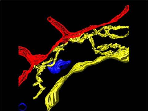
3400: Small blood vessels in a mouse retina
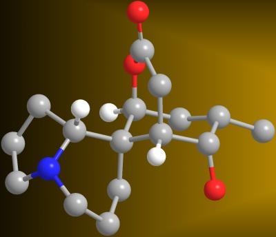
2687: Serratezomine A
2418: Genetic imprinting in Arabidopsis
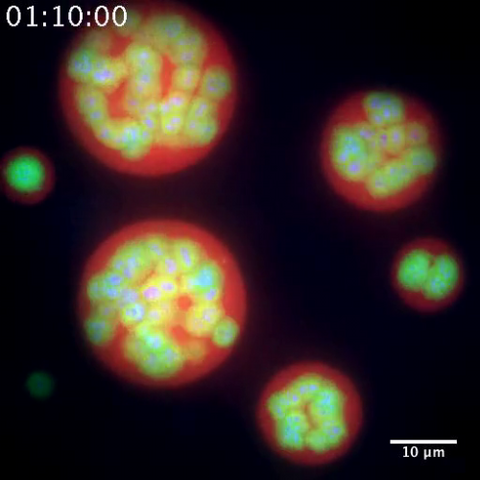
3791: Nucleolus subcompartments spontaneously self-assemble 2
However, how the nucleolus grows and maintains its structure has puzzled scientists for some time. It turns out that even though it looks like a simple liquid blob, it's rather well-organized, consisting of three distinct layers: the fibrillar center, where the RNA polymerase is active; the dense fibrillar component, which is enriched in the protein fibrillarin; and the granular component, which contains a protein called nucleophosmin. Researchers have now discovered that this multilayer structure of the nucleolus arises from differences in how the proteins in each compartment mix with water and with each other. These differences let the proteins readily separate from each other into the three nucleolus compartments.
This video of nucleoli in the eggs of a commonly used lab animal, the frog Xenopus laevis, shows how each of the compartments (the granular component is shown in red, the fibrillarin in yellow-green, and the fibrillar center in blue) spontaneously fuse with each other on encounter without mixing with the other compartments.
For more details on this research, see this press release from Princeton. Related to video 3789, image 3792 and image 3793.