Switch to Gallery View
Image and Video Gallery
This is a searchable collection of scientific photos, illustrations, and videos. The images and videos in this gallery are licensed under Creative Commons Attribution Non-Commercial ShareAlike 3.0. This license lets you remix, tweak, and build upon this work non-commercially, as long as you credit and license your new creations under identical terms.

The Structure of Cilia’s Doublet Microtubules
6549
Cilia (cilium in singular) are complex molecular machines found on many of our cells. Brown Lab, Harvard Medical School and Veronica Falconieri Hays View Media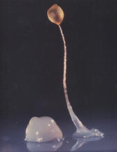
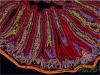
Dicty fruit
2684
Dictyostelium discoideum is a microscopic amoeba. A group of 100,000 form a mound as big as a grain of sand. Featured in The New Genetics. View Media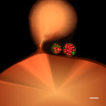
Multivesicular bodies containing intralumenal vesicles assemble at the vacuole 1
5769
Collecting and transporting cellular waste and sorting it into recylable and nonrecylable pieces is a complex business in the cell. Matthew West and Greg Odorizzi, University of Colorado View Media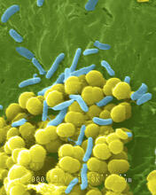
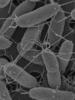
Bacteria shapes
1158
A colorized scanning electron micrograph of bacteria. Scanning electron microscopes allow scientists to see the three-dimensional surface of their samples. Tina Weatherby Carvalho, University of Hawaii at Manoa View Media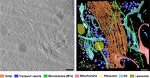
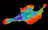
Cryo-ET cross-section of the Golgi apparatus
6606
On the left, a cross-section slice of a rat pancreas cell captured using cryo-electron tomography (cryo-ET). On the right, a 3D, color-coded version of the image highlighting cell structures. Xianjun Zhang, University of Southern California. View Media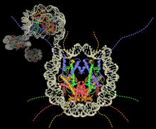
Nucleosome
2741
Like a strand of white pearls, DNA wraps around an assembly of special proteins called histones (colored) to form the nucleosome, a structure responsible for regulating genes and condensing DNA strand Karolin Luger, Colorado State University View Media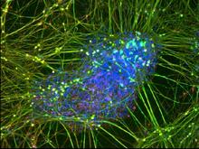
Peripheral nerve cell derived from ES cells
3264
A peripheral nerve cell made from human embryonic stem cell-derived neural crest stem cells. Stephen Dalton, University of Georgia View Media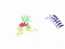
A molecular switch strips transcription factor from DNA
3729
In this video, Rice University scientists used molecular modeling with a mathematical algorithm called AWSEM (for associative memory, water-mediated, structure and energy model) and structural data to Davit Potoyan and Peter Wolynes View Media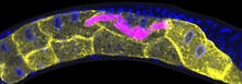
Microsporidia in roundworm 1
5777
Many disease-causing microbes manipulate their host’s metabolism and cells for their own ends. Keir Balla and Emily Troemel, University of California San Diego View Media
Nerve cell
1338
Nerve cells have long, invisibly thin fibers that carry electrical impulses throughout the body. Some of these fibers extend about 3 feet from the spinal cord to the toes. Judith Stoffer View Media
Introduction to Genome Editing Using CRISPR/Cas9
5815
Genome editing using CRISPR/Cas9 is a rapidly expanding field of scientific research with emerging applications in disease treatment, medical therapeutics and bioenergy, just to name a few. Janet Iwasa View Media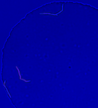
Genetically identical mycobacteria respond differently to antibiotic 2
5752
Antibiotic resistance in microbes is a serious health concern. So researchers have turned their attention to how bacteria undo the action of some antibiotics. Bree Aldridge, Tufts University View Media
Brains of sleep-deprived and well-rested fruit flies
3490
On top, the brain of a sleep-deprived fly glows orange because of Bruchpilot, a communication protein between brain cells. These bright orange brain areas are associated with learning. Chiara Cirelli, University of Wisconsin-Madison View Media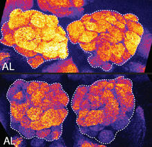
Sleep and the fly brain
2596
In the top snapshots, the brain of a sleep-deprived fruit fly glows orange, marking high concentrations of a synaptic protein called Bruchpilot (BRP) involved in communication between neurons. Chiara Cirelli, University of Wisconsin-Madison View Media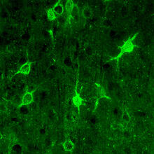
Confocal microscopy of perineuronal nets in the brain 1
3741
The photo shows a confocal microscopy image of perineuronal nets (PNNs), which are specialized extracellular matrix (ECM) structures in the brain. Tom Deerinck, National Center for Microscopy and Imaging Research (NCMIR) View Media
Neural tube development
2328
Proteins in the neural tissues of this zebrafish embryo direct cells to line up and form the neural tube, which will become the spinal cord and brain. Alexander Schier, Harvard University View Media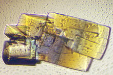
RNase A (2)
2402
A crystal of RNase A protein created for X-ray crystallography, which can reveal detailed, three-dimensional protein structures. Alex McPherson, University of California, Irvine View Media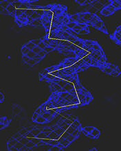
Section of an electron density map
2354
Electron density maps such as this one are generated from the diffraction patterns of X-rays passing through protein crystals. The Southeast Collaboratory for Structural Genomics View Media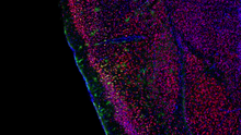
Video of Calling Cards in a mouse brain
6781
The green spots in this mouse brain are cells labeled with Calling Cards, a technology that records molecular events in brain cells as they mature. NIH Director's Blog View Media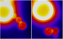
Dynamin Fission
3448
Time lapse series shows short dynamin assemblies (not visible) constricting a lipid tube to make a "beads on a string" appearance, then cutting off one of the beads i.e., catalyzing membrane fission). Ramachandran, Pucadyil et al. , The Scripps Research Institute View Media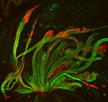
Fruit fly spermatids
3590
Developing spermatids (precursors of mature sperm cells) begin as small, round cells and mature into long-tailed, tadpole-shaped ones. Lacramioara Fabian, The Hospital for Sick Children, Toronto, Canada View Media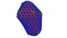
Atomic-level structure of the HIV capsid
6601
This animation shows atoms of the HIV capsid, the shell that encloses the virus's genetic material. Juan R. Perilla and the Theoretical and Computational Biophysics Group, University of Illinois at Urbana-Champaign View Media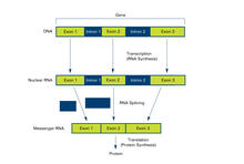
Introns (with labels)
2551
Genes are often interrupted by stretches of DNA (introns, blue) that do not contain instructions for making a protein. Crabtree + Company View Media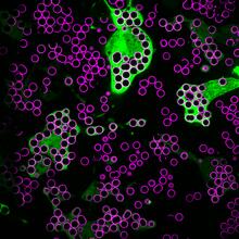
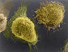
Hungry, hungry macrophages
7009
Macrophages (green) are the professional eaters of our immune system. Meghan Morrissey, University of California, Santa Barbara. View Media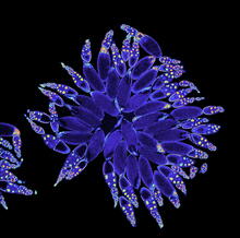
Fruit fly ovary
3607
A fruit fly ovary, shown here, contains as many as 20 eggs. Fruit flies are not merely tiny insects that buzz around overripe fruit—they are a venerable scientific tool. Denise Montell, Johns Hopkins University and University of California, Santa Barbara View Media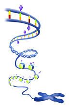
Epigenetic code
2562
The "epigenetic code" controls gene activity with chemical tags that mark DNA (purple diamonds) and the "tails" of histone proteins (purple triangles). Crabtree + Company View Media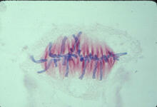
Lily mitosis 07
1017
A light microscope image of a cell from the endosperm of an African globe lily (Scadoxus katherinae). This is one frame of a time-lapse sequence that shows cell division in action. Andrew S. Bajer, University of Oregon, Eugene View Media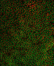
Retinal pigment epithelium derived from human ES cells 02
3287
This image shows a layer of retinal pigment epithelium cells derived from human embryonic stem cells, highlighting the nuclei (red) and cell surfaces (green). David Buckholz and Sherry Hikita, University of California, Santa Barbara, via CIRM View Media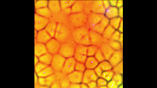
Cell-like compartments emerging from scrambled frog eggs
6587
Cell-like compartments spontaneously emerge from scrambled frog eggs, with nuclei (blue) from frog sperm. Endoplasmic reticulum (red) and microtubules (green) are also visible. Xianrui Cheng, Stanford University School of Medicine. View Media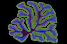
Fluorescent microscopy of kidney tissue
3723
Serum albumin (SA) is the most abundant protein in the blood plasma of mammals. SA has a characteristic heart-shape structure and is a highly versatile protein. Tom Deerinck , National Center for Microscopy and Imaging Research View Media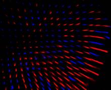
Fruit fly retina 02
2434
Section of a fruit fly retina showing the light-sensing molecules rhodopsin-5 (blue) and rhodopsin-6 (red). Hermann Steller, Rockefeller University View Media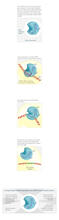
CRISPR illustration
3719
This illustration shows, in simplified terms, how the CRISPR-Cas9 system can be used as a gene-editing tool. National Institute of General Medical Sciences. View Media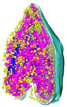
Soft X-ray tomography of a pancreatic beta cell
6605
A color-coded, 3D model of a rat pancreatic β cell. This type of cell produces insulin, a hormone that helps regulate blood sugar. Carolyn Larabell, University of California, San Francisco. View Media
Yeast art depicting the New York City skyline
6521
This skyline of New York City was created by “printing” nanodroplets containing yeast (Saccharomyces cerevisiae) onto a large plate. Each dot is a separate yeast colony. Michael Shen, Ph.D., Jasmine Temple, Leslie Mitchell, Ph.D., and Jef Boeke, Ph.D., New York University School of Medicine; and Nick Phillips, James Chuang, Ph.D., and Jiarui Wang, Johns Hopkins University. View Media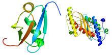
Ubiquitin-fold modifier 1 from C. elegans
2388
Solution NMR structure of protein target WR41 (left) from C. elegans. Northeast Structural Genomics Consortium View Media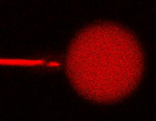
Bee venom toxin destroying a cell
3583
This video condenses 6.5 minutes into less than a minute to show how the toxin in bee venom, called melittin, destroys an animal or bacterial cell. Huey Huang, Rice University View Media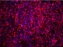
Neurons from human ES cells
3284
These neural precursor cells were derived from human embryonic stem cells. The neural cell bodies are stained red, and the nuclei are blue. Xianmin Zeng lab, Buck Institute for Age Research, via CIRM View Media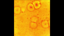
Cell-like compartments emerging from scrambled frog eggs 2
6588
Cell-like compartments spontaneously emerge from scrambled frog eggs, with nuclei (blue) from frog sperm. Endoplasmic reticulum (red) and microtubules (green) are also visible. Xianrui Cheng, Stanford University School of Medicine. View Media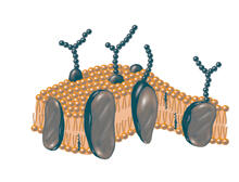
Plasma membrane
2523
The plasma membrane is a cell's protective barrier. See image 2524 for a labeled version of this illustration. Featured in The Chemistry of Health. Crabtree + Company View Media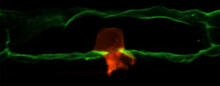
Anchor cell in basement membrane
2707
An anchor cell (red) pushes through the basement membrane (green) that surrounds it. Elliott Hagedorn, Duke University. View Media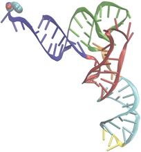
Phenylalanine tRNA molecule
3406
Phenylalanine tRNA showing the anticodon (yellow) and the amino acid, phenylalanine (blue and red spheres). Patrick O'Donoghue and Dieter Soll, Yale University View Media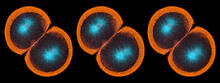
Sea urchin embryo 06
1052
Stereo triplet of a sea urchin embryo stained to reveal actin filaments (orange) and microtubules (blue). George von Dassow, University of Washington View Media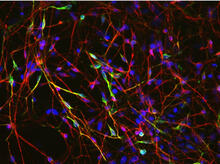
Neurons from human ES cells 02
3285
These neurons were derived from human embryonic stem cells. The neural cell bodies with axonal projections are visible in red, and the nuclei in blue. Xianmin Zeng lab, Buck Institute for Age Research, via CIRM View Media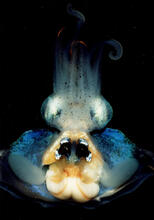
An adult Hawaiian bobtail squid
7013
An adult female Hawaiian bobtail squid, Euprymna scolopes, with its mantle cavity exposed from the underside. Margaret J. McFall-Ngai, Carnegie Institution for Science/California Institute of Technology, and Edward G. Ruby, California Institute of Technology. View Media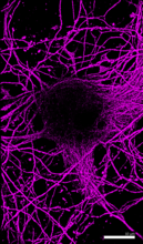
Microtubules in hippocampal neurons
6890
Microtubules (magenta) in neurons of the hippocampus, a part of the brain involved in learning and memory. Microtubules are strong, hollow fibers that provide structural support to cells. Melike Lakadamyali, Perelman School of Medicine at the University of Pennsylvania. View Media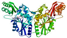
PanC from M. tuberculosis
2383
Model of an enzyme, PanC, that is involved in the last step of vitamin B5 biosynthesis in Mycobacterium tuberculosis. PanC is essential for the growth of M. Mycobacterium Tuberculosis Center, PSI View Media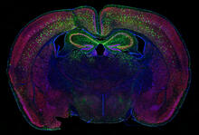
Calling Cards in a mouse brain
6780
The green spots in this mouse brain are cells labeled with Calling Cards, a technology that records molecular events in brain cells as they mature. Allen Yen, Lab of Joseph Dougherty, Washington University School of Medicine in St. Louis. View Media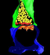
3-D Architecture of a Synapse
5885
This image shows the structure of a synapse, or junction between two nerve cells in three dimensions. From the brain of a mouse. Anton Maximov, The Scripps Research Institute, La Jolla, CA View Media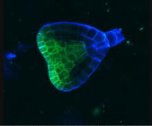
Early development in Arabidopsis
2733
Early on, this Arabidopsis plant embryo picks sides: While one end will form the shoot, the other will take root underground. Zachery R. Smith, Jeff Long lab at the Salk Institute for Biological Studies View Media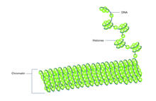
Histones in chromatin (with labels)
2561
Histone proteins loop together with double-stranded DNA to form a structure that resembles beads on a string. Crabtree + Company View Media