Switch to Gallery View
Image and Video Gallery
This is a searchable collection of scientific photos, illustrations, and videos. The images and videos in this gallery are licensed under Creative Commons Attribution Non-Commercial ShareAlike 3.0. This license lets you remix, tweak, and build upon this work non-commercially, as long as you credit and license your new creations under identical terms.
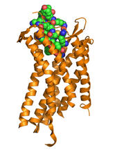
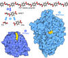
Chemokine CXCR4 receptor
3365
The receptor is shown bound to a small molecule peptide called CVX15. Raymond Stevens, The Scripps Research Institute View Media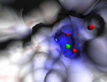
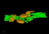
Active site of sulfite oxidase
2746
Sulfite oxidase is an enzyme that is essential for normal neurological development in children. John Enemark, University of Arizona View Media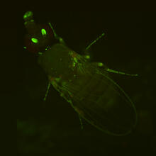
Fly by night
2417
This fruit fly expresses green fluorescent protein (GFP) in the same pattern as the period gene, a gene that regulates circadian rhythm and is expressed in all sensory neurons on the surface of the fl Jay Hirsh, University of Virginia View Media
Telomerase illustration
1335
Reactivating telomerase in our cells does not appear to be a good way to extend the human lifespan. Cancer cells reactivate telomerase. Judith Stoffer View Media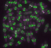
Yeast cells entering mitosis
6791
Yeast cells entering mitosis, also known as cell division. The green and magenta dots are two proteins that play important roles in mitosis. They show where the cells will split. Alaina Willet, Kathy Gould’s lab, Vanderbilt University. View Media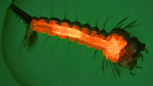
Culex quinquefasciatus mosquito larva
6769
A mosquito larva with genes edited by CRISPR. The red-orange glow is a fluorescent protein used to track the edits. Valentino Gantz, University of California, San Diego. View Media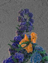
Flu virus proteins during self-replication
3434
Influenza (flu) virus proteins in the act of self-replication. Viral nucleoprotein (blue) encapsidates [encapsulates] the RNA genome (green). Scripps Research Institute in La Jolla, CA View Media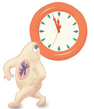
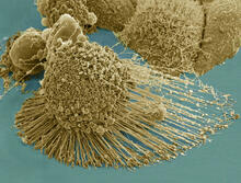
HeLa cells
3519
Scanning electron micrograph of an apoptotic HeLa cell. Zeiss Merlin HR-SEM. National Center for Microscopy and Imaging Research View Media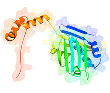
Hsp33 figure 1
3354
Featured in the March 15, 2012 issue of Biomedical Beat. Related to Hsp33 Figure 2, image 3355. Ursula Jakob and Dana Reichmann, University of Michigan View Media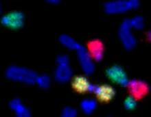
Painted chromosomes
2764
Like a paint-by-numbers picture, painted probes tint individual human chromosomes by targeting specific DNA sequences. Beth A. Sullivan, Duke University View Media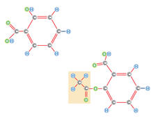
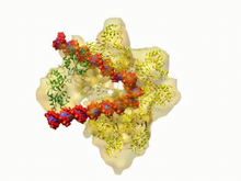
DNA replication origin recognition complex (ORC)
3307
A study published in March 2012 used cryo-electron microscopy to determine the structure of the DNA replication origin recognition complex (ORC), a semi-circular, protein complex (yellow) that recogni Huilin Li, Brookhaven National Laboratory View Media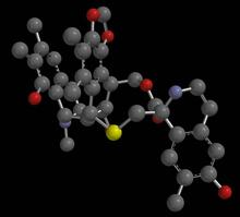
Anti-tumor drug ecteinascidin 743 (ET-743), structure without hydrogens 03
2796
Ecteinascidin 743 (ET-743, brand name Yondelis), was discovered and isolated from a sea squirt, Ecteinascidia turbinata, by NIGMS grantee Kenneth Rinehart at the University of Illinois. Timothy Jamison, Massachusetts Institute of Technology View Media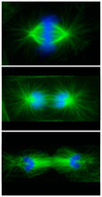
Cell division phases in Xenopus frog cells
3442
These images show three stages of cell division in Xenopus XL177 cells, which are derived from tadpole epithelial cells. They are (from top): metaphase, anaphase and telophase. Claire Walczak, who took them while working as a postdoc in the laboratory of Timothy Mitchison View Media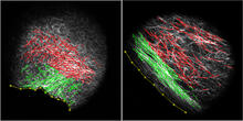
DDR2 Receptors Attach to Collagen in Breast Tumor
3478
On the left, the boundary of a breast tumor (yellow) attaches to collagen fibers that are closest to it (green) using DDR2. On the right, a tumor without DDR2 remains disconnected from the collagen. Callie Corsa and Suzanne Ponik, Washington University School of Medicine in St. Louis View Media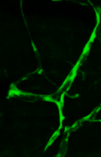
Disrupted and restored vasculature development in frog embryos
3405
Disassembly of vasculature and reassembly after addition and then washout of 250 µM TBZ in kdr:GFP frogs. Hye Ji Cha, University of Texas at Austin View Media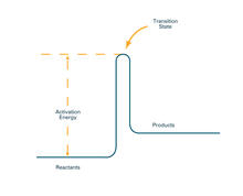
Activation energy (with labels)
2526
To become products, reactants must overcome an energy hill. See image 2525 for an unlabeled version of this illustration. Crabtree + Company View Media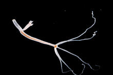
Hydra 06
2442
Hydra magnipapillata is an invertebrate animal used as a model organism to study developmental questions, for example the formation of the body axis. Hiroshi Shimizu, National Institute of Genetics in Mishima, Japan View Media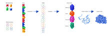
From DNA to Protein (labeled)
2510
The genetic code in DNA is transcribed into RNA, which is translated into proteins with specific sequences. Crabtree + Company View Media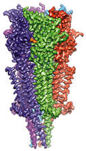
Full-length serotonin receptor (ion channel)
6579
A 3D reconstruction, created using cryo-electron microscopy, of an ion channel known as the full-length serotonin receptor in complex with the antinausea drug granisetron (orange). Sudha Chakrapani, Case Western Reserve University School of Medicine. View Media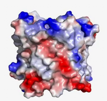
VDAC video 03
2572
This video shows the structure of the pore-forming protein VDAC-1 from humans. Gerhard Wagner, Harvard Medical School View Media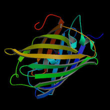
Fruitful dyes
2317
These colorful, computer-generated ribbons show the backbone of a molecule that glows a fluorescent red. Roger Y. Tsien, University of California, San Diego View Media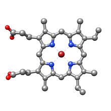
Structure of heme, top view
3539
Molecular model of the struture of heme. Heme is a small, flat molecule with an iron ion (dark red) at its center. Rachel Kramer Green, RCSB Protein Data Bank View Media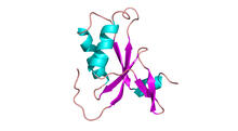
Antitoxin GhoS (Illustration 1)
3427
Structure of the bacterial antitoxin protein GhoS. GhoS inhibits the production of a bacterial toxin, GhoT, which can contribute to antibiotic resistance. Rebecca Page and Wolfgang Peti, Brown University and Thomas K. Wood, Pennsylvania State University View Media
Fluorescence in situ hybridization (FISH) in mouse ES cells shows DNA interactions
3296
Researchers used fluorescence in situ hybridization (FISH) to confirm the presence of long range DNA-DNA interactions in mouse embryonic stem cells. Kathrin Plath, University of California, Los Angeles View Media
Early life of a protein
2740
This illustration represents the early life of a protein—specifically, apomyoglobin—as it is synthesized by a ribosome and emerges from the ribosomal tunnel, which contains the newly formed protein's Silvia Cavagnero, University of Wisconsin, Madison View Media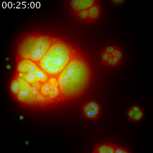
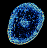
Nucleolus subcompartments spontaneously self-assemble 1
3789
The nucleolus is a small but very important protein complex located in the cell's nucleus. Nilesh Vaidya, Princeton University View Media
Mitochondrion from insect flight muscle
3662
This is a tomographic reconstruction of a mitochondrion from an insect flight muscle. National Center for Microscopy and Imaging Research View Media
Fruit fly ovary_2
3656
A fruit fly ovary, shown here, contains as many as 20 eggs. Fruit flies are not merely tiny insects that buzz around overripe fruit--they are a venerable scientific tool. Denise Montell, University of California, Santa Barbara View Media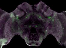
Honeybee brain
6755
Insect brains, like the honeybee brain shown here, are very different in shape from human brains. Gene Robinson, University of Illinois at Urbana-Champaign. View Media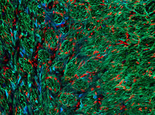
Optic nerve astrocytes
5852
Astrocytes in the cross section of a human optic nerve head Tom Deerinck and Keunyoung (“Christine”) Kim, NCMIR View Media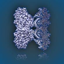
Aldolase
6350
2.5Å resolution reconstruction of rabbit muscle aldolase collected on a FEI/Thermo Fisher Titan Krios with energy filter and image corrector. National Resource for Automated Molecular Microscopy http://nramm.nysbc.org/nramm-images/ Source: Bridget Carragher View Media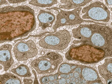
Transmission electron microscopy of myelinated axons with ECM between the axons
3736
The extracellular matrix (ECM) is most prevalent in connective tissues but also is present between the stems (axons) of nerve cells, as shown here. Tom Deerinck, National Center for Microscopy and Imaging Research (NCMIR) View Media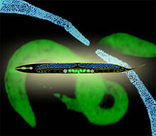
Worms and human infertility
2333
This montage of tiny, transparent C. elegans--or roundworms--may offer insight into understanding human infertility. Abby Dernburg, Lawrence Berkeley National Laboratory View Media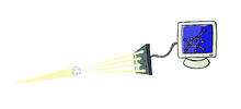
X-ray crystallography
2511
X-ray crystallography allows researchers to see structures too small to be seen by even the most powerful microscopes. Crabtree + Company View Media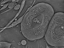
Crane fly spermatocyte undergoing meiosis
6898
A crane fly spermatocyte during metaphase of meiosis-I, a step in the production of sperm. Michael Shribak, Marine Biological Laboratory/University of Chicago. View Media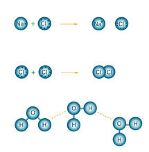
Bond types
2519
Ionic and covalent bonds hold molecules, like sodium chloride and chlorine gas, together. Hydrogen bonds among molecules, notably involving water, also play an important role in biology. Crabtree + Company View Media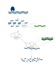
RNA interference
2558
RNA interference or RNAi is a gene-silencing process in which double-stranded RNAs trigger the destruction of specific RNAs. Crabtree + Company View Media
Rabbit GPDA
2405
A crystal of rabbit GPDA protein created for X-ray crystallography, which can reveal detailed, three-dimensional protein structures. Alex McPherson, University of California, Irvine View Media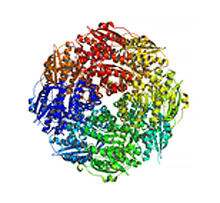
Mandelate racemase from B. subtilis
2350
Model of the mandelate racemase enzyme from Bacillus subtilis, a bacterium commonly found in soil. New York Structural GenomiX Research Consortium, PSI View Media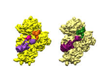
Bacterial ribosome assembly
6578
3D reconstructions of two stages in the assembly of the bacterial ribosome created from time-resolved cryo-electron microscopy images. Ribosomes translate genetic instructions into proteins. Joachim Frank, Columbia University. View Media
Dense tubular matrices in the peripheral endoplasmic reticulum (ER) 1
5855
Superresolution microscopy work on endoplasmic reticulum (ER) in the peripheral areas of the cell showing details of the structure and arrangement in a complex web of tubes. Jennifer Lippincott-Schwartz, Howard Hughes Medical Institute Janelia Research Campus, Virginia View Media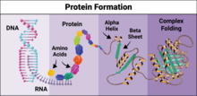
Protein formation
6603
Proteins are 3D structures made up of smaller units. DNA is transcribed to RNA, which in turn is translated into amino acids. NIGMS, with the folded protein illustration adapted from Jane Richardson, Duke University Medical Center View Media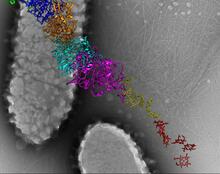
Bacterial nanowire model
6580
A model of a Geobacter sulfurreducens nanowire created from cryo-electron microscopy images. Edward Egelman, University of Virginia. View Media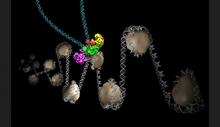
Intasome
6346
Salk researchers captured the structure of a protein complex called an intasome (center) that lets viruses similar to HIV establish permanent infection in their hosts. National Resource for Automated Molecular Microscopy http://nramm.nysbc.org/nramm-images/ Source: Bridget Carragher View Media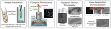
Correlative imaging by annotation with single molecules (CIASM) process
6568
These images illustrate a technique combining cryo-electron tomography and super-resolution fluorescence microscopy called correlative imaging by annotation with single molecules (CIASM). Peter Dahlberg, Stanford University. View Media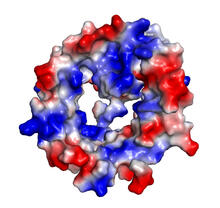
VDAC-1 (4)
2495
The structure of the pore-forming protein VDAC-1 from humans. Gerhard Wagner, Harvard Medical School View Media
TEM cross-section of C. elegans (roundworm)
5759
The worm Caenorhabditis elegans is a popular laboratory animal because its small size and fairly simple body make it easy to study. Piali Sengupta, Brandeis University View Media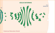
Cisternae maturation model
1307
Animation for the cisternae maturation model of Golgi transport. Judith Stoffer View Media