Switch to Gallery View
Image and Video Gallery
This is a searchable collection of scientific photos, illustrations, and videos. The images and videos in this gallery are licensed under Creative Commons Attribution Non-Commercial ShareAlike 3.0. This license lets you remix, tweak, and build upon this work non-commercially, as long as you credit and license your new creations under identical terms.
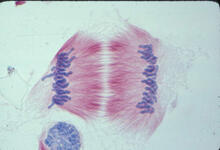
Lily mitosis 11
1011
A light microscope image of cells from the endosperm of an African globe lily (Scadoxus katherinae). This is one frame of a time-lapse sequence that shows cell division in action. Andrew S. Bajer, University of Oregon, Eugene View Media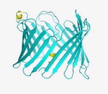
VDAC video 01
2570
This video shows the structure of the pore-forming protein VDAC-1 from humans. Gerhard Wagner, Harvard Medical School View Media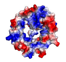
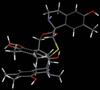
VDAC-1 (4)
2495
The structure of the pore-forming protein VDAC-1 from humans. Gerhard Wagner, Harvard Medical School View Media
Petri dish
6752
The white circle in this image is a Petri dish, named for its inventor, Julius Richard Petri. H. Robert Horvitz and Dipon Ghosh, Massachusetts Institute of Technology. View Media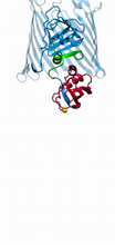
Bacteria working to eat
2304
Gram-negative bacteria perform molecular acrobatics just to eat. Because they're encased by two membranes, they must haul nutrients across both. Emad Tajkhorshid, University of Illinois at Urbana-Champaign View Media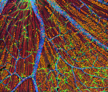
Mouse Retina
3309
A genetic disorder of the nervous system, neurofibromatosis causes tumors to form on nerves throughout the body, including a type of tumor called an optic nerve glioma that can result in childhood bli Tom Deerinck, NCMIR View MediaCone cell
1271
The cone cell of the eye allows you to see in color. Appears in the NIGMS booklet Inside the Cell. Judith Stoffer View Media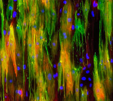
Mouse heart muscle cells 02
3283
This image shows neonatal mouse heart cells. These cells were grown in the lab on a chip that aligns the cells in a way that mimics what is normally seen in the body. Kara McCloskey lab, University of California, Merced, via CIRM View Media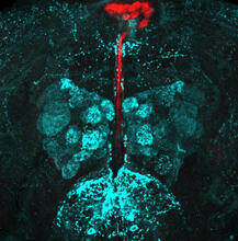
Fruit fly brain responds to adipokines
6985
Drosophila adult brain showing that an adipokine (fat hormone) generates a response from neurons (aqua) and regulates insulin-producing neurons (red).Akhila Rajan, Fred Hutchinson Cancer Center View Media
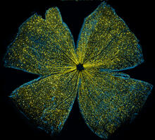
Mouse retina
5793
What looks like the gossamer wings of a butterfly is actually the retina of a mouse, delicately snipped to lay flat and sparkling with fluorescent molecules. Tom Deerinck and Keunyoung (“Christine”) Kim, NCMIR View Media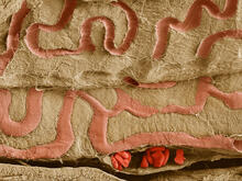
Scanning electron microscopy of the ECM on the surface of a calf muscle
3739
This image shows the extracellular matrix (ECM) on the surface of a soleus (lower calf) muscle in light brown and blood vessels in pink. Tom Deerinck, National Center for Microscopy and Imaging Research (NCMIR) View Media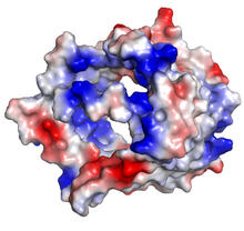
VDAC-1 (3)
2494
The structure of the pore-forming protein VDAC-1 from humans. Gerhard Wagner, Harvard Medical School View Media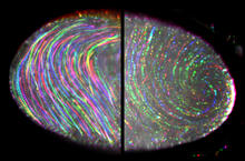
Fruit fly egg ooplasmic streaming
6809
Two fruit fly (Drosophila melanogaster) egg cells, one on each side of the central black line. Vladimir I. Gelfand, Feinberg School of Medicine, Northwestern University. View Media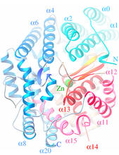
Oligoendopeptidase F from B. stearothermophilus
2373
Crystal structure of oligoendopeptidase F, a protein slicing enzyme from Bacillus stearothermophilus, a bacterium that can cause food products to spoil. Accelerated Technologies Center for Gene to 3D Structure/Midwest Center for Structural Genomics View Media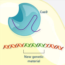
CRISPR Illustration Frame 4
6488
This illustration shows, in simplified terms, how the CRISPR-Cas9 system can be used as a gene-editing tool. National Institute of General Medical Sciences. View Media
Pathways: What is Basic Science?
6539
Learn about basic science, sometimes called “pure” or “fundamental” science, and how it contributes to the development of medical treatments. National Institute of General Medical Sciences View Media
Abnormal, spiky fibroblast
3613
This is a fibroblast, a connective tissue cell that plays an important role in wound healing. Normal fibroblasts have smooth edges. Praveen Suraneni, Stowers Institute for Medical Research, Kansas City, Mo. View Media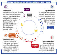
Ciclo circadiano de un adolescente típico
6612
Los ritmos circadianos son cambios físicos, mentales y conductuales que siguen un ciclo de 24 horas. NIGMS View Media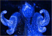
Pores on the surface of the Hawaiian bobtail squid light organ
7016
The light organ (~0.5 mm across) of a juvenile Hawaiian bobtail squid, Euprymna scolopes, stained blue. Margaret J. McFall-Ngai, Carnegie Institution for Science/California Institute of Technology, and Edward G. Ruby, California Institute of Technology. View Media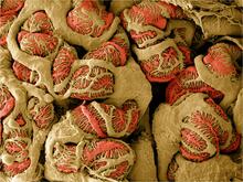
NCMIR Kidney Glomeruli
3392
Stained glomeruli in the kidney. The kidney is an essential organ responsible for disposing wastes from the body and for maintaining healthy ion levels in the blood. Tom Deerinck, National Center for Microscopy and Imaging Research (NCMIR) View Media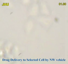
Precisely Delivering Chemical Cargo to Cells
3779
Moving protein or other molecules to specific cells to treat or examine them has been a major biological challenge. Nature Nanotechnology View Media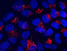
Suicidal Stem Cells
3341
Embryonic stem cells store pre-activated Bax (red) in the Golgi, near the nucleus (blue). Featured in the June 21, 2012, issue of Biomedical Beat. Mohanish Deshmukh View Media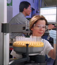
Protein purification robot
2375
Irina Dementieva, a biochemist, and Youngchang Kim, a biophysicist and crystallographer, work with the first robot of its type in the U.S. to automate protein purification. Midwest Center for Structural Genomics View Media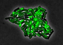
Pulsating response to stress in bacteria
3253
By attaching fluorescent proteins to the genetic circuit responsible for B. subtilis's stress response, researchers can observe the cells' pulses as green flashes. Michael Elowitz, Caltech University View Media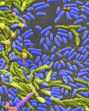
Vibrio bacteria
1160
Vibrio, a type (genus) of rod-shaped bacteria. Some Vibrio species cause cholera in humans. Tina Weatherby Carvalho, University of Hawaii at Manoa View Media
CRISPR surveillance complex
6352
This image shows how the CRISPR surveillance complex is disabled by two copies of anti-CRISPR protein AcrF1 (red) and one AcrF2 (light green). NRAMM National Resource for Automated Molecular Microscopy http://nramm.nysbc.org/nramm-images/ Source: Bridget Carragher View Media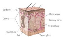
Skin cross-section
1056
Cross-section of skin anatomy shows layers and different tissue types. National Institutes of Health Medical Arts View Media
Seeing signaling protein activation in cells 04
2454
Cdc42, a member of the Rho family of small guanosine triphosphatase (GTPase) proteins, regulates multiple cell functions, including motility, proliferation, apoptosis, and cell morphology. Klaus Hahn, University of North Carolina, Chapel Hill Medical School View Media
PanB from M. tuberculosis (2)
2382
Model of an enzyme, PanB, from Mycobacterium tuberculosis, the bacterium that causes most cases of tuberculosis. This enzyme is an attractive drug target. Mycobacterium Tuberculosis Center, PSI-1 View Media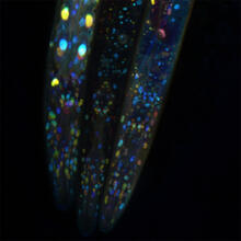
Mosaicism in C. elegans (Black Background)
6532
In the worm C. elegans, double-stranded RNA made in neurons can silence matching genes in a variety of cell types through the transport of RNA between cells. Snusha Ravikumar, Ph.D., University of Maryland, College Park, and Antony M. Jose, Ph.D., University of Maryland, College Park View Media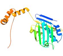
Hsp33 figure 2
3355
Featured in the March 15, 2012 issue of Biomedical Beat. Related to Hsp33 Figure 1, image 3354. Ursula Jakob and Dana Reichmann, University of Michigan View Media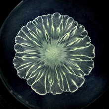
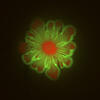
Floral pattern in a mixture of two bacterial species, Acinetobacter baylyi and Escherichia coli, grown on a semi-solid agar for 24 hours
6557
Floral pattern emerging as two bacterial species, motile Acinetobacter baylyi and non-motile Escherichia coli (green), are grown together for 24 hours on 0.75% agar surface from a small L. Xiong et al, eLife 2020;9: e48885 View Media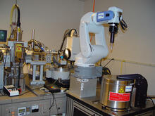
Automated crystal screening system
2362
Automated crystal screening systems such as the one shown here are becoming a common feature at synchrotron and other facilities where high-throughput crystal structure determination is being carried Southeast Collaboratory for Structural Genomics View Media
V. Cholerae Biofilm
3580
Industrious V. cholerae bacteria (yellow) tend to thrive in denser biofilms (left) while moochers (red) thrive in weaker biofilms (right). View Media
Human embryonic stem cells on feeder cells
3274
This fluorescent microscope image shows human embryonic stem cells whose nuclei are stained green. Blue staining shows the surrounding supportive feeder cells. Michael Longaker lab, Stanford University School of Medicine, via CIRM View Media
How cilia do the wave
3494
Thin, hair-like biological structures called cilia are tiny but mighty. Zvonimir Dogic, Brandeis University View Media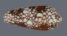
Cone snail shell
2576
A shell from the venomous cone snail Conus omaria, which lives in the Pacific and Indian oceans and eats other snails. Kerry Matz, University of Utah View Media
Natcher Building 07
1087
NIGMS staff are located in the Natcher Building on the NIH campus. Alisa Machalek, National Institute of General Medical Sciences View Media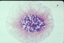
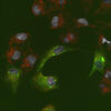
Lily mitosis 04
1014
A light microscope image of a cell from the endosperm of an African globe lily (Scadoxus katherinae). This is one frame of a time-lapse sequence that shows cell division in action. Andrew S. Bajer, University of Oregon, Eugene View Media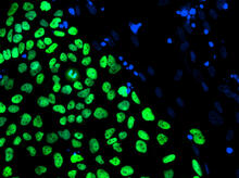
Human embryonic stem cells on feeder cells
3275
The nuclei stained green highlight human embryonic stem cells grown under controlled conditions in a laboratory. Blue represents the DNA of surrounding, supportive feeder cells. Julie Baker lab, Stanford University School of Medicine, via CIRM View Media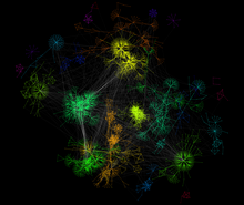
A molecular interaction network in yeast 2
3732
The image visualizes a part of the yeast molecular interaction network. Keiichiro Ono, UCSD View MediaCentral dogma, illustrated (with labels)
2548
DNA encodes RNA, which encodes protein. DNA is transcribed to make messenger RNA (mRNA). The mRNA sequence (dark red strand) is complementary to the DNA sequence (blue strand). Crabtree + Company View Media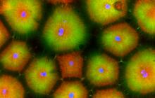
Cell-like compartments from frog eggs 5
6592
Cell-like compartments that spontaneously emerged from scrambled frog eggs, with nuclei (blue) from frog sperm. Endoplasmic reticulum (red) and microtubules (green) are also visible. Xianrui Cheng, Stanford University School of Medicine. View Media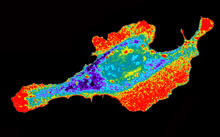
Seeing signaling protein activation in cells 03
2453
Cdc42, a member of the Rho family of small guanosine triphosphatase (GTPase) proteins, regulates multiple cell functions, including motility, proliferation, apoptosis, and cell morphology. Klaus Hahn, University of North Carolina, Chapel Hill Medical School View Media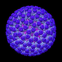
Rotavirus structure
3584
This image shows a computer-generated, three-dimensional map of the rotavirus structure. This virus infects humans and other animals and causes severe diarrhea in infants and young children. Bridget Carragher, The Scripps Research Institute, La Jolla, CA View Media
Zebrafish pigment cell
5754
Pigment cells are cells that give skin its color. David Parichy, University of Washington View Media
Induced stem cells from adult skin 03
2605
The human skin cells pictured contain genetic modifications that make them pluripotent, essentially equivalent to embryonic stem cells. James Thomson, University of Wisconsin-Madison View Media
Cytonemes in developing fruit fly cells
3574
Scientists have long known that multicellular organisms use biological molecules produced by one cell and sensed by another to transmit messages that, for instance, guide proper development of organs Sougata Roy, University of California, San Francisco View Media
Yeast cells with nuclear envelopes and tubulin
6798
Yeast cells with nuclear envelopes shown in magenta and tubulin shown in light blue. The nuclear envelope defines the borders of the nucleus, which houses DNA. Alaina Willet, Kathy Gould’s lab, Vanderbilt University. View Media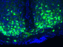
Master clock of the mouse brain
3547
An image of the area of the mouse brain that serves as the 'master clock,' which houses the brain's time-keeping neurons. The nuclei of the clock cells are shown in blue. Erik Herzog, Washington University in St. Louis View Media