Switch to Gallery View
Image and Video Gallery
This is a searchable collection of scientific photos, illustrations, and videos. The images and videos in this gallery are licensed under Creative Commons Attribution Non-Commercial ShareAlike 3.0. This license lets you remix, tweak, and build upon this work non-commercially, as long as you credit and license your new creations under identical terms.
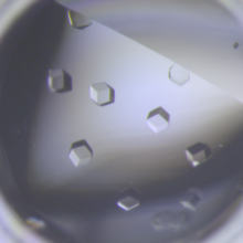
Crystals of CCD-1 in complex with cefotaxime
6764
CCD-1 is an enzyme produced by the bacterium Clostridioides difficile that helps it resist antibiotics. Keith Hodgson, Stanford University. View Media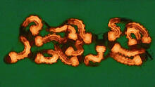
Group of Culex quinquefasciatus mosquito larvae
6770
Mosquito larvae with genes edited by CRISPR. Valentino Gantz, University of California, San Diego. View Media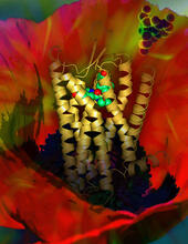
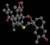
Human opioid receptor structure superimposed on poppy
3314
Opioid receptors on the surfaces of brain cells are involved in pleasure, pain, addiction, depression, psychosis, and other conditions. Raymond Stevens, The Scripps Research Institute View Media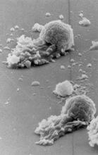
Worm sperm
3489
To develop a system for studying cell motility in unnatrual conditions -- a microscope slide instead of the body -- Tom Roberts and Katsuya Shimabukuro at Florida State University disassembled and rec Tom Roberts, Florida State University View Media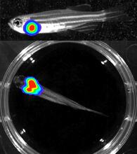
Bioluminescent imaging in adult zebrafish - lateral and overhead view
3556
Luciferase-based imaging enables visualization and quantification of internal organs and transplanted cells in live adult zebrafish. Kenneth Poss, Duke University View Media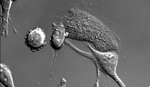
Dying melanoma cells
6966
Melanoma (skin cancer) cells undergoing programmed cell death, also called apoptosis. This process was triggered by raising the pH of the medium that the cells were growing in. Dylan T. Burnette, Vanderbilt University School of Medicine. View Media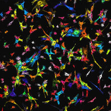
Single-cell “radios” image
7021
Individual cells are color-coded based on their identity and signaling activity using a protein circuit technology developed by the Coyle Lab. Scott Coyle, University of Wisconsin-Madison. View Media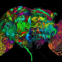
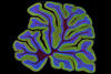
Color coding of the Drosophila brain - black background
5868
This image results from a research project to visualize which regions of the adult fruit fly (Drosophila) brain derive from each neural stem cell. Yong Wan from Charles Hansen’s lab, University of Utah. Data preparation and visualization by Masayoshi Ito in the lab of Kei Ito, University of Tokyo. View Media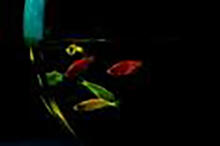
Glowing fish
2667
Professor Marc Zimmer's family pets, including these fish, glow in the dark in response to blue light. Featured in the September 2009 issue of Findings. View Media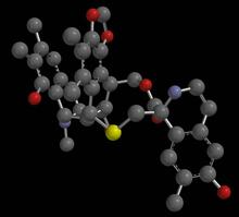
Anti-tumor drug ecteinascidin 743 (ET-743), structure without hydrogens 03
2796
Ecteinascidin 743 (ET-743, brand name Yondelis), was discovered and isolated from a sea squirt, Ecteinascidia turbinata, by NIGMS grantee Kenneth Rinehart at the University of Illinois. Timothy Jamison, Massachusetts Institute of Technology View Media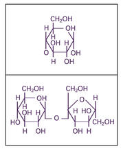
Glucose and sucrose
2500
Glucose (top) and sucrose (bottom) are sugars made of carbon, hydrogen, and oxygen atoms. Carbohydrates include simple sugars like these and are the main source of energy for the human body. Crabtree + Company View Media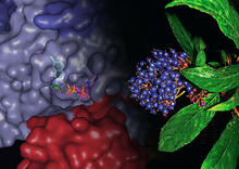
Chang Shan
3483
For thousands of years, Chinese herbalists have treated malaria using Chang Shan, a root extract from a type of hydrangea that grows in Tibet and Nepal. Paul Schimmel Lab, Scripps Research Institute View Media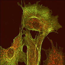
Endothelial cell
1102
This image shows two components of the cytoskeleton, microtubules (green) and actin filaments (red), in an endothelial cell derived from a cow lung. Tina Weatherby Carvalho, University of Hawaii at Manoa View Media
Multivesicular bodies containing intralumenal vesicles assemble at the vacuole 3
5767
Collecting and transporting cellular waste and sorting it into recylable and nonrecylable pieces is a complex business in the cell. Matthew West and Greg Odorizzi, University of Colorado View Media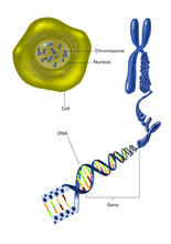
Chromosome inside nucleus (with labels)
2540
The long, stringy DNA that makes up genes is spooled within chromosomes inside the nucleus of a cell. Crabtree + Company View Media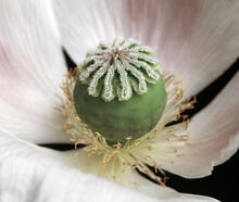
White Poppy (cropped)
3423
A cropped image of a white poppy. View poppy uncropped here 3424. Judy Coyle, Donald Danforth Plant Science Center View Media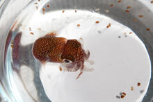
Adult and juvenile Hawaiian bobtail squids
7010
An adult Hawaiian bobtail squid, Euprymna scolopes, (~4 cm) surrounded by newly hatched juveniles (~2 mm) in a bowl of seawater.Margaret J. McFall-Ngai, Carnegie Institution for Science/California Institute of Technology, and Edward G. Ruby, California Institute of Technology. View Media
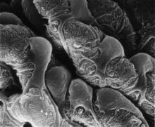
Podocytes from a chronically diseased kidney
3565
This scanning electron microscope (SEM) image shows podocytes--cells in the kidney that play a vital role in filtering waste from the bloodstream--from a patient with chronic kidney disease. Olga Troyanskaya, Princeton University and Matthias Kretzler, University of Michigan View Media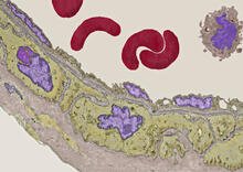
Transmission electron microscopy of coronary artery wall with elastin-rich ECM pseudocolored in light brown
3738
Elastin is a fibrous protein in the extracellular matrix (ECM). It is abundant in artery walls like the one shown here. As its name indicates, elastin confers elasticity. Tom Deerinck, National Center for Microscopy and Imaging Research (NCMIR) View Media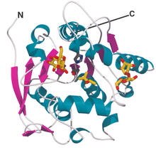
Most abundant protein in M. tuberculosis
2378
Model of a protein, antigen 85B, that is the most abundant protein exported by Mycobacterium tuberculosis, which causes most cases of tuberculosis. Mycobacterium Tuberculosis Center, PSI View Media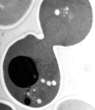
EM of yeast cell division
5770
Cell division is an incredibly coordinated process. Matthew West and Greg Odorizzi, University of Colorado View Media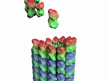
How a microtubule builds and deconstructs
3650
A microtubule, part of the cell's skeleton, builds and deconstructs. View Media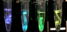
Bioluminescence in a Tube
5895
Details about the basic biology and chemistry of the ingredients that produce bioluminescence are allowing scientists to harness it as an imaging tool. Credit: Nathan Shaner, Scintillon Institute. Nathan Shaner, Scintillon Institute View Media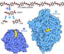
Plastic-eating enzymes
7000
PETase enzyme degrades polyester plastic (polyethylene terephthalate, or PET) into monohydroxyethyl terephthalate (MHET). Amy Wu and Christine Zardecki, RCSB Protein Data Bank. View Media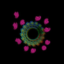
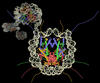
Actin filaments bundled around the dynamin helical polymer
6571
Multiple actin filaments (magenta) are organized around a dynamin helical polymer (rainbow colored) in this model derived from cryo-electron tomography. Elizabeth Chen, University of Texas Southwestern Medical Center. View Media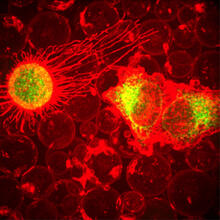
Plasma-Derived Membrane Vesicles
5887
This fiery image doesn’t come from inside a bubbling volcano. Instead, it shows animal cells caught in the act of making bubbles, or blebbing. Jeanne Stachowiak, University of Texas at Austin View Media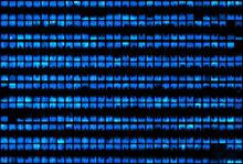
Biopixels
3266
Bioengineers were able to coax bacteria to blink in unison on microfluidic chips. This image shows a small chip with about 500 blinking bacterial colonies or biopixels. Jeff Hasty Lab, UC San Diego View Media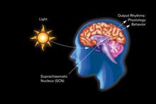
Circadian rhythm
2841
The human body keeps time with a master clock called the suprachiasmatic nucleus or SCN. Crabtree + Company View Media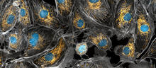
Panorama view of golden mitochondria
5762
Mitochondria are the powerhouses of the cells, generating the energy the cells need to do their tasks and to stay alive. Torsten Wittmann, University of California, San Francisco View Media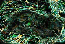
Larvae from the parasitic worm that causes schistosomiasis
3627
The parasitic worm that causes schistosomiasis hatches in water and grows up in a freshwater snail, as shown here. Bo Wang and Phillip A. Newmark, University of Illinois at Urbana-Champaign, 2013 FASEB BioArt winner View Media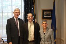
Lorsch Swearing In
3530
Jon Lorsch at his swearing in as NIGMS director in August 2013. Also shown are Francis Collins, NIH Director, and Judith Greenberg, former NIGMS Acting Director. View Media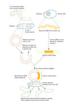
Recombinant DNA (with labels)
2565
To splice a human gene (in this case, the one for insulin) into a plasmid, scientists take the plasmid out of an E. Crabtree + Company View Media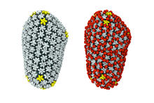
Cryo-EM reveals how the HIV capsid attaches to a human protein to evade immune detection
3755
The illustration shows the capsid of human immunodeficiency virus (HIV) whose molecular features were resolved with cryo-electron microscopy (cryo-EM). Juan R. Perilla, University of Illinois at Urbana-Champaign View Media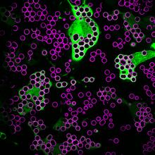
Hungry, hungry macrophages
7009
Macrophages (green) are the professional eaters of our immune system. Meghan Morrissey, University of California, Santa Barbara. View Media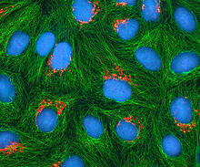
HeLa cells
3522
Multiphoton fluorescence image of cultured HeLa cells with a fluorescent protein targeted to the Golgi apparatus (orange), microtubules (green) and counterstained for DNA (cyan). National Center for Microscopy and Imaging Research (NCMIR) View Media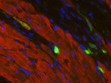
Heart muscle with reprogrammed skin cells
3273
Skins cells were reprogrammed into heart muscle cells. The cells highlighted in green are remaining skin cells. Red indicates a protein that is unique to heart muscle. Deepak Srivastava, Gladstone Institute of Cardiovascular Disease, via CIRM View Media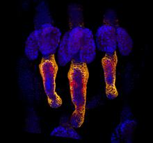
Growing hair follicle stem cells
3499
Wound healing requires the action of stem cells. Hermann Steller, Rockefeller University View Media
Nucleolinus
2762
The nucleolinus is a cellular compartment that has been a lonely bystander in scientific endeavors. Mary Anne Alliegro, Marine Biological Laboratory View Media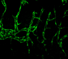
Normal vascular development in frog embryos
3404
In vivo vascular development in kdr:GFP frogs. Related to images 3403 and 3405. Hye Ji Cha, University of Texas at Austin View Media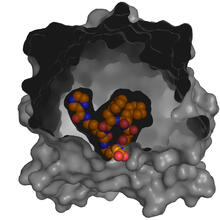
X-ray co-crystal structure of Src kinase bound to a DNA-templated macrocycle inhibitor 5
3417
X-ray co-crystal structure of Src kinase bound to a DNA-templated macrocycle inhibitor. Markus A. Seeliger, Stony Brook University Medical School and David R. Liu, Harvard University View Media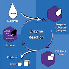
Enzyme reaction
6604
Enzymes speed up chemical reactions by reducing the amount of energy needed for the reactions. NIGMS View Media
Myelinated axons 2
3397
Top view of myelinated axons in a rat spinal root. Tom Deerinck, National Center for Microscopy and Imaging Research (NCMIR) View Media
Protein clumping in zinc-deficient yeast cells
3550
The green spots in this image are clumps of protein inside yeast cells that are deficient in both zinc and a protein called Tsa1 that prevents clumping. Colin MacDiarmid and David Eide, University of Wisconsin--Madison View Media
Color coding of the Drosophila brain - video
5843
This video results from a research project to visualize which regions of the adult fruit fly (Drosophila) brain derive from each neural stem cell. Yong Wan from Charles Hansen’s lab, University of Utah. Data preparation and visualization by Masayoshi Ito in the lab of Kei Ito, University of Tokyo. View Media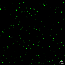
Staphylococcus aureus aggregating upon contact with synovial fluid
6805
Staphylococcus aureus bacteria (green) grouping together upon contact with synovial fluid—a viscous substance found in joints. Paul Stoodley, The Ohio State University. View Media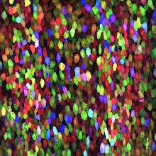
A multicolored fish scale 1
3782
Each of the colored specs in this image is a cell on the surface of a fish scale. Chen-Hui Chen and Kenneth Poss, Duke University View Media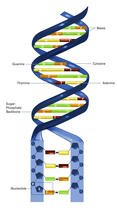
Nucleotides make up DNA (with labels)
2542
DNA consists of two long, twisted chains made up of nucleotides. Each nucleotide contains one base, one phosphate molecule, and the sugar molecule deoxyribose. Crabtree + Company View Media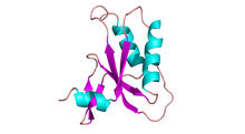
Antitoxin GhoS (Illustration 2)
3428
Structure of the bacterial antitoxin protein GhoS. GhoS inhibits the production of a bacterial toxin, GhoT, which can contribute to antibiotic resistance. Rebecca Page and Wolfgang Peti, Brown University and Thomas K. Wood, Pennsylvania State University View Media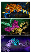
HIV enzyme
6999
These images model the molecular structures of three enzymes with critical roles in the life cycle of the human immunodeficiency virus (HIV). Amy Wu and Christine Zardecki, RCSB Protein Data Bank. View Media
Sea urchin embryo 03
1049
Stereo triplet of a sea urchin embryo stained to reveal actin filaments (orange) and microtubules (blue). George von Dassow, University of Washington View Media