Switch to Gallery View
Image and Video Gallery
This is a searchable collection of scientific photos, illustrations, and videos. The images and videos in this gallery are licensed under Creative Commons Attribution Non-Commercial ShareAlike 3.0. This license lets you remix, tweak, and build upon this work non-commercially, as long as you credit and license your new creations under identical terms.
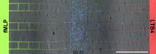
Neutrophil-like cells migrating in a microfluidic chip
6886
Neutrophil-like cells (blue) in a microfluidic chip preferentially migrating toward LTB4 over fMLP. Caroline Jones, University of Texas at Dallas. View Media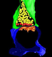
3-D Architecture of a Synapse
5885
This image shows the structure of a synapse, or junction between two nerve cells in three dimensions. From the brain of a mouse. Anton Maximov, The Scripps Research Institute, La Jolla, CA View Media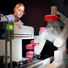
Student overseeing protein cloning robot
2356
Student Christina Hueneke of the Midwest Center for Structural Genomics is overseeing a protein cloning robot. Midwest Center for Structural Genomics View Media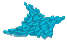
Smooth ER
1292
The endoplasmic reticulum comes in two types: Rough ER is covered with ribosomes and prepares newly made proteins; smooth ER specializes in making lipids and breaking down toxic molecules. Judith Stoffer View Media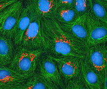
HeLa cells
3522
Multiphoton fluorescence image of cultured HeLa cells with a fluorescent protein targeted to the Golgi apparatus (orange), microtubules (green) and counterstained for DNA (cyan). National Center for Microscopy and Imaging Research (NCMIR) View Media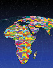
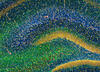
Mapping human genetic variation
2443
This map paints a colorful portrait of human genetic variation around the world. Noah Rosenberg and Martin Soave, University of Michigan View Media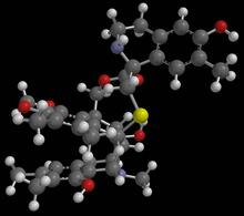
Anti-tumor drug ecteinascidin 743 (ET-743) with hydrogens 01
2790
Ecteinascidin 743 (ET-743, brand name Yondelis), was discovered and isolated from a sea squirt, Ecteinascidia turbinata, by NIGMS grantee Kenneth Rinehart at the University of Illinois. Timothy Jamison, Massachusetts Institute of Technology View Media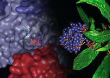
Chang Shan
3483
For thousands of years, Chinese herbalists have treated malaria using Chang Shan, a root extract from a type of hydrangea that grows in Tibet and Nepal. Paul Schimmel Lab, Scripps Research Institute View Media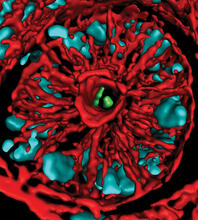
Mouse colon with gut bacteria
3566
A section of mouse colon with gut bacteria (center, in green) residing within a protective pocket. Sarkis K. Mazmanian, California Institute of Technology View Media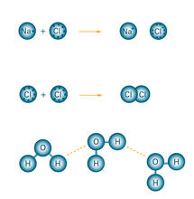
Bond types
2519
Ionic and covalent bonds hold molecules, like sodium chloride and chlorine gas, together. Hydrogen bonds among molecules, notably involving water, also play an important role in biology. Crabtree + Company View Media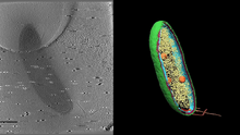
Cryo-electron tomography of a Caulobacter bacterium
6569
3D image of Caulobacter bacterium with various components highlighted: cell membranes (red and blue), protein shell (green), protein factories known as ribosomes (yellow), and storage granules Peter Dahlberg, Stanford University. View Media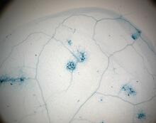
Disease-resistant Arabidopsis leaf
2781
This is a magnified view of an Arabidopsis thaliana leaf a few days after being exposed to the pathogen Hyaloperonospora arabidopsidis. Jeff Dangl, University of North Carolina, Chapel Hill View Media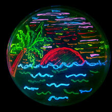
Glowing bacteria make a pretty postcard
3492
This tropical scene, reminiscent of a postcard from Key West, is actually a petri dish containing an artistic arrangement of genetically engineered bacteria. Nathan C. Shaner, The Scintillon Institute View Media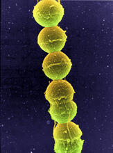
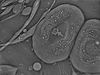
Streptococcus bacteria
1157
Image of Streptococcus, a type (genus) of spherical bacteria that can colonize the throat and back of the mouth. Stroptococci often occur in pairs or in chains, as shown here. Tina Weatherby Carvalho, University of Hawaii at Manoa View Media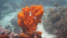
Sponge
2728
Many of today's medicines come from products found in nature, such as this sponge found off the coast of Palau in the Pacific Ocean. Phil Baran, Scripps Research Institute View Media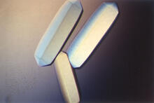
Fungal lipase (2)
2411
Crystals of fungal lipase protein created for X-ray crystallography, which can reveal detailed, three-dimensional protein structures. Alex McPherson, University of California, Irvine View Media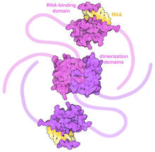
SARS-CoV-2 nucleocapsid dimer
6991
In SARS-CoV-2, the virus that causes COVID-19, nucleocapsid is a complex molecule with many functional parts. Amy Wu and Christine Zardecki, RCSB Protein Data Bank. View Media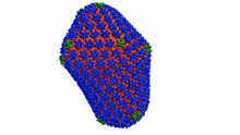
Atomic-level structure of the HIV capsid
6601
This animation shows atoms of the HIV capsid, the shell that encloses the virus's genetic material. Juan R. Perilla and the Theoretical and Computational Biophysics Group, University of Illinois at Urbana-Champaign View Media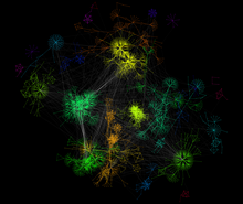
A molecular interaction network in yeast 2
3732
The image visualizes a part of the yeast molecular interaction network. Keiichiro Ono, UCSD View Media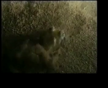
Adult Hawaiian bobtail squid burying in the sand
7012
Each morning, the nocturnal Hawaiian bobtail squid, Euprymna scolopes, hides from predators by digging into the sand. At dusk, it leaves the sand again to hunt. Margaret J. McFall-Ngai, Carnegie Institution for Science/California Institute of Technology, and Edward G. Ruby, California Institute of Technology. View Media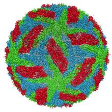
Protective membrane and membrane proteins of the dengue virus visualized with cryo-EM
3756
Dengue virus is a mosquito-borne illness that infects millions of people in the tropics and subtropics each year. Like many viruses, dengue is enclosed by a protective membrane. Hong Zhou, UCLA View Media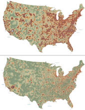
Mapping disease spread
2320
How far and fast an infectious disease spreads across a community depends on many factors, including transportation. These U.S. David Chrest, RTI International View Media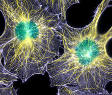
Colorful cells
2428
Actin (purple), microtubules (yellow), and nuclei (green) are labeled in these cells by immunofluorescence. This image won first place in the Nikon 2003 Small World photo competition. Torsten Wittmann, Scripps Research Institute View Media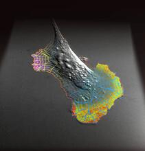
Biosensors illustration
2802
A rendering of an activity biosensor image overlaid with a cell-centered frame of reference used for image analysis of signal transduction. Gaudenz Danuser, Harvard Medical School View Media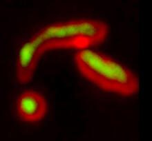
Misfolded proteins within in the mitochondria
5878
Misfolded proteins (green) within mitochondria (red). Related to video 5877. Rong Li rong@jhu.edu Department of Chemical and Biomolecular Engineering, Whiting School of Engineering, Johns Hopkins University, Baltimore, Maryland 21218, USA. View Media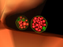
Multivesicular bodies containing intralumenal vesicles assemble at the vacuole 3
5767
Collecting and transporting cellular waste and sorting it into recylable and nonrecylable pieces is a complex business in the cell. Matthew West and Greg Odorizzi, University of Colorado View Media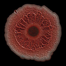
Biofilm formed by a pathogen
6518
A biofilm is a highly organized community of microorganisms that develops naturally on certain surfaces. Scott Chimileski, Ph.D., and Roberto Kolter, Ph.D., Harvard Medical School. View Media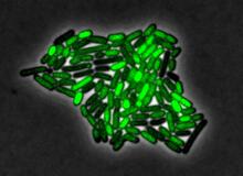
Pulsating response to stress in bacteria
3253
By attaching fluorescent proteins to the genetic circuit responsible for B. subtilis's stress response, researchers can observe the cells' pulses as green flashes. Michael Elowitz, Caltech University View Media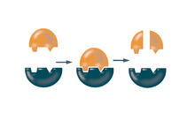
Enzymes convert subtrates into products
2521
Enzymes convert substrates into products very quickly. See image 2522 for a labeled version of this illustration. Featured in The Chemistry of Health. Crabtree + Company View Media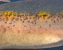
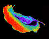
Pigment cells in fish skin
5756
Pigment cells are cells that give skin its color. David Parichy, University of Washington View Media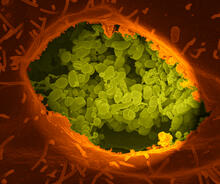
Q fever bacteria in an infected cell
3621
This image shows Q fever bacteria (yellow), which infect cows, sheep, and goats around the world and can infect humans, as well. When caught early, Q fever can be cured with antibiotics. Robert Heinzen, Elizabeth Fischer, and Anita Mora, National Institute of Allergy and Infectious Diseases, National Institutes of Health View Media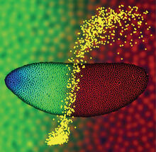
Precise development in the fruit fly embryo
2593
This 2-hour-old fly embryo already has a blueprint for its formation, and the process for following it is so precise that the difference of just a few key molecules can change the plans. Thomas Gregor, Princeton University View Media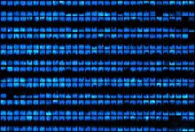
Biopixels
3266
Bioengineers were able to coax bacteria to blink in unison on microfluidic chips. This image shows a small chip with about 500 blinking bacterial colonies or biopixels. Jeff Hasty Lab, UC San Diego View Media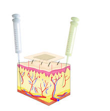
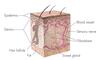
Drugs enter skin
2531
Drugs enter different layers of skin via intramuscular, subcutaneous, or transdermal delivery methods. See image 2532 for a labeled version of this illustration. Crabtree + Company View Media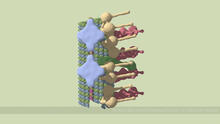
Partial Model of a Cilium’s Doublet Microtubule
6548
Cilia (cilium in singular) are complex molecular machines found on many of our cells. Brown Lab, Harvard Medical School and Veronica Falconieri Hays. View Media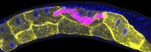
Microsporidia in roundworm 1
5777
Many disease-causing microbes manipulate their host’s metabolism and cells for their own ends. Keir Balla and Emily Troemel, University of California San Diego View Media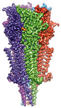
Full-length serotonin receptor (ion channel)
6579
A 3D reconstruction, created using cryo-electron microscopy, of an ion channel known as the full-length serotonin receptor in complex with the antinausea drug granisetron (orange). Sudha Chakrapani, Case Western Reserve University School of Medicine. View Media
Cross section of a Drosophila melanogaster pupa lacking Draper
2759
In the absence of the engulfment receptor Draper, salivary gland cells (light blue) persist in the thorax of a developing Drosophila melanogaster pupa. Christina McPhee and Eric Baehrecke, University of Massachusetts Medical School View Media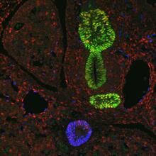
Transcription factor Sox17 controls embryonic development of certain internal organs
3440
During embryonic development, transcription factors (proteins that regulate gene expression) govern the differentiation of cells into separate tissues and organs. James M. Wells, Cincinnati Children's Hospital Medical Center View Media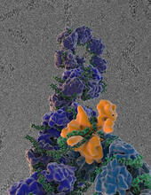
Flu virus proteins during self-replication
3434
Influenza (flu) virus proteins in the act of self-replication. Viral nucleoprotein (blue) encapsidates [encapsulates] the RNA genome (green). Scripps Research Institute in La Jolla, CA View Media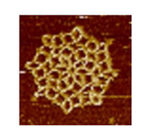
Snowflake DNA origami
3724
An atomic force microscopy image shows DNA folded into an intricate, computer-designed structure. The image is featured on Biomedical Beat blog post Cool Images: A Holiday-Themed Collection. Hao Yan, Arizona State University View Media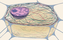
Cytoskeleton
1272
The three fibers of the cytoskeleton--microtubules in blue, intermediate filaments in red, and actin in green--play countless roles in the cell. Judith Stoffer View Media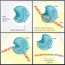
CRISPR Illustration
7036
This illustration shows, in simplified terms, how the CRISPR-Cas9 system can be used as a gene-editing tool. National Institute of General Medical Sciences. View Media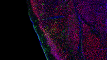
Video of Calling Cards in a mouse brain
6781
The green spots in this mouse brain are cells labeled with Calling Cards, a technology that records molecular events in brain cells as they mature. NIH Director's Blog View Media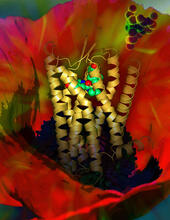
Human opioid receptor structure superimposed on poppy
3314
Opioid receptors on the surfaces of brain cells are involved in pleasure, pain, addiction, depression, psychosis, and other conditions. Raymond Stevens, The Scripps Research Institute View Media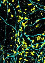
Lysosomes and microtubules
6889
Lysosomes (yellow) and detyrosinated microtubules (light blue). Lysosomes are bubblelike organelles that take in molecules and use enzymes to break them down. Melike Lakadamyali, Perelman School of Medicine at the University of Pennsylvania. View Media
Life of an AIDS virus (with labels)
2514
HIV is a retrovirus, a type of virus that carries its genetic material not as DNA but as RNA. Crabtree + Company View Media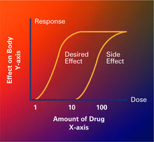
Dose response curves
2533
Dose-response curves determine how much of a drug (X-axis) causes a particular effect, or a side effect, in the body (Y-axis). Featured in Medicines By Design. Crabtree + Company View Media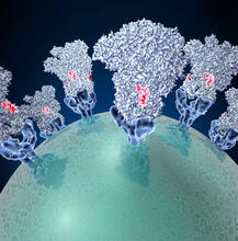
Coronavirus spike protein structure
3753
Coronaviruses are enveloped viruses responsible for 30 percent of mild respiratory infections and atypical deadly pneumonia in humans worldwide. Melody Campbell, UCSF View Media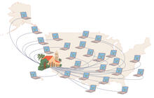
Folding@Home
1276
Stanford University scientist Vijay Pande decided to couple the power of computers with the help of the public. Judith Stoffer View Media