Switch to Gallery View
Image and Video Gallery
This is a searchable collection of scientific photos, illustrations, and videos. The images and videos in this gallery are licensed under Creative Commons Attribution Non-Commercial ShareAlike 3.0. This license lets you remix, tweak, and build upon this work non-commercially, as long as you credit and license your new creations under identical terms.
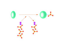
Kinases
2534
Kinases are enzymes that add phosphate groups (red-yellow structures) to proteins (green), assigning the proteins a code. Crabtree + Company View Media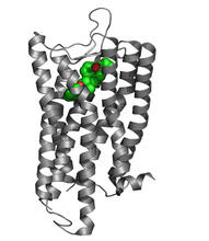
Nociceptin/orphanin FQ peptide opioid receptor
3364
The receptor is shown bound to an antagonist, compound-24 Raymond Stevens, The Scripps Research Institute View Media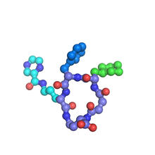
X-ray co-crystal structure of Src kinase bound to a DNA-templated macrocycle inhibitor 7
3419
X-ray co-crystal structure of Src kinase bound to a DNA-templated macrocycle inhibitor. Markus A. Seeliger, Stony Brook University Medical School and David R. Liu, Harvard University View Media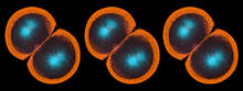
Sea urchin embryo 06
1052
Stereo triplet of a sea urchin embryo stained to reveal actin filaments (orange) and microtubules (blue). George von Dassow, University of Washington View Media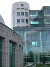
Natcher Building 07
1087
NIGMS staff are located in the Natcher Building on the NIH campus. Alisa Machalek, National Institute of General Medical Sciences View Media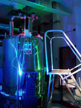
Superconducting magnet
1120
Superconducting magnet for NMR research, from the February 2003 profile of Dorothee Kern in Findings. Mike Lovett View Media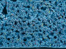
STORM image of axonal cytoskeleton
3678
This image shows the long, branched structures (axons) of nerve cells. Xiaowei Zhuang Laboratory, Howard Hughes Medical Institute, Harvard University View Media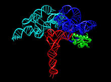
Ribonuclease P structure
3660
Ribbon diagram showing the structure of Ribonuclease P with tRNA. PDB entry 3Q1Q, molecular modeling by Fred Friedman, NIGMS View Media
Bovine milk alpha-lactalbumin (1)
2397
A crystal of bovine milk alpha-lactalbumin protein created for X-ray crystallography, which can reveal detailed, three-dimensional protein structures. Alex McPherson, University of California, Irvine View Media
Mouse heart muscle cells 02
3283
This image shows neonatal mouse heart cells. These cells were grown in the lab on a chip that aligns the cells in a way that mimics what is normally seen in the body. Kara McCloskey lab, University of California, Merced, via CIRM View Media
Mosaicism in C. elegans (White Background)
6534
In the worm C. elegans, double-stranded RNA made in neurons can silence matching genes in a variety of cell types through the transport of RNA between cells. Snusha Ravikumar, Ph.D., University of Maryland, College Park, and Antony M. Jose, Ph.D., University of Maryland, College Park View Media
Histones in chromatin
2560
Histone proteins loop together with double-stranded DNA to form a structure that resembles beads on a string. Crabtree + Company View Media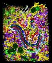
Cellular metropolis
2308
Like a major city, a cell teems with specialized workers that carry out its daily operations--making energy, moving proteins, or helping with other tasks. Kathryn Howell, University of Colorado Health Sciences Center View Media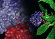
Chang Shan
3483
For thousands of years, Chinese herbalists have treated malaria using Chang Shan, a root extract from a type of hydrangea that grows in Tibet and Nepal. Paul Schimmel Lab, Scripps Research Institute View Media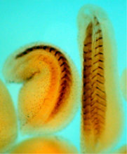
Xenopus laevis embryos
2756
Xenopus laevis, the African clawed frog, has long been used as a model organism for studying embryonic development. The frog embryo on the left lacks the developmental factor Sizzled. Michael Klymkowsky, University of Colorado, Boulder View Media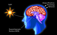
Circadian rhythm (with labels)
2569
The human body keeps time with a master clock called the suprachiasmatic nucleus or SCN. Crabtree + Company View Media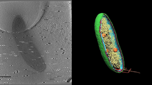
Cryo-electron tomography of a Caulobacter bacterium
6569
3D image of Caulobacter bacterium with various components highlighted: cell membranes (red and blue), protein shell (green), protein factories known as ribosomes (yellow), and storage granules Peter Dahlberg, Stanford University. View Media
Fruit fly ovary
3607
A fruit fly ovary, shown here, contains as many as 20 eggs. Fruit flies are not merely tiny insects that buzz around overripe fruit—they are a venerable scientific tool. Denise Montell, Johns Hopkins University and University of California, Santa Barbara View Media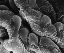
Podocytes from a chronically diseased kidney
3565
This scanning electron microscope (SEM) image shows podocytes--cells in the kidney that play a vital role in filtering waste from the bloodstream--from a patient with chronic kidney disease. Olga Troyanskaya, Princeton University and Matthias Kretzler, University of Michigan View Media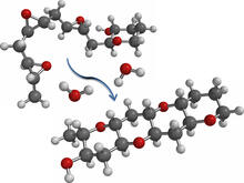
Cascade reaction promoted by water
2490
This illustration of an epoxide-opening cascade promoted by water emulates the proposed biosynthesis of some of the Red Tide toxins. Tim Jamison, Massachusetts Institute of Technology View Media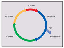
Cell cycle (with labels)
2499
Cells progress through a cycle that consists of phases for growth (G1, S, and G2) and division (M). Cells become quiescent when they exit this cycle (G0). Crabtree + Company View Media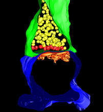
3-D Architecture of a Synapse
5885
This image shows the structure of a synapse, or junction between two nerve cells in three dimensions. From the brain of a mouse. Anton Maximov, The Scripps Research Institute, La Jolla, CA View Media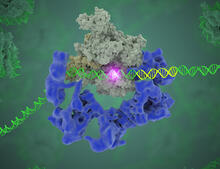
TFIID complex binds DNA to start gene transcription
3766
Gene transcription is a process by which the genetic information encoded in DNA is transcribed into RNA. Eva Nogales, Berkeley Lab View Media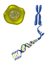
Chromosome inside nucleus
2539
The long, stringy DNA that makes up genes is spooled within chromosomes inside the nucleus of a cell. Crabtree + Company View Media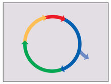
Cell cycle
2498
Cells progress through a cycle that consists of phases for growth (blue, green, yellow) and division (red). Cells become quiescent when they exit this cycle (purple). Crabtree + Company View Media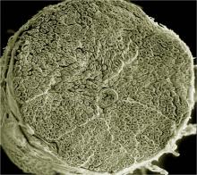
NCMIR human spinal nerve
3387
Spinal nerves are part of the peripheral nervous system. They run within the spinal column to carry nerve signals to and from all parts of the body. Tom Deerinck, National Center for Microscopy and Imaging Research (NCMIR) View Media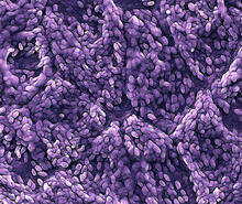
Retinal pigment epithelium derived from human ES cells
3286
This color-enhanced image is a scanning electron microscope image of retinal pigment epithelial (RPE) cells derived from human embryonic stem cells. David Hinton lab, University of Southern California, via CIRM View Media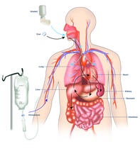
A drug's life in the body (with labels)
2528
A drug's life in the body. Medicines taken by mouth (oral) pass through the liver before they are absorbed into the bloodstream. Crabtree + Company View Media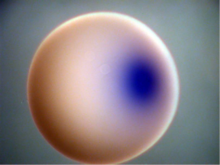
Xenopus laevis egg
2753
Xenopus laevis, the African clawed frog, has long been used as a model organism for studying embryonic development. Michael Klymkowsky, University of Colorado, Boulder View Media
Cross section of a Drosophila melanogaster pupa lacking Draper
2759
In the absence of the engulfment receptor Draper, salivary gland cells (light blue) persist in the thorax of a developing Drosophila melanogaster pupa. Christina McPhee and Eric Baehrecke, University of Massachusetts Medical School View Media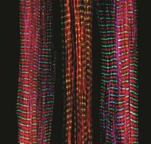
Three muscle fibers; the middle has a defect found in some neuromuscular diseases
3630
Of the three muscle fibers shown here, the one on the right and the one on the left are normal. The middle fiber is deficient a large protein called nebulin (blue). Christopher Pappas and Carol Gregorio, University of Arizona View Media
Fluorescence in situ hybridization (FISH) in mouse ES cells shows DNA interactions
3296
Researchers used fluorescence in situ hybridization (FISH) to confirm the presence of long range DNA-DNA interactions in mouse embryonic stem cells. Kathrin Plath, University of California, Los Angeles View Media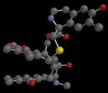
Anti-tumor drug ecteinascidin 743 (ET-743), structure without hydrogens 01
2794
Ecteinascidin 743 (ET-743, brand name Yondelis), was discovered and isolated from a sea squirt, Ecteinascidia turbinata, by NIGMS grantee Kenneth Rinehart at the University of Illinois. Timothy Jamison, Massachusetts Institute of Technology View Media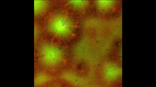
Cell-like compartments emerging from scrambled frog eggs 4
6590
Cell-like compartments that spontaneously emerged from scrambled frog eggs, with nuclei (blue) from frog sperm. Endoplasmic reticulum (red) and microtubules (green) are also visible. Xianrui Cheng, Stanford University School of Medicine. View Media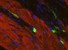
Heart muscle with reprogrammed skin cells
3273
Skins cells were reprogrammed into heart muscle cells. The cells highlighted in green are remaining skin cells. Red indicates a protein that is unique to heart muscle. Deepak Srivastava, Gladstone Institute of Cardiovascular Disease, via CIRM View Media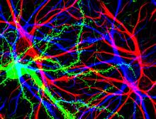
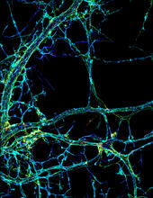
Brain cells in the hippocampus
3688
Hippocampal cells in culture with a neuron in green, showing hundreds of the small protrusions known as dendritic spines. Shelley Halpain, UC San Diego View Media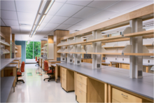
iPS cell facility at the Coriell Institute for Medical Research
2723
This lab space was designed for work on the induced pluripotent stem (iPS) cell collection, part of the NIGMS Human Genetic Cell Repository at the Coriell Institute for Medical Research. Courtney Sill, Coriell Institute for Medical Research View Media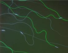
GFP sperm
2683
Fruit fly sperm cells glow bright green when they express the gene for green fluorescent protein (GFP). View Media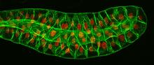
Salivary gland in the developing fruit fly
3603
For fruit flies, the salivary gland is used to secrete materials for making the pupal case, the protective enclosure in which a larva transforms into an adult fly. Richard Fehon, University of Chicago View Media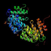
Protein from E. faecalis
2342
X-ray structure of a DNA repair enzyme superfamily representative from the human gastrointestinal bacterium Enterococcus faecalis. Midwest Center for Structural Genomics View Media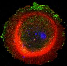
mDia1 antibody staining-01
3330
Cells move forward with lamellipodia and filopodia supported by networks and bundles of actin filaments. Proper, controlled cell movement is a complex process. Rong Li and Praveen Suraneni, Stowers Institute for Medical Research View Media
Neural tube development
2328
Proteins in the neural tissues of this zebrafish embryo direct cells to line up and form the neural tube, which will become the spinal cord and brain. Alexander Schier, Harvard University View Media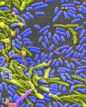
Vibrio bacteria
1160
Vibrio, a type (genus) of rod-shaped bacteria. Some Vibrio species cause cholera in humans. Tina Weatherby Carvalho, University of Hawaii at Manoa View Media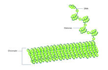
Histones in chromatin (with labels)
2561
Histone proteins loop together with double-stranded DNA to form a structure that resembles beads on a string. Crabtree + Company View Media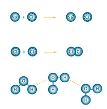
Bond types
2519
Ionic and covalent bonds hold molecules, like sodium chloride and chlorine gas, together. Hydrogen bonds among molecules, notably involving water, also play an important role in biology. Crabtree + Company View Media
Antibodies in silica honeycomb
2750
Antibodies are among the most promising therapies for certain forms of cancer, but patients must take them intravenously, exposing healthy tissues to the drug and increasing the risk of side effects. Chenghong Lei, Pacific Northwest National Laboratory & Karl Erik Hellstrom, University of Washington View Media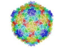
Bacteriophage P22 capsid
5874
Cryo-electron microscopy (cryo-EM) has the power to capture details of proteins and other small biological structures at the molecular level. This image shows proteins in the capsid, or outer co Dr. Wah Chiu, Baylor College of Medicine View Media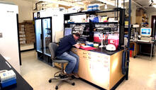
Protein purification facility
2376
The Center for Eukaryotic Structural Genomics protein purification facility is responsible for purifying all recombinant proteins produced by the center. Center for Eukaryotic Structural Genomics View Media
Lily mitosis 06
1016
A light microscope image of a cell from the endosperm of an African globe lily (Scadoxus katherinae). This is one frame of a time-lapse sequence that shows cell division in action. Andrew S. Bajer, University of Oregon, Eugene View Media