Switch to Gallery View
Image and Video Gallery
This is a searchable collection of scientific photos, illustrations, and videos. The images and videos in this gallery are licensed under Creative Commons Attribution Non-Commercial ShareAlike 3.0. This license lets you remix, tweak, and build upon this work non-commercially, as long as you credit and license your new creations under identical terms.
Chang Shan
3483
For thousands of years, Chinese herbalists have treated malaria using Chang Shan, a root extract from a type of hydrangea that grows in Tibet and Nepal. Paul Schimmel Lab, Scripps Research Institute View Media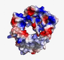
VDAC video 02
2571
This video shows the structure of the pore-forming protein VDAC-1 from humans. Gerhard Wagner, Harvard Medical School View Media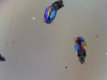
Young squids
6903
Real-time movie of young squids. Michael Shribak, Marine Biological Laboratory/University of Chicago. View Media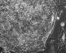
Induced stem cells from adult skin 01
2603
These cells are induced stem cells made from human adult skin cells that were genetically reprogrammed to mimic embryonic stem cells. James Thomson, University of Wisconsin-Madison View Media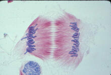
Lily mitosis 11
1011
A light microscope image of cells from the endosperm of an African globe lily (Scadoxus katherinae). This is one frame of a time-lapse sequence that shows cell division in action. Andrew S. Bajer, University of Oregon, Eugene View Media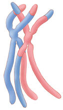
Chromosomes after crossing over
1314
Duplicated pair of chromosomes have exchanged material. Judith Stoffer View Media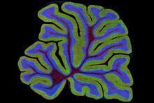
Fluorescent microscopy of kidney tissue
3723
Serum albumin (SA) is the most abundant protein in the blood plasma of mammals. SA has a characteristic heart-shape structure and is a highly versatile protein. Tom Deerinck , National Center for Microscopy and Imaging Research View Media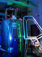
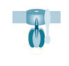
Superconducting magnet
1120
Superconducting magnet for NMR research, from the February 2003 profile of Dorothee Kern in Findings. Mike Lovett View Media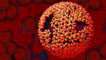
Molecular model of freshly made Rous sarcoma virus (RSV)
3771
Viruses have been the foes of animals and other organisms for time immemorial. Boon Chong Goh, University of Illinois at Urbana-Champaign View Media
Natcher Building 04
1084
NIGMS staff are located in the Natcher Building on the NIH campus. Alisa Machalek, National Institute of General Medical Sciences View Media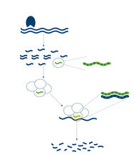
RNA interference
2558
RNA interference or RNAi is a gene-silencing process in which double-stranded RNAs trigger the destruction of specific RNAs. Crabtree + Company View Media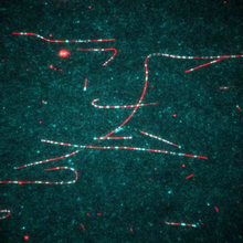
Dynein moving along microtubules
7023
Dynein (green) is a motor protein that “walks” along microtubules (red, part of the cytoskeleton) and carries its cargo along with it. This video was captured through fluorescence microscopy. Morgan DeSantis, University of Michigan. View Media
Nerve cell
1338
Nerve cells have long, invisibly thin fibers that carry electrical impulses throughout the body. Some of these fibers extend about 3 feet from the spinal cord to the toes. Judith Stoffer View Media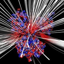
Electrostatic map of human spermine synthase
3658
From PDB entry 3c6k, Crystal structure of human spermine synthase in complex with spermidine and 5-methylthioadenosine. Emil Alexov, Clemson University View Media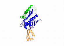
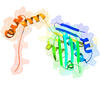
Hsp33 Heat Shock Protein Inactive to Active
3402
When the heat shock protein hsp33 is folded, it is inactive and contains a zinc ion, stabilizing the redox sensitive domain (orange). Dana Reichmann, University of Michigan View Media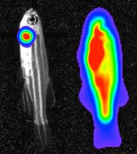
Bioluminescent imaging in adult zebrafish 04
3559
Luciferase-based imaging enables visualization and quantification of internal organs and transplanted cells in live adult zebrafish. View Media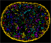
Chromatin in human fibroblast
6888
The nucleus of a human fibroblast cell with chromatin—a substance made up of DNA and proteins—shown in various colors. Melike Lakadamyali, Perelman School of Medicine at the University of Pennsylvania. View Media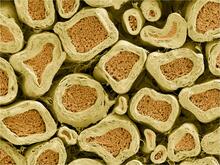
Myelinated axons 2
3397
Top view of myelinated axons in a rat spinal root. Tom Deerinck, National Center for Microscopy and Imaging Research (NCMIR) View Media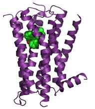
H1 histamine receptor
3360
The receptor is shown bound to an inverse agonist, doxepin. Raymond Stevens, The Scripps Research Institute View Media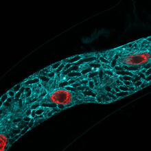
Microsporidia in roundworm 2
5778
Many disease-causing microbes manipulate their host’s metabolism and cells for their own ends. Keir Balla and Emily Troemel, University of California San Diego View Media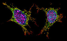
Two mouse fibroblast cells
6789
Two mouse fibroblasts, one of the most common types of cells in mammalian connective tissue. They play a key role in wound healing and tissue repair. Dylan T. Burnette, Vanderbilt University School of Medicine. View Media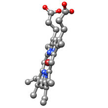
Structure of heme, side view
3540
Molecular model of the struture of heme. Heme is a small, flat molecule with an iron ion (dark red) at its center. Rachel Kramer Green, RCSB Protein Data Bank View Media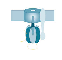
ATP synthase
2517
The world's smallest motor, ATP synthase, generates energy for the cell. See image 2518 for a labeled version of this illustration. Crabtree + Company View Media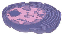
Nucleus and rough ER
1290
The nucleus contains the DNA of eukaryotic cells. Judith Stoffer View Media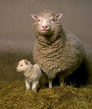
Dolly the sheep
2690
Scientists in Scotland were the first to clone an animal, this sheep named Dolly. She later gave birth to Bonnie, the lamb next to her. View Media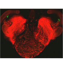
Bacterial cells aggregating above the light organ of the Hawaiian bobtail squid
7018
A light organ (~0.5 mm across) of a juvenile Hawaiian bobtail squid, Euprymna scolopes. Margaret J. McFall-Ngai, Carnegie Institution for Science/California Institute of Technology, and Edward G. Ruby, California Institute of Technology. View Media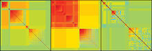
Genetic patchworks
2588
Each point in these colorful patchworks represents the correlation between two sleep-associated genes in fruit flies. Susan Harbison and Trudy Mackay, North Carolina State University View Media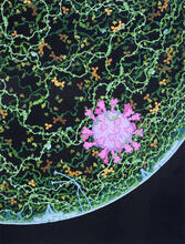
Respiratory droplet
6994
This painting shows a cross section of a small respiratory droplet, like the ones that are thought to transmit SARS-CoV-2, the virus that causes COVID-19. Amy Wu and Christine Zardecki, RCSB Protein Data Bank. View Media
Hsp33 figure 1
3354
Featured in the March 15, 2012 issue of Biomedical Beat. Related to Hsp33 Figure 2, image 3355. Ursula Jakob and Dana Reichmann, University of Michigan View Media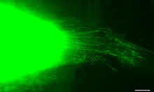
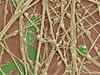
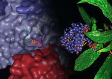
Cytonemes in developing fruit fly cells
3574
Scientists have long known that multicellular organisms use biological molecules produced by one cell and sensed by another to transmit messages that, for instance, guide proper development of organs Sougata Roy, University of California, San Francisco View Media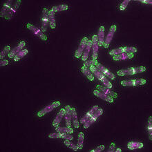
Yeast cells with endocytic actin patches
6793
Yeast cells with endocytic actin patches (green). These patches help cells take in outside material. When a cell is in interphase, patches concentrate at its ends. Alaina Willet, Kathy Gould’s lab, Vanderbilt University. View Media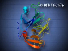
See how immune cell acid destroys bacterial proteins
6602
This animation shows the effect of exposure to hypochlorous acid, which is found in certain types of immune cells, on bacterial proteins. American Chemistry Council View Media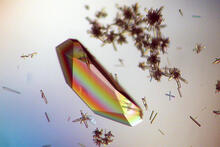
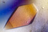
Hen egg lysozyme (1)
2396
Crystals of hen egg lysozyme protein created for X-ray crystallography, which can reveal detailed, three-dimensional protein structures. Alex McPherson, University of California, Irvine View Media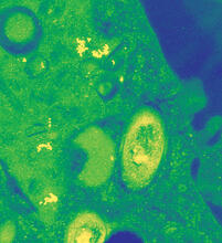
Mycobacterium tuberculosis
2716
Mycobacterium tuberculosis, the bacterium that causes tuberculosis, has infected one-quarter of the world's population and causes more than one million deaths each year, according to the Reuben Peters, Iowa State University View Media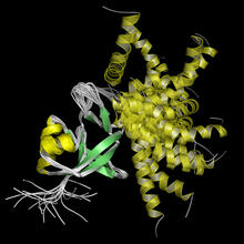
Protein from Arabidopsis thaliana
2339
NMR solution structure of a plant protein that may function in host defense. This protein was expressed in a convenient and efficient wheat germ cell-free system. Center for Eukaryotic Structural Genomics View Media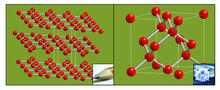
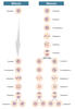
Carbon building blocks (with examples)
2507
The arrangement of identical molecular components can make a dramatic difference. For example, carbon atoms can be arranged into dull graphite (left) or sparkly diamonds (right). Crabtree + Company View MediaAssembly of the HIV capsid
5729
The HIV capsid is a pear-shaped structure that is made of proteins the virus needs to mature and become infective. John Grime and Gregory Voth, The University of Chicago View Media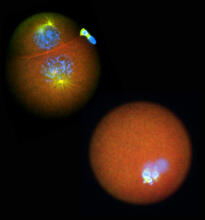
Nucleolinus
2762
The nucleolinus is a cellular compartment that has been a lonely bystander in scientific endeavors. Mary Anne Alliegro, Marine Biological Laboratory View Media
NCMIR mouse tail
3395
Stained cross section of a mouse tail. Tom Deerinck, National Center for Microscopy and Imaging Research (NCMIR) View Media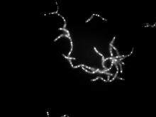
Bacillus anthracis being killed
3481
Bacillus anthracis (anthrax) cells being killed by a fluorescent trans-translation inhibitor, which disrupts bacterial protein synthesis. John Alumasa, Keiler Laboratory, Pennsylvania State University View Media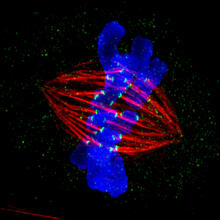
Dividing cell in metaphase
3445
This image of a mammalian epithelial cell, captured in metaphase, was the winning image in the high- and super-resolution microscopy category of the 2012 GE Healthcare Life Sciences Cell Imaging Compe Jane Stout in the laboratory of Claire Walczak, Indiana University, GE Healthcare 2012 Cell Imaging Competition View Media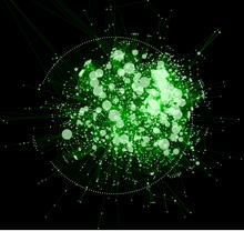
Cytoscape network diagram 1
2737
Molecular biologists are increasingly relying on bioinformatics software to visualize molecular interaction networks and to integrate these networks with data such as gene expression profiles. Keiichiro Ono, Trey Ideker lab, University of California, San Diego View Media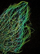
Tiny strands of tubulin, a protein in a cell's skeleton
3611
Just as our bodies rely on bones for structural support, our cells rely on a cellular skeleton. Pakorn Kanchanawong, National University of Singapore and National Heart, Lung, and Blood Institute, National Institutes of Health; and Clare Waterman, National Heart, Lung, and Blood Institute, National Institutes of Health View Media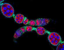
Confocal microscopy image of two Drosophila ovarioles
5772
Ovarioles in female insects are tubes in which egg cells (called oocytes) form at one end and complete their development as they reach the other end of the tube. 2004 Olympus BioScapes Competition View Media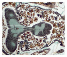
Cross section of a Drosophila melanogaster pupa
2758
This photograph shows a magnified view of a Drosophila melanogaster pupa in cross section. Compare this normal pupa to one that lacks an important receptor, shown in image 2759. Christina McPhee and Eric Baehrecke, University of Massachusetts Medical School View Media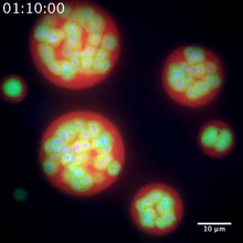
Nucleolus subcompartments spontaneously self-assemble 2
3791
The nucleolus is a small but very important protein complex located in the cell's nucleus. Nilesh Vaidya, Princeton University View Media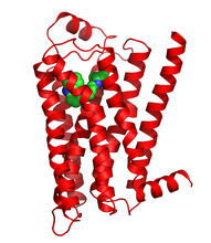
Beta 2-adrenergic receptor
3358
The receptor is shown bound to a partial inverse agonist, carazolol. Raymond Stevens, The Scripps Research Institute View Media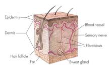
Skin cross-section
1056
Cross-section of skin anatomy shows layers and different tissue types. National Institutes of Health Medical Arts View Media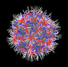
Electrostatic map of the adeno-associated virus
3374
The new highly efficient parallelized DelPhi software was used to calculate the potential map distribution of an entire virus, the adeno-associated virus, which is made up of more than 484,000 atoms. Emil Alexov, Clemson University View Media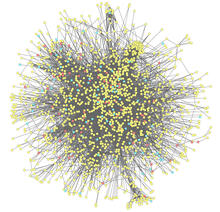
Molecular interactions
2743
This network map shows molecular interactions (yellow) associated with a congenital condition that causes heart arrhythmias and the targets for drugs that alter these interactions (red and blue). Ravi Iyengar, Mount Sinai School of Medicine View Media