Switch to Gallery View
Image and Video Gallery
This is a searchable collection of scientific photos, illustrations, and videos. The images and videos in this gallery are licensed under Creative Commons Attribution Non-Commercial ShareAlike 3.0. This license lets you remix, tweak, and build upon this work non-commercially, as long as you credit and license your new creations under identical terms.
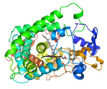
Cytochrome structure with anticancer drug
3326
This image shows the structure of the CYP17A1 enzyme (ribbons colored from blue N-terminus to red C-terminus), with the associated heme colored black. Emily Scott, University of Kansas View Media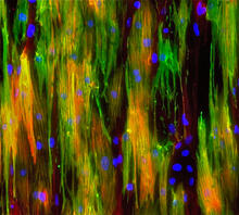
Mouse heart muscle cells 02
3283
This image shows neonatal mouse heart cells. These cells were grown in the lab on a chip that aligns the cells in a way that mimics what is normally seen in the body. Kara McCloskey lab, University of California, Merced, via CIRM View Media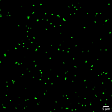
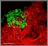
Staphylococcus aureus aggregating upon contact with synovial fluid
6805
Staphylococcus aureus bacteria (green) grouping together upon contact with synovial fluid—a viscous substance found in joints. Paul Stoodley, The Ohio State University. View Media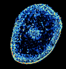
Chromatin in human tenocyte
6893
The nucleus of a degenerating human tendon cell, also known as a tenocyte. It has been color-coded based on the density of chromatin—a substance made up of DNA and proteins. Melike Lakadamyali, Perelman School of Medicine at the University of Pennsylvania. View Media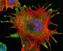
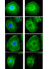
mDia1 antibody staining- 02
3331
Cells move forward with lamellipodia and filopodia supported by networks and bundles of actin filaments. Proper, controlled cell movement is a complex process. Rong Li and Praveen Suraneni, Stowers Institute for Medical Research View Media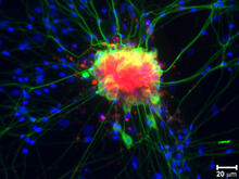
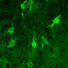
Spinal nerve cells
3251
Neurons (green) and glial cells from isolated dorsal root ganglia express COX-2 (red) after exposure to an inflammatory stimulus (cell nuclei are blue). Lawrence Marnett, Vanderbilt University View Media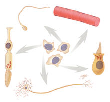
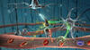
Stem cell differentiation
1294
Undifferentiated embryonic stem cells cease to exist a few days after conception. In this image, ES cells are shown to differentiate into sperm, muscle fiber, hair cells, nerve cells, and cone cells. Judith Stoffer View Media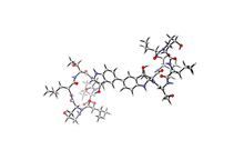
Himastatin
6848
A model of the molecule himastatin, which was first isolated from the bacterium Streptomyces himastatinicus. Himastatin shows antibiotic activity. Mohammad Movassaghi, Massachusetts Institute of Technology. View Media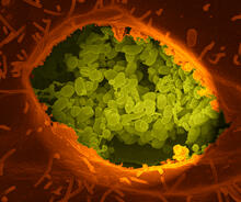
Q fever bacteria in an infected cell
3621
This image shows Q fever bacteria (yellow), which infect cows, sheep, and goats around the world and can infect humans, as well. When caught early, Q fever can be cured with antibiotics. Robert Heinzen, Elizabeth Fischer, and Anita Mora, National Institute of Allergy and Infectious Diseases, National Institutes of Health View Media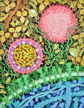
Zika virus
6998
Zika virus is shown in cross section at center left. On the outside, it includes envelope protein (red) and membrane protein (magenta) embedded in a lipid membrane (light purple). Amy Wu and Christine Zardecki, RCSB Protein Data Bank. View Media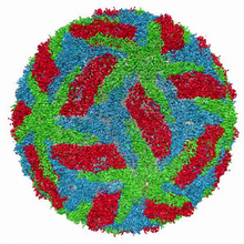
Cryo-electron microscopy of the dengue virus showing protective membrane and membrane proteins
3748
Dengue virus is a mosquito-borne illness that infects millions of people in the tropics and subtropics each year. Like many viruses, dengue is enclosed by a protective membrane. Hong Zhou, UCLA View Media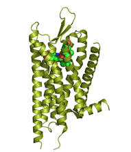
Kappa opioid receptor
3359
The receptor is shown bound to an antagonist, JDTic. Raymond Stevens, The Scripps Research Institute View Media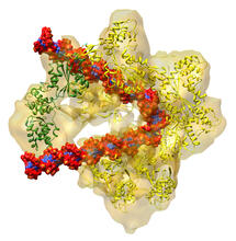
DNA replication origin recognition complex (ORC)
3597
A study published in March 2012 used cryo-electron microscopy to determine the structure of the DNA replication origin recognition complex (ORC), a semi-circular, protein complex (yellow) that recogni Huilin Li, Brookhaven National Laboratory View Media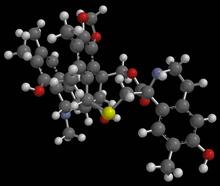
Anti-tumor drug ecteinascidin 743 (ET-743) with hydrogens 03
2792
Ecteinascidin 743 (ET-743, brand name Yondelis), was discovered and isolated from a sea squirt, Ecteinascidia turbinata, by NIGMS grantee Kenneth Rinehart at the University of Illinois. Timothy Jamison, Massachusetts Institute of Technology View Media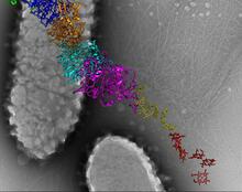
Bacterial nanowire model
6580
A model of a Geobacter sulfurreducens nanowire created from cryo-electron microscopy images. Edward Egelman, University of Virginia. View Media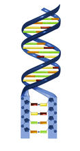
Nucleotides make up DNA
2541
DNA consists of two long, twisted chains made up of nucleotides. Each nucleotide contains one base, one phosphate molecule, and the sugar molecule deoxyribose. Crabtree + Company View Media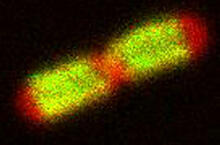
TonB protein in gram-negative bacteria
3549
The green in this image highlights a protein called TonB, which is produced by many gram-negative bacteria, including those that cause typhoid fever, meningitis and dysentery. Phillip Klebba, Kansas State University View Media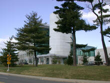
Natcher Building 02
1082
NIGMS staff are located in the Natcher Building on the NIH campus. Alisa Machalek, National Institute of General Medical Sciences View Media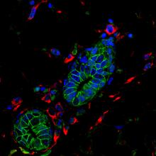
Mouse mammary cells lacking anti-cancer protein
3432
Shortly after a pregnant woman gives birth, her breasts start to secrete milk. This process is triggered by hormonal and genetic cues, including the protein Elf5. Nature Cell Biology, November 2012, Volume 14 No 11 pp1113-1231 View Media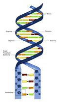
Nucleotides make up DNA (with labels)
2542
DNA consists of two long, twisted chains made up of nucleotides. Each nucleotide contains one base, one phosphate molecule, and the sugar molecule deoxyribose. Crabtree + Company View Media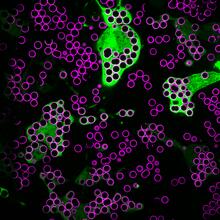
Hungry, hungry macrophages
7009
Macrophages (green) are the professional eaters of our immune system. Meghan Morrissey, University of California, Santa Barbara. View Media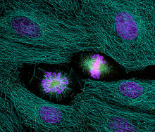
Highlighted cells
2429
The cytoskeleton (green) and DNA (purple) are highlighed in these cells by immunofluorescence. Torsten Wittmann, Scripps Research Institute View Media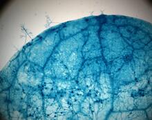
Disease-susceptible Arabidopsis leaf
2782
This is a magnified view of an Arabidopsis thaliana leaf after several days of infection with the pathogen Hyaloperonospora arabidopsidis. Jeff Dangl, University of North Carolina, Chapel Hill View Media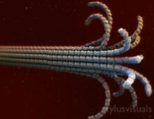
Microtubule breakdown
2321
Like a building supported by a steel frame, a cell contains its own sturdy internal scaffolding made up of proteins, including microtubules. Eva Nogales, University of California, Berkeley View Media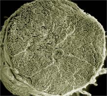
NCMIR human spinal nerve
3387
Spinal nerves are part of the peripheral nervous system. They run within the spinal column to carry nerve signals to and from all parts of the body. Tom Deerinck, National Center for Microscopy and Imaging Research (NCMIR) View Media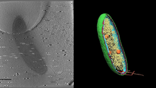
Cryo-electron tomography of a Caulobacter bacterium
6569
3D image of Caulobacter bacterium with various components highlighted: cell membranes (red and blue), protein shell (green), protein factories known as ribosomes (yellow), and storage granules Peter Dahlberg, Stanford University. View Media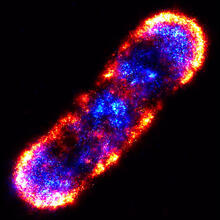
Self-organizing proteins
2771
Under the microscope, an E. coli cell lights up like a fireball. Each bright dot marks a surface protein that tells the bacteria to move toward or away from nearby food and toxins. View Media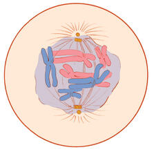
Mitosis - prometaphase
1331
A cell in prometaphase during mitosis: The nuclear membrane breaks apart, and the spindle starts to interact with the chromosomes. Judith Stoffer View Media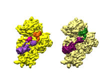
Bacterial ribosome assembly
6578
3D reconstructions of two stages in the assembly of the bacterial ribosome created from time-resolved cryo-electron microscopy images. Ribosomes translate genetic instructions into proteins. Joachim Frank, Columbia University. View Media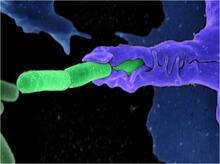
Anthrax bacteria (green) being swallowed by an immune system cell
3612
Multiple anthrax bacteria (green) being enveloped by an immune system cell (purple). Anthrax bacteria live in soil and form dormant spores that can survive for decades. Camenzind G. Robinson, Sarah Guilman, and Arthur Friedlander, United States Army Medical Research Institute of Infectious Diseases View Media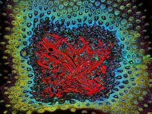
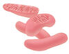
Tongue 1
5810
Microscopy image of tongue. One in a series of two, see image 5811 National Center for Microscopy and Imaging Research (NCMIR) View Media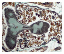
Cross section of a Drosophila melanogaster pupa
2758
This photograph shows a magnified view of a Drosophila melanogaster pupa in cross section. Compare this normal pupa to one that lacks an important receptor, shown in image 2759. Christina McPhee and Eric Baehrecke, University of Massachusetts Medical School View Media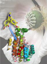
Atomic Structure of Poppy Enzyme
3422
The atomic structure of the morphine biosynthetic enzyme salutaridine reductase bound to the cofactor NADPH. The substrate salutaridine is shown entering the active site. Judy Coyle, Donald Danforth Plant Science Center View Media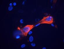
Smooth muscle from mouse stem cells
3289
These smooth muscle cells were derived from mouse neural crest stem cells. Red indicates smooth muscle proteins, blue indicates nuclei. Deepak Srivastava, Gladstone Institutes, via CIRM View Media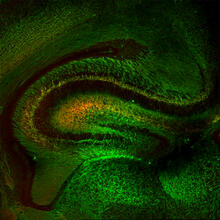
Mouse Brain Cross Section
5886
The brain sections are treated with fluorescent antibodies specific to a particular protein and visualized using serial electron microscopy (SEM). Anton Maximov, The Scripps Research Institute, La Jolla, CA View Media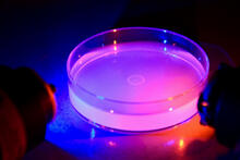
Petri dish containing C. elegans
6751
This Petri dish contains microscopic roundworms called Caenorhabditis elegans. Researchers used these particular worms to study how C. H. Robert Horvitz and Dipon Ghosh, Massachusetts Institute of Technology. View Media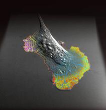
Biosensors illustration
2802
A rendering of an activity biosensor image overlaid with a cell-centered frame of reference used for image analysis of signal transduction. Gaudenz Danuser, Harvard Medical School View Media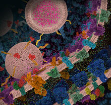
Molecular view of glutamatergic synapse
6992
This illustration highlights spherical pre-synaptic vesicles that carry the neurotransmitter glutamate. Amy Wu and Christine Zardecki, RCSB Protein Data Bank. View Media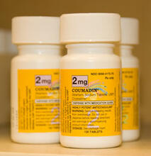
Bottles of warfarin
2579
In 2007, the FDA modified warfarin's label to indicate that genetic makeup may affect patient response to the drug. The widely used blood thinner is sold under the brand name Coumadin®. Alisa Machalek, NIGMS/NIH View Media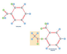
Aspirin (with labels)
2530
Acetylsalicylate (bottom) is the aspirin of today. Crabtree + Company View Media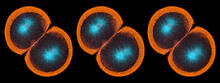
Sea urchin embryo 06
1052
Stereo triplet of a sea urchin embryo stained to reveal actin filaments (orange) and microtubules (blue). George von Dassow, University of Washington View Media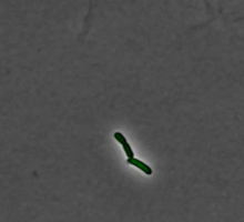
Pulsating response to stress in bacteria - video
3254
By attaching fluorescent proteins to the genetic circuit responsible for B. subtilis's stress response, researchers can observe the cells' pulses as green flashes. Michael Elowitz, Caltech University View Media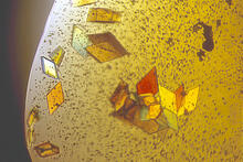
Jack bean concanavalin A
2407
Crystals of jack bean concanavalin A protein created for X-ray crystallography, which can reveal detailed, three-dimensional protein structures. Alex McPherson, University of California, Irvine View Media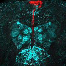
Fruit fly brain responds to adipokines
6985
Drosophila adult brain showing that an adipokine (fat hormone) generates a response from neurons (aqua) and regulates insulin-producing neurons (red).Akhila Rajan, Fred Hutchinson Cancer Center View Media
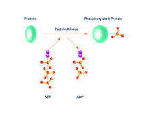
Kinases (with labels)
2535
Kinases are enzymes that add phosphate groups (red-yellow structures) to proteins (green), assigning the proteins a code. Crabtree + Company View Media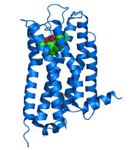
Dopamine D3 receptor
3363
The receptor is shown bound to an antagonist, eticlopride Raymond Stevens, The Scripps Research Institute View Media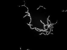
Bacillus anthracis being killed
3481
Bacillus anthracis (anthrax) cells being killed by a fluorescent trans-translation inhibitor, which disrupts bacterial protein synthesis. John Alumasa, Keiler Laboratory, Pennsylvania State University View Media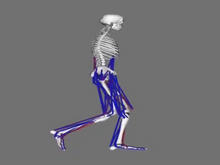
Simulation of leg muscles moving
6598
When we walk, muscles and nerves interact in intricate ways. This simulation, which is based on data from a six-foot-tall man, shows these interactions. Chand John and Eran Guendelman, Stanford University View Media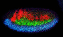
Neural development
2327
Using techniques that took 4 years to design, a team of developmental biologists showed that certain proteins can direct the subdivision of fruit fly and chicken nervous system tissue into the regions Mieko Mizutani and Ethan Bier, University of California, San Diego, and Henk Roelink, University of Washington View Media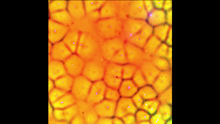
Cell-like compartments emerging from scrambled frog eggs
6587
Cell-like compartments spontaneously emerge from scrambled frog eggs, with nuclei (blue) from frog sperm. Endoplasmic reticulum (red) and microtubules (green) are also visible. Xianrui Cheng, Stanford University School of Medicine. View Media