Switch to Gallery View
Image and Video Gallery
This is a searchable collection of scientific photos, illustrations, and videos. The images and videos in this gallery are licensed under Creative Commons Attribution Non-Commercial ShareAlike 3.0. This license lets you remix, tweak, and build upon this work non-commercially, as long as you credit and license your new creations under identical terms.
HIV Infected Cell
3386
The human immunodeficiency virus (HIV), shown here as tiny purple spheres, causes the disease known as AIDS (for acquired immunodeficiency syndrome). Tom Deerinck, National Center for Microscopy and Imaging Research (NCMIR) View Media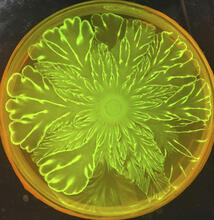
Floral pattern in a mixture of two bacterial species, Acinetobacter baylyi and Escherichia coli, grown on a semi-solid agar for 72 hour
6556
Floral pattern emerging as two bacterial species, motile Acinetobacter baylyi and non-motile Escherichia coli (green), are grown together for 72 hours on 0.5% agar surface from a small i L. Xiong et al, eLife 2020;9: e48885 View Media
Centrioles anchor cilia in planaria
3292
Centrioles (green) anchor cilia (red), which project on the surface of pharynx cells of the freshwater planarian Schmidtea mediterranea. Juliette Azimzadeh, University of California, San Francisco View Media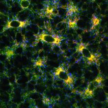
Endoplasmic reticulum
2649
Fluorescent markers show the interconnected web of tubes and compartments in the endoplasmic reticulum. The protein atlastin helps build and maintain this critical part of cells. Andrea Daga, Eugenio Medea Scientific Institute (Conegliano, Italy) View MediaBeta-galactosidase montage showing cryo-EM improvement--transparent background
5882
Composite image of beta-galactosidase showing how cryo-EM’s resolution has improved dramatically in recent years. Older images to the left, more recent to the right. Veronica Falconieri, Sriram Subramaniam Lab, National Cancer Institute View Media
Mapping human genetic variation
2443
This map paints a colorful portrait of human genetic variation around the world. Noah Rosenberg and Martin Soave, University of Michigan View Media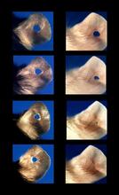
Regeneration of Mouse Ears
3426
Normal mice, like the B6 breed pictured on the left, develop scars when their ears are pierced. Ellen Heber-Katz, The Wistar Institute View Media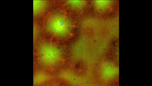
Cell-like compartments emerging from scrambled frog eggs 4
6590
Cell-like compartments that spontaneously emerged from scrambled frog eggs, with nuclei (blue) from frog sperm. Endoplasmic reticulum (red) and microtubules (green) are also visible. Xianrui Cheng, Stanford University School of Medicine. View Media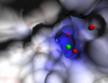
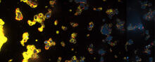
V. Cholerae Biofilm
3580
Industrious V. cholerae bacteria (yellow) tend to thrive in denser biofilms (left) while moochers (red) thrive in weaker biofilms (right). View Media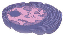
Nucleus and rough ER
1290
The nucleus contains the DNA of eukaryotic cells. Judith Stoffer View Media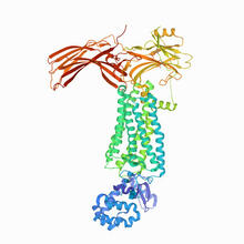
Rhodopsin bound to visual arrestin
6768
Rhodopsin is a pigment in the rod cells of the retina (back of the eye). It is extremely light-sensitive, supporting vision in low-light conditions. Protein Data Bank. View Media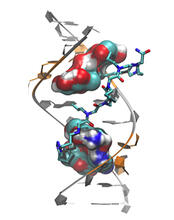
Myotonic dystrophy type 2 genetic defect
3573
Scientists revealed a detailed image of the genetic defect that causes myotonic dystrophy type 2 and used that information to design drug candidates to counteract the disease. Matthew Disney, Scripps Research Institute and Ilyas Yildirim, Northwestern University View Media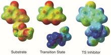
Enzyme transition states
3429
The molecule on the left is an electrostatic potential map of the van der Waals surface of the transition state for human purine nucleoside phosphorylase. Vern Schramm, Albert Einstein College of Medicine of Yeshiva University View Media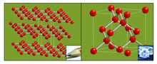
Carbon building blocks (with examples)
2507
The arrangement of identical molecular components can make a dramatic difference. For example, carbon atoms can be arranged into dull graphite (left) or sparkly diamonds (right). Crabtree + Company View Media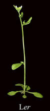
Mature, flowering Arabidopsis
2779
This is an adult flowering Arabidopsis thaliana plant with the inbred designation L-er. Arabidopsis is the most widely used model organism for researchers who study plant genetics. Jeff Dangl, University of North Carolina, Chapel Hill View Media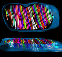
Mitochondrion from insect flight muscle
3662
This is a tomographic reconstruction of a mitochondrion from an insect flight muscle. National Center for Microscopy and Imaging Research View Media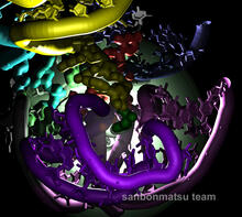
Natural nanomachine in action
2336
Using a supercomputer to simulate the movement of atoms in a ribosome, researchers looked into the core of this protein-making nanomachine and took snapshots. Kevin Sanbonmatsu, Los Alamos National Laboratory View Media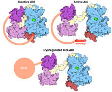
Protein kinases as cancer chemotherapy targets
7004
Protein kinases—enzymes that add phosphate groups to molecules—are cancer chemotherapy targets because they play significant roles in almost all aspects of cell function, are tightly regulated, and co Amy Wu and Christine Zardecki, RCSB Protein Data Bank. View Media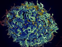
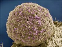
HIV, the AIDS virus, infecting a human cell
3638
This human T cell (blue) is under attack by HIV (yellow), the virus that causes AIDS. Seth Pincus, Elizabeth Fischer, and Austin Athman, National Institute of Allergy and Infectious Diseases, National Institutes of Health View Media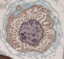
Transmission electron microscopy showing cross-section of the node of Ranvier
3740
Nodes of Ranvier are short gaps in the myelin sheath surrounding myelinated nerve cells (axons). Tom Deerinck, National Center for Microscopy and Imaging Research (NCMIR) View Media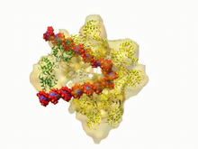
DNA replication origin recognition complex (ORC)
3307
A study published in March 2012 used cryo-electron microscopy to determine the structure of the DNA replication origin recognition complex (ORC), a semi-circular, protein complex (yellow) that recogni Huilin Li, Brookhaven National Laboratory View Media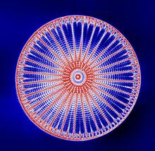
Arachnoidiscus diatom
6902
An Arachnoidiscus diatom with a diameter of 190µm. Michael Shribak, Marine Biological Laboratory/University of Chicago. View Media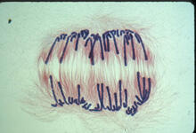
Lily mitosis 10
1010
A light microscope image of a cell from the endosperm of an African globe lily (Scadoxus katherinae). This is one frame of a time-lapse sequence that shows cell division in action. Andrew S. Bajer, University of Oregon, Eugene View Media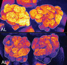
Sleep and the fly brain
2596
In the top snapshots, the brain of a sleep-deprived fruit fly glows orange, marking high concentrations of a synaptic protein called Bruchpilot (BRP) involved in communication between neurons. Chiara Cirelli, University of Wisconsin-Madison View Media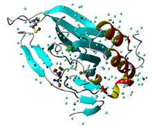
Cysteine dioxygenase from mouse
2347
Model of the mammalian iron enzyme cysteine dioxygenase from a mouse. Center for Eukaryotic Structural Genomics, PSI View Media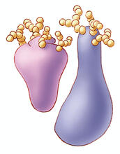
Glycoproteins
1270
About half of all human proteins include chains of sugar molecules that are critical for the proteins to function properly. Appears in the NIGMS booklet Inside the Cell. Judith Stoffer View Media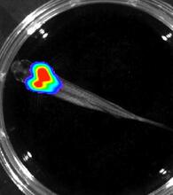
Bioluminescent imaging in adult zebrafish - overhead view
3557
Luciferase-based imaging enables visualization and quantification of internal organs and transplanted cells in live adult zebrafish. Kenneth Poss, Duke University View Media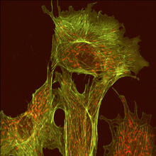
Endothelial cell
1102
This image shows two components of the cytoskeleton, microtubules (green) and actin filaments (red), in an endothelial cell derived from a cow lung. Tina Weatherby Carvalho, University of Hawaii at Manoa View Media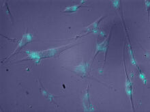
Molecules blocking Huntington's protein production
2600
The molecules that glow blue in these cultured cells prevent the expression of the mutant proteins that cause Huntington's disease. Jiaxin Hu, David W. Dodd and Robert H. E. Hudson, UT Southwestern Medical Center View Media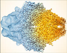
Beta-galactosidase montage showing cryo-EM improvement--gradient background
5883
Composite image of beta-galactosidase showing how cryo-EM’s resolution has improved dramatically in recent years. Older images to the left, more recent to the right. Veronica Falconieri, Sriram Subramaniam Lab, National Cancer Institute View Media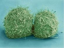
HeLa cells
3518
Scanning electron micrograph of just-divided HeLa cells. Zeiss Merlin HR-SEM. National Center for Microscopy and Imaging Research View Media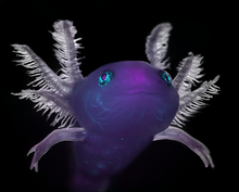
Axolotl
6932
An axolotl—a type of salamander—that has been genetically modified so that its developing nervous system glows purple and its Schwann cell nuclei appear light blue. Prayag Murawala, MDI Biological Laboratory and Hannover Medical School. View Media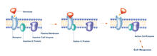
G switch (with labels)
2537
The G switch allows our bodies to respond rapidly to hormones. G proteins act like relay batons to pass messages from circulating hormones into cells. Crabtree + Company View Media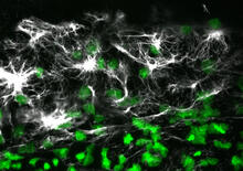
Vimentin in a quail embryo
2807
Confocal image showing high levels of the protein vimentin (white) at the edge zone of a quail embryo. Cell nuclei are labeled green. Andrés Garcia, Georgia Tech View Media
Proteins related to myotonic dystrophy
2727
Myotonic dystrophy is thought to be caused by the binding of a protein called Mbnl1 to abnormal RNA repeats. Manuel Ares, University of California, Santa Cruz View Media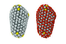
Cryo-EM reveals how the HIV capsid attaches to a human protein to evade immune detection
3755
The illustration shows the capsid of human immunodeficiency virus (HIV) whose molecular features were resolved with cryo-electron microscopy (cryo-EM). Juan R. Perilla, University of Illinois at Urbana-Champaign View Media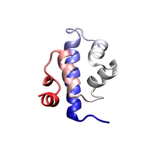
Protein folding video
3391
Proteins are long chains of amino acids. Each protein has a unique amino acid sequence. It is still a mystery how a protein folds into the proper shape based on its sequence. Theoretical and Computational Biophysics Group View Media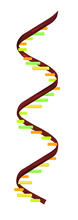
RNA strand
2554
Ribonucleic acid (RNA) has a sugar-phosphate backbone and the bases adenine (A), cytosine (C), guanine (G), and uracil (U). Crabtree + Company View Media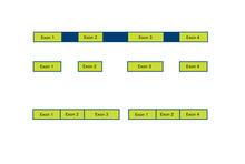
Alternative splicing
2552
Arranging exons in different patterns, called alternative splicing, enables cells to make different proteins from a single gene. Crabtree + Company View Media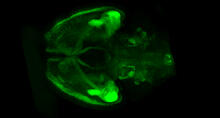
Mouse brain 1
6929
A mouse brain that was genetically modified so that subpopulations of its neurons glow. Prayag Murawala, MDI Biological Laboratory and Hannover Medical School. View Media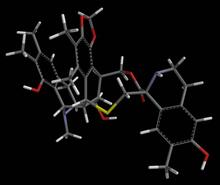
Anti-tumor drug ecteinascidin 743 (ET-743) with hydrogens 04
2793
Ecteinascidin 743 (ET-743, brand name Yondelis), was discovered and isolated from a sea squirt, Ecteinascidia turbinata, by NIGMS grantee Kenneth Rinehart at the University of Illinois. Timothy Jamison, Massachusetts Institute of Technology View Media
Circadian rhythm neurons in the fruit fly brain
3754
Some nerve cells (neurons) in the brain keep track of the daily cycle. This time-keeping mechanism, called the circadian clock, is found in all animals including us. Justin Blau, New York University View Media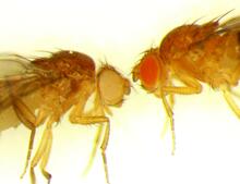
Drosophila
6344
Two adult fruit flies (Drosophila) Dr. Vicki Losick, MDI Biological Laboratory, www.mdibl.org View Media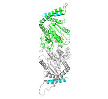
Dynamin structure
2744
When a molecule arrives at a cell's outer membrane, the membrane creates a pouch around the molecule that protrudes inward. Josh Chappie, National Institute of Diabetes and Digestive and Kidney Diseases, NIH View Media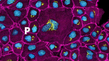
Pathways: The Fascinating Cells of Research Organisms
6538
Learn how research organisms, such as fruit flies and mice, can help us understand and treat human diseases. National Institute of General Medical Sciences View Media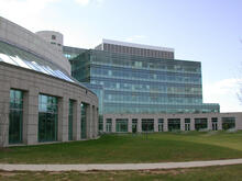
Natcher Building 05
1085
NIGMS staff are located in the Natcher Building on the NIH campus. Alisa Machalek, National Institute of General Medical Sciences View Media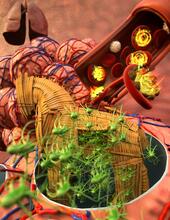
Host infection stimulates antibiotic resistance
5764
This illustration shows pathogenic bacteria behave like a Trojan horse: switching from antibiotic susceptibility to resistance during infection. View Media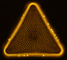
Trigonium diatom
6962
A Trigonium diatom imaged by a quantitative orientation-independent differential interference contrast (OI-DIC) microscope. Michael Shribak, Marine Biological Laboratory/University of Chicago. View Media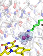
O2 reacting with a flavin-dependent enzyme
3411
Department of Biological Chemistry, University of Michigan View Media