Image Gallery: Dividing cell
Video file
ID
6965
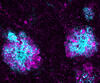
As this cell was undergoing cell division, it was imaged with two microscopy techniques: differential interference contrast (DIC) and confocal. The DIC view appears in blue and shows the entire cell. The confocal view appears in pink and shows the chromosomes.
Source
Dylan T. Burnette, Vanderbilt University School of Medicine.
Topics