Switch to Gallery View
Image and Video Gallery
This is a searchable collection of scientific photos, illustrations, and videos. The images and videos in this gallery are licensed under Creative Commons Attribution Non-Commercial ShareAlike 3.0. This license lets you remix, tweak, and build upon this work non-commercially, as long as you credit and license your new creations under identical terms.
Multivesicular bodies containing intralumenal vesicles assemble at the vacuole 1
5769
Collecting and transporting cellular waste and sorting it into recylable and nonrecylable pieces is a complex business in the cell. Matthew West and Greg Odorizzi, University of Colorado View Media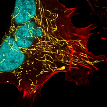
Multinucleated cancer cell
6967
A cancer cell with three nuclei, shown in turquoise. The abnormal number of nuclei indicates that the cell failed to go through cell division, probably more than once. Dylan T. Burnette, Vanderbilt University School of Medicine. View Media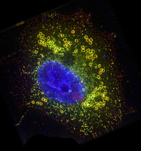
Cell Nucleus and Lipid Droplets
6547
A cell nucleus (blue) surrounded by lipid droplets (yellow). James Olzmann, University of California, Berkeley View Media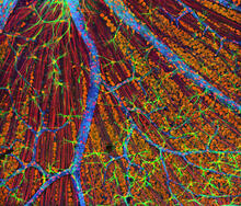
Mouse Retina
3309
A genetic disorder of the nervous system, neurofibromatosis causes tumors to form on nerves throughout the body, including a type of tumor called an optic nerve glioma that can result in childhood bli Tom Deerinck, NCMIR View Media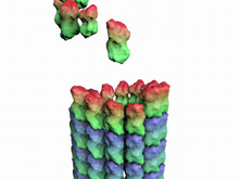
How a microtubule builds and deconstructs
3650
A microtubule, part of the cell's skeleton, builds and deconstructs. View Media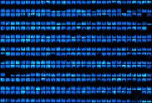
Biopixels
3266
Bioengineers were able to coax bacteria to blink in unison on microfluidic chips. This image shows a small chip with about 500 blinking bacterial colonies or biopixels. Jeff Hasty Lab, UC San Diego View Media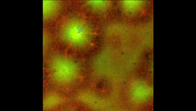
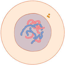
Cell-like compartments emerging from scrambled frog eggs 4
6590
Cell-like compartments that spontaneously emerged from scrambled frog eggs, with nuclei (blue) from frog sperm. Endoplasmic reticulum (red) and microtubules (green) are also visible. Xianrui Cheng, Stanford University School of Medicine. View Media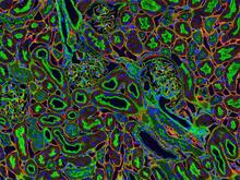
NCMIR kidney-1
3675
Stained kidney tissue. The kidney is an essential organ responsible for disposing wastes from the body and for maintaining healthy ion levels in the blood. Tom Deerinck, National Center for Microscopy and Imaging Research (NCMIR) View Media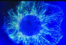
Lily mitosis 01
1058
A light microscope image shows the chromosomes, stained dark blue, in a dividing cell of an African globe lily (Scadoxus katherinae). Andrew S. Bajer, University of Oregon, Eugene View Media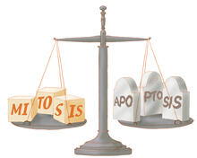
Life in balance
1336
Mitosis creates cells, and apoptosis kills them. The processes often work together to keep us healthy. Judith Stoffer View Media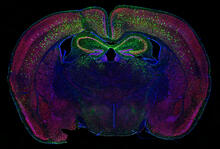
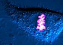
Calling Cards in a mouse brain
6780
The green spots in this mouse brain are cells labeled with Calling Cards, a technology that records molecular events in brain cells as they mature. Allen Yen, Lab of Joseph Dougherty, Washington University School of Medicine in St. Louis. View Media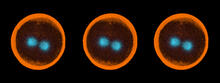
Sea urchin embryo 04
1050
Stereo triplet of a sea urchin embryo stained to reveal actin filaments (orange) and microtubules (blue). George von Dassow, University of Washington View Media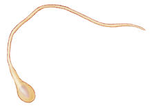
Mitosis - interphase
1316
A cell in interphase, at the start of mitosis: Chromosomes duplicate, and the copies remain attached to each other. Judith Stoffer View Media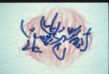
Lily mitosis 06
1016
A light microscope image of a cell from the endosperm of an African globe lily (Scadoxus katherinae). This is one frame of a time-lapse sequence that shows cell division in action. Andrew S. Bajer, University of Oregon, Eugene View Media
Dividing cell
6965
As this cell was undergoing cell division, it was imaged with two microscopy techniques: differential interference contrast (DIC) and confocal. The DIC view appears in blue and shows the entire cell. Dylan T. Burnette, Vanderbilt University School of Medicine. View Media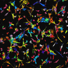
Single-cell “radios” image
7021
Individual cells are color-coded based on their identity and signaling activity using a protein circuit technology developed by the Coyle Lab. Scott Coyle, University of Wisconsin-Madison. View Media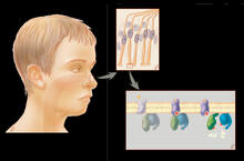
Olfactory system
1291
Sensory organs have cells equipped for detecting signals from the environment, such as odors. Judith Stoffer View Media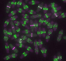
Yeast cells entering mitosis
6791
Yeast cells entering mitosis, also known as cell division. The green and magenta dots are two proteins that play important roles in mitosis. They show where the cells will split. Alaina Willet, Kathy Gould’s lab, Vanderbilt University. View Media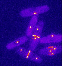
Dividing yeast cells with spindle pole bodies and contractile rings
6796
During cell division, spindle pole bodies (glowing dots) move toward the ends of yeast cells to separate copied genetic information. Alaina Willet, Kathy Gould’s lab, Vanderbilt University. View Media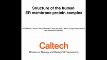
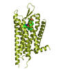
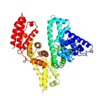
Human endoplasmic reticulum membrane protein complex
6777
A 3D model of the human endoplasmic reticulum membrane protein complex (EMC) that identifies its nine essential subunits. Rebecca Voorhees, California Institute of Technology. View Media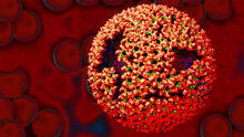
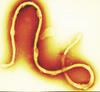
Molecular model of freshly made Rous sarcoma virus (RSV)
3771
Viruses have been the foes of animals and other organisms for time immemorial. Boon Chong Goh, University of Illinois at Urbana-Champaign View Media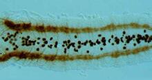
Developing fruit fly nerve cord
2435
The glial cells (black dots) and nerve cells (brown bands) in this developing fruit fly nerve cord formed normally despite the absence of the SPITZ protein, which blocks their impending suicide. Hermann Steller, Rockefeller University View Media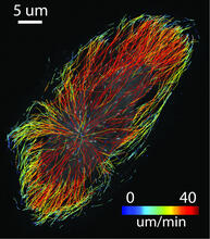
Microtubule growth
2800
Map of microtubule growth rates. Rates are color coded. This is an example of NIH-supported research on single-cell analysis. Gaudenz Danuser, Harvard Medical School View Media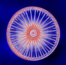
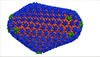
Arachnoidiscus diatom
6902
An Arachnoidiscus diatom with a diameter of 190µm. Michael Shribak, Marine Biological Laboratory/University of Chicago. View Media
“Two-faced” Janus particle activating a macrophage
6801
A macrophage—a type of immune cell that engulfs invaders—“eats” and is activated by a “two-faced” Janus particle. Yan Yu, Indiana University, Bloomington. View Media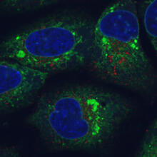
Endoplasmic reticulum abnormalities
6773
Human cells with the gene that codes for the protein FIT2 deleted. Green indicates an endoplasmic reticulum (ER) resident protein. Michel Becuwe, Harvard University. View Media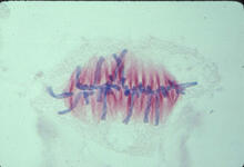
Lily mitosis 07
1017
A light microscope image of a cell from the endosperm of an African globe lily (Scadoxus katherinae). This is one frame of a time-lapse sequence that shows cell division in action. Andrew S. Bajer, University of Oregon, Eugene View Media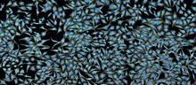
A panorama view of cells
5761
This photograph shows a panoramic view of HeLa cells, a cell line many researchers use to study a large variety of important research questions. Tom Deerinck, National Center for Microscopy and Imaging Research View Media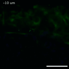
Vimentin in a quail embryo
2809
Video of high-resolution confocal images depicting vimentin immunofluorescence (green) and nuclei (blue) at the edge of a quail embryo yolk. Andrés Garcia, Georgia Tech View Media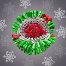
H1N1 Influenza Virus
6355
CellPack image of the H1N1 influenza virus, with hemagglutinin and neuraminidase glycoproteins in green and red, respectively, on the outer envelope (white); matrix protein in gray, and ribonucleoprot Dr. Rommie Amaro, University of California, San Diego View Media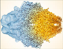
Beta-galactosidase montage showing cryo-EM improvement--gradient background
5883
Composite image of beta-galactosidase showing how cryo-EM’s resolution has improved dramatically in recent years. Older images to the left, more recent to the right. Veronica Falconieri, Sriram Subramaniam Lab, National Cancer Institute View Media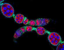
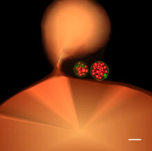
Confocal microscopy image of two Drosophila ovarioles
5772
Ovarioles in female insects are tubes in which egg cells (called oocytes) form at one end and complete their development as they reach the other end of the tube. 2004 Olympus BioScapes Competition View MediaBeta-galactosidase montage showing cryo-EM improvement--transparent background
5882
Composite image of beta-galactosidase showing how cryo-EM’s resolution has improved dramatically in recent years. Older images to the left, more recent to the right. Veronica Falconieri, Sriram Subramaniam Lab, National Cancer Institute View Media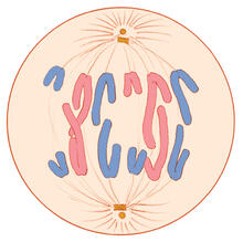
Mitosis - anaphase
1328
A cell in anaphase during mitosis: Chromosomes separate into two genetically identical groups and move to opposite ends of the spindle. Judith Stoffer View Media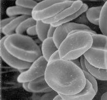
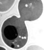
Red blood cells
1101
This image of human red blood cells was obtained with the help of a scanning electron microscope, an instrument that uses a finely focused electron beam to yield detailed images of the surface of a sa Tina Weatherby Carvalho, University of Hawaii at Manoa View Media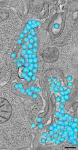
HIV-1 virus in the colon
3571
A tomographic reconstruction of the colon shows the location of large pools of HIV-1 virus particles (in blue) located in the spaces between adjacent cells. Mark Ladinsky, California Institute of Technology View Media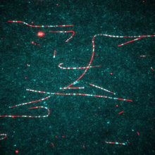
Dynein moving along microtubules
7023
Dynein (green) is a motor protein that “walks” along microtubules (red, part of the cytoskeleton) and carries its cargo along with it. This video was captured through fluorescence microscopy. Morgan DeSantis, University of Michigan. View Media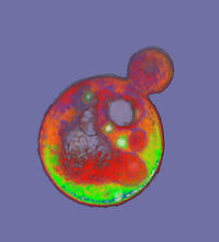
Cells frozen in time
2307
The fledgling field of X-ray microscopy lets researchers look inside whole cells rapidly frozen to capture their actions at that very moment. Here, a yeast cell buds before dividing into two. Carolyn Larabell, University of California, San Francisco, and the Lawrence Berkeley National Laboratory View Media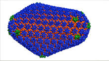
HIV Capsid
3477
This image is a computer-generated model of the approximately 4.2 million atoms of the HIV capsid, the shell that contains the virus' genetic material. Juan R. Perilla and the Theoretical and Computational Biophysics Group, University of Illinois at Urbana-Champaign View Media
Blinking bacteria
2724
Like a pulsing blue shower, E. coli cells flash in synchrony. Genes inserted into each cell turn a fluorescent protein on and off at regular intervals. Jeff Hasty, University of California, San Diego View Media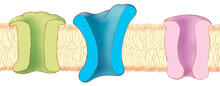
Ion channels
1284
The body uses a variety of ion channels to transport small molecules across cell membranes. Judith Stoffer View Media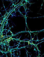
STORM image of axonal cytoskeleton
3678
This image shows the long, branched structures (axons) of nerve cells. Xiaowei Zhuang Laboratory, Howard Hughes Medical Institute, Harvard University View Media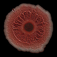
Biofilm formed by a pathogen
6518
A biofilm is a highly organized community of microorganisms that develops naturally on certain surfaces. Scott Chimileski, Ph.D., and Roberto Kolter, Ph.D., Harvard Medical School. View Media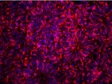
Neurons from human ES cells
3284
These neural precursor cells were derived from human embryonic stem cells. The neural cell bodies are stained red, and the nuclei are blue. Xianmin Zeng lab, Buck Institute for Age Research, via CIRM View Media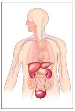
Body toxins
2496
Body organs such as the liver and kidneys process chemicals and toxins. These "target" organs are susceptible to damage caused by these substances. Crabtree + Company View Media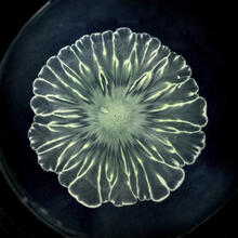
Floral pattern in a mixture of two bacterial species, Acinetobacter baylyi and Escherichia coli, grown on a semi-solid agar for 24 hours
6557
Floral pattern emerging as two bacterial species, motile Acinetobacter baylyi and non-motile Escherichia coli (green), are grown together for 24 hours on 0.75% agar surface from a small L. Xiong et al, eLife 2020;9: e48885 View Media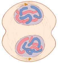
Mitosis - telophase
1332
Telophase during mitosis: Nuclear membranes form around each of the two sets of chromosomes, the chromosomes begin to spread out, and the spindle begins to break down. Judith Stoffer View Media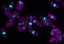
Yeast cells with accumulated cell wall material
6797
Yeast cells that abnormally accumulate cell wall material (blue) at their ends and, when preparing to divide, in their middles. This image was captured using wide-field microscopy with deconvolution. Alaina Willet, Kathy Gould’s lab, Vanderbilt University. View Media