Switch to Gallery View
Image and Video Gallery
This is a searchable collection of scientific photos, illustrations, and videos. The images and videos in this gallery are licensed under Creative Commons Attribution Non-Commercial ShareAlike 3.0. This license lets you remix, tweak, and build upon this work non-commercially, as long as you credit and license your new creations under identical terms.
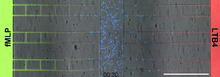
Neutrophil-like cells migrating in a microfluidic chip
6886
Neutrophil-like cells (blue) in a microfluidic chip preferentially migrating toward LTB4 over fMLP. Caroline Jones, University of Texas at Dallas. View Media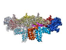
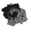
Dengue virus membrane protein structure
3758
Dengue virus is a mosquito-borne illness that infects millions of people in the tropics and subtropics each year. Like many viruses, dengue is enclosed by a protective membrane. Hong Zhou, UCLA View Media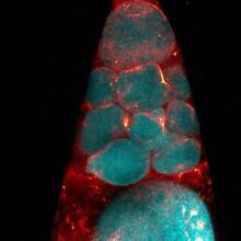
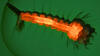
Fruit fly nurse cells during egg development
6753
In many animals, the egg cell develops alongside sister cells. Adam C. Martin, Massachusetts Institute of Technology. View Media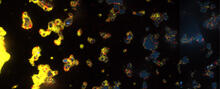
V. Cholerae Biofilm
3580
Industrious V. cholerae bacteria (yellow) tend to thrive in denser biofilms (left) while moochers (red) thrive in weaker biofilms (right). View Media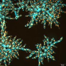
Snowflake yeast 3
6971
Multicellular yeast called snowflake yeast that researchers created through many generations of directed evolution from unicellular yeast. William Ratcliff, Georgia Institute of Technology. View Media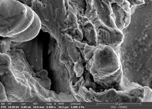
Staphylococcus aureus in the porous coating of a femoral hip stem
6804
Staphylococcus aureus bacteria (blue) on the porous coating of a femoral hip stem used in hip replacement surgery. Paul Stoodley, The Ohio State University. View Media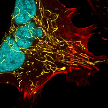
Multinucleated cancer cell
6967
A cancer cell with three nuclei, shown in turquoise. The abnormal number of nuclei indicates that the cell failed to go through cell division, probably more than once. Dylan T. Burnette, Vanderbilt University School of Medicine. View Media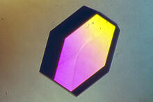
Hen egg lysozyme (2)
2406
A crystal of hen egg lysozyme protein created for X-ray crystallography, which can reveal detailed, three-dimensional protein structures. Alex McPherson, University of California, Irvine View Media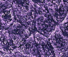
Retinal pigment epithelium derived from human ES cells
3286
This color-enhanced image is a scanning electron microscope image of retinal pigment epithelial (RPE) cells derived from human embryonic stem cells. David Hinton lab, University of Southern California, via CIRM View Media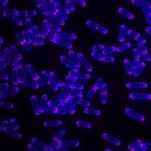
Yeast cells with Fimbrin Fim1
6794
Yeast cells with the protein Fimbrin Fim1 shown in magenta. This protein plays a role in cell division. This image was captured using wide-field microscopy with deconvolution.Alaina Willet, Kathy Gould’s lab, Vanderbilt University. View Media
Developing zebrafish fin
3598
Originally from the waters of India, Nepal, and neighboring countries, zebrafish can now be found swimming in science labs (and home aquariums) throughout the world. Jessica Plavicki View Media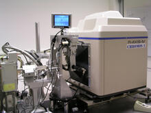
Chromium X-ray source
2361
In the determination of protein structures by X-ray crystallography, this unique soft (l = 2.29Å) X-ray source is used to collect anomalous scattering data from protein crystals containing light atoms The Southeast Collaboratory for Structural Genomics View Media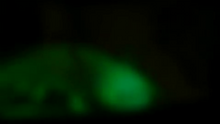
Glow-in-the-dark salamanders
2715
These six-month-old axolotls, a kind of salamander, glow green and blue under ultraviolet light. That's because they were genetically modified to make harmless green fluorescent protein, or GFP. View Media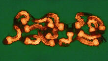
Group of Culex quinquefasciatus mosquito larvae
6770
Mosquito larvae with genes edited by CRISPR. Valentino Gantz, University of California, San Diego. View Media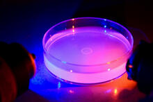
Petri dish containing C. elegans
6751
This Petri dish contains microscopic roundworms called Caenorhabditis elegans. Researchers used these particular worms to study how C. H. Robert Horvitz and Dipon Ghosh, Massachusetts Institute of Technology. View Media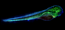
Zebrafish embryo showing vasculature
6661
A zebrafish embryo. The blue areas are cell bodies, the green lines are blood vessels, and the red glow is blood. Kevin Eliceiri, University of Wisconsin-Madison. View Media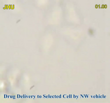
Precisely Delivering Chemical Cargo to Cells
3779
Moving protein or other molecules to specific cells to treat or examine them has been a major biological challenge. Nature Nanotechnology View Media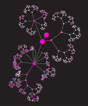
Network diagram of genes, cellular components and processes (unlabeled)
3436
This image shows the hierarchical ontology of genes, cellular components and processes derived from large genomic datasets. From Dutkowski et al. Janusz Dutkowski and Trey Ideker View Media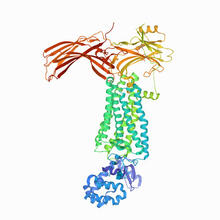
Rhodopsin bound to visual arrestin
6768
Rhodopsin is a pigment in the rod cells of the retina (back of the eye). It is extremely light-sensitive, supporting vision in low-light conditions. Protein Data Bank. View Media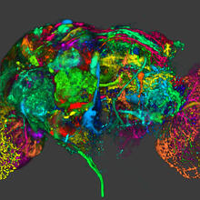
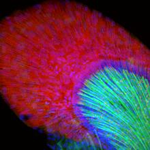
Color coding of the Drosophila brain - image
5838
This image results from a research project to visualize which regions of the adult fruit fly (Drosophila) brain derive from each neural stem cell. Yong Wan from Charles Hansen’s lab, University of Utah. Data preparation and visualization by Masayoshi Ito in the lab of Kei Ito, University of Tokyo. View Media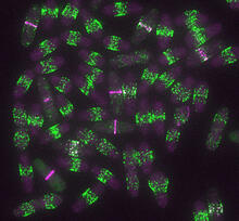
Yeast cells entering mitosis
6791
Yeast cells entering mitosis, also known as cell division. The green and magenta dots are two proteins that play important roles in mitosis. They show where the cells will split. Alaina Willet, Kathy Gould’s lab, Vanderbilt University. View Media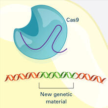
CRISPR Illustration Frame 4
6488
This illustration shows, in simplified terms, how the CRISPR-Cas9 system can be used as a gene-editing tool. National Institute of General Medical Sciences. View Media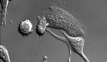
Dying melanoma cells
6966
Melanoma (skin cancer) cells undergoing programmed cell death, also called apoptosis. This process was triggered by raising the pH of the medium that the cells were growing in. Dylan T. Burnette, Vanderbilt University School of Medicine. View Media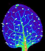
Zinc levels in a plant leaf
3727
Zinc is required for the function of more than 300 enzymes, including those that help regulate gene expression, in various organisms including humans. Suzana Car, Dartmouth College View Media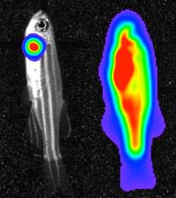
Bioluminescent imaging in adult zebrafish 04
3559
Luciferase-based imaging enables visualization and quantification of internal organs and transplanted cells in live adult zebrafish. View Media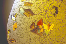
Jack bean concanavalin A
2407
Crystals of jack bean concanavalin A protein created for X-ray crystallography, which can reveal detailed, three-dimensional protein structures. Alex McPherson, University of California, Irvine View Media
Fluorescence in situ hybridization (FISH) in mouse ES cells shows DNA interactions
3296
Researchers used fluorescence in situ hybridization (FISH) to confirm the presence of long range DNA-DNA interactions in mouse embryonic stem cells. Kathrin Plath, University of California, Los Angeles View Media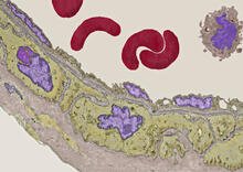
Transmission electron microscopy of coronary artery wall with elastin-rich ECM pseudocolored in light brown
3738
Elastin is a fibrous protein in the extracellular matrix (ECM). It is abundant in artery walls like the one shown here. As its name indicates, elastin confers elasticity. Tom Deerinck, National Center for Microscopy and Imaging Research (NCMIR) View Media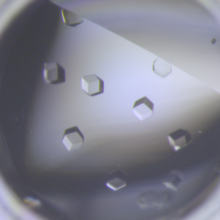
Crystals of CCD-1 in complex with cefotaxime
6764
CCD-1 is an enzyme produced by the bacterium Clostridioides difficile that helps it resist antibiotics. Keith Hodgson, Stanford University. View Media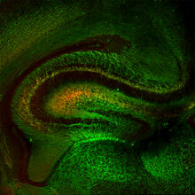
Mouse Brain Cross Section
5886
The brain sections are treated with fluorescent antibodies specific to a particular protein and visualized using serial electron microscopy (SEM). Anton Maximov, The Scripps Research Institute, La Jolla, CA View Media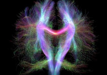
Microtubule breakdown
2321
Like a building supported by a steel frame, a cell contains its own sturdy internal scaffolding made up of proteins, including microtubules. Eva Nogales, University of California, Berkeley View Media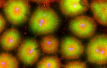
Cell-like compartments from frog eggs 4
6591
Cell-like compartments that spontaneously emerged from scrambled frog eggs, with nuclei (blue) from frog sperm. Endoplasmic reticulum (red) and microtubules (green) are also visible. Xianrui Cheng, Stanford University School of Medicine. View Media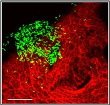
Bacterial cells aggregated above a light-organ pore of the Hawaiian bobtail squid
7019
The beating of cilia on the outside of the Hawaiian bobtail squid’s light organ concentrates Vibrio fischeri cells (green) present in the seawater into aggregates near the pore-containing tis Margaret J. McFall-Ngai, Carnegie Institution for Science/California Institute of Technology, and Edward G. Ruby, California Institute of Technology. View Media
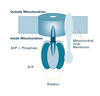
Human endoplasmic reticulum membrane protein complex
6777
A 3D model of the human endoplasmic reticulum membrane protein complex (EMC) that identifies its nine essential subunits. Rebecca Voorhees, California Institute of Technology. View Media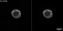
Breast cancer cells change migration phenotypes
6986
Cancer cells can change their migration phenotype, which includes their shape and the way that they move to invade different tissues. Bo Sun, Oregon State University. View Media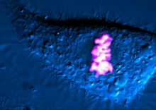
Dividing cell
6965
As this cell was undergoing cell division, it was imaged with two microscopy techniques: differential interference contrast (DIC) and confocal. The DIC view appears in blue and shows the entire cell. Dylan T. Burnette, Vanderbilt University School of Medicine. View Media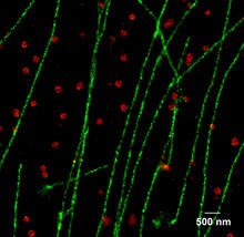
Multicolor STORM
2325
In 2006, scientists developed an optical microscopy technique enabling them to clearly see individual molecules within cells. In 2007, they took the technique, abbreviated STORM, a step further. Xiaowei Zhuang, Harvard University View Media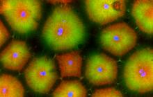
Cell-like compartments from frog eggs 5
6592
Cell-like compartments that spontaneously emerged from scrambled frog eggs, with nuclei (blue) from frog sperm. Endoplasmic reticulum (red) and microtubules (green) are also visible. Xianrui Cheng, Stanford University School of Medicine. View Media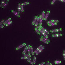
Yeast cells with endocytic actin patches
6793
Yeast cells with endocytic actin patches (green). These patches help cells take in outside material. When a cell is in interphase, patches concentrate at its ends. Alaina Willet, Kathy Gould’s lab, Vanderbilt University. View Media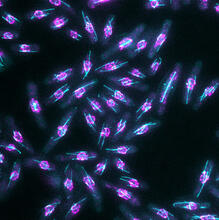
Yeast cells with nuclear envelopes and tubulin
6798
Yeast cells with nuclear envelopes shown in magenta and tubulin shown in light blue. The nuclear envelope defines the borders of the nucleus, which houses DNA. Alaina Willet, Kathy Gould’s lab, Vanderbilt University. View Media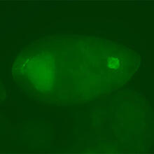
Fly cells
3594
If a picture is worth a thousand words, what's a movie worth? Denise Montell, Johns Hopkins University School of Medicine View Media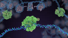
Cas9 protein involved in the CRISPR gene-editing technology
5816
In the gene-editing tool CRISPR, a small strand of RNA identifies a specific chunk of DNA. Janet Iwasa View Media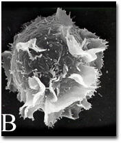
Activated mast cell surface
2637
A scanning electron microscope image of an activated mast cell. This image illustrates the interesting topography of the cell membrane, which is populated with receptors. Bridget Wilson, University of New Mexico View Media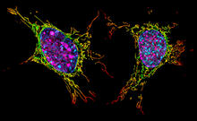
Two mouse fibroblast cells
6789
Two mouse fibroblasts, one of the most common types of cells in mammalian connective tissue. They play a key role in wound healing and tissue repair. Dylan T. Burnette, Vanderbilt University School of Medicine. View Media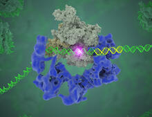
TFIID complex binds DNA to start gene transcription
3766
Gene transcription is a process by which the genetic information encoded in DNA is transcribed into RNA. Eva Nogales, Berkeley Lab View Media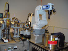
Automated crystal screening system
2362
Automated crystal screening systems such as the one shown here are becoming a common feature at synchrotron and other facilities where high-throughput crystal structure determination is being carried Southeast Collaboratory for Structural Genomics View Media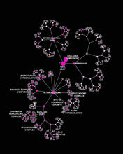
Network diagram of genes, cellular components and processes (labeled)
3437
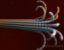
This image shows the hierarchical ontology of genes, cellular components and processes derived from large genomic datasets. From Dutkowski et al. Janusz Dutkowski and Trey Ideker, University of California, San Diego View MediaAssembly of the HIV capsid
5729
The HIV capsid is a pear-shaped structure that is made of proteins the virus needs to mature and become infective. John Grime and Gregory Voth, The University of Chicago View Media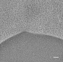
Multivesicular bodies containing intralumenal vesicles assemble at the vacuole 2
5768
Collecting and transporting cellular waste and sorting it into recylable and nonrecylable pieces is a complex business in the cell. Matthew West and Greg Odorizzi, University of Colorado View Media