Switch to Gallery View
Image and Video Gallery
This is a searchable collection of scientific photos, illustrations, and videos. The images and videos in this gallery are licensed under Creative Commons Attribution Non-Commercial ShareAlike 3.0. This license lets you remix, tweak, and build upon this work non-commercially, as long as you credit and license your new creations under identical terms.
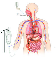
A drug's life in the body
2527
A drug's life in the body. Medicines taken by mouth pass through the liver before they are absorbed into the bloodstream. Crabtree + Company View Media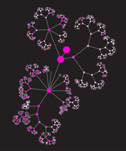
Network diagram of genes, cellular components and processes (unlabeled)
3436
This image shows the hierarchical ontology of genes, cellular components and processes derived from large genomic datasets. From Dutkowski et al. Janusz Dutkowski and Trey Ideker View Media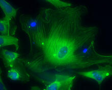
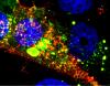
Smooth muscle from human ES cells
3288
These smooth muscle cells were derived from human embryonic stem cells. The nuclei are stained blue, and the proteins of the cytoskeleton are stained green. Alexey Terskikh lab, Burnham Institute for Medical Research, via CIRM View Media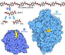
Plastic-eating enzymes
7000
PETase enzyme degrades polyester plastic (polyethylene terephthalate, or PET) into monohydroxyethyl terephthalate (MHET). Amy Wu and Christine Zardecki, RCSB Protein Data Bank. View Media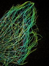
Tiny strands of tubulin, a protein in a cell's skeleton
3611
Just as our bodies rely on bones for structural support, our cells rely on a cellular skeleton. Pakorn Kanchanawong, National University of Singapore and National Heart, Lung, and Blood Institute, National Institutes of Health; and Clare Waterman, National Heart, Lung, and Blood Institute, National Institutes of Health View Media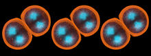
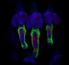
Sea urchin embryo 01
1047
Stereo triplet of a sea urchin embryo stained to reveal actin filaments (orange) and microtubules (blue). George von Dassow, University of Washington View Media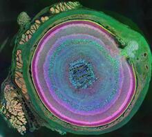
A mammalian eye has approximately 70 different cell types
3641
The incredible complexity of a mammalian eye (in this case from a mouse) is captured here. Each color represents a different type of cell. Bryan William Jones and Robert E. Marc, University of Utah View Media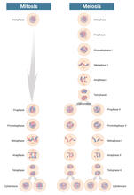
Mitosis and meiosis compared-labeled
6788
Meiosis is used to make sperm and egg cells. During meiosis, a cell's chromosomes are copied once, but the cell divides twice. Judith Stoffer View Media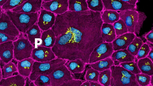
Pathways: The Fascinating Cells of Research Organisms
6538
Learn how research organisms, such as fruit flies and mice, can help us understand and treat human diseases. National Institute of General Medical Sciences View Media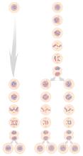
Mitosis and meiosis compared
1333
Meiosis is used to make sperm and egg cells. During meiosis, a cell's chromosomes are copied once, but the cell divides twice. Judith Stoffer View Media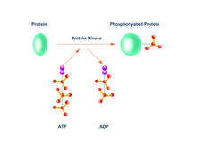
Kinases (with labels)
2535
Kinases are enzymes that add phosphate groups (red-yellow structures) to proteins (green), assigning the proteins a code. Crabtree + Company View Media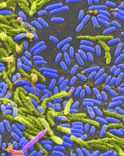
Vibrio bacteria
1160
Vibrio, a type (genus) of rod-shaped bacteria. Some Vibrio species cause cholera in humans. Tina Weatherby Carvalho, University of Hawaii at Manoa View Media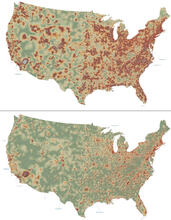
Mapping disease spread
2320
How far and fast an infectious disease spreads across a community depends on many factors, including transportation. These U.S. David Chrest, RTI International View Media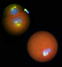
Nucleolinus
2762
The nucleolinus is a cellular compartment that has been a lonely bystander in scientific endeavors. Mary Anne Alliegro, Marine Biological Laboratory View Media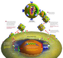
Life of an AIDS virus (with labels and stages)
2515
HIV is a retrovirus, a type of virus that carries its genetic material not as DNA but as RNA. Crabtree + Company View Media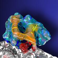
Kinesin moves cellular cargo
3491
A protein called kinesin (blue) is in charge of moving cargo around inside cells and helping them divide. Charles Sindelar, Yale University View Media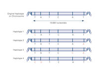
Haplotypes (with labels)
2567
Haplotypes are combinations of gene variants that are likely to be inherited together within the same chromosomal region. Crabtree + Company View Media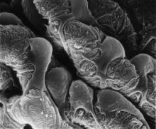
Podocytes from a chronically diseased kidney
3565
This scanning electron microscope (SEM) image shows podocytes--cells in the kidney that play a vital role in filtering waste from the bloodstream--from a patient with chronic kidney disease. Olga Troyanskaya, Princeton University and Matthias Kretzler, University of Michigan View Media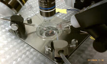
Electrode probe on mouse Huntington's muscle cell
3479
Using an electrode, researchers apply an electrical pulse onto a piece of muscle tissue affected by Huntington's disease. Grigor Varuzhanyan and Andrew A. Voss, California State Polytechnic University View Media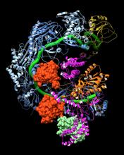
CRISPR surveillance complex
6352
This image shows how the CRISPR surveillance complex is disabled by two copies of anti-CRISPR protein AcrF1 (red) and one AcrF2 (light green). NRAMM National Resource for Automated Molecular Microscopy http://nramm.nysbc.org/nramm-images/ Source: Bridget Carragher View Media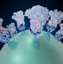
Coronavirus spike protein structure
3753
Coronaviruses are enveloped viruses responsible for 30 percent of mild respiratory infections and atypical deadly pneumonia in humans worldwide. Melody Campbell, UCSF View Media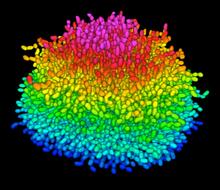
A Growing Bacterial Biofilm
5825
A growing Vibrio cholerae (cholera) biofilm. Cholera bacteria form colonies called biofilms that enable them to resist antibiotic therapy within the body and other challenges to their growth. Jing Yan, Ph.D., and Bonnie Bassler, Ph.D., Department of Molecular Biology, Princeton University, Princeton, NJ. View Media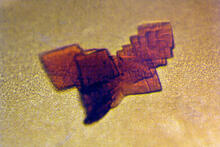
Sheep hemoglobin crystal
2392
A crystal of sheep hemoglobin protein created for X-ray crystallography, which can reveal detailed, three-dimensional protein structures. Alex McPherson, University of California, Irvine View Media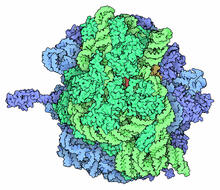
Ribosome illustration from PDB
5780
Ribosomes are complex machines made up of more than 50 proteins and three or four strands of genetic material called ribosomal RNA (rRNA). From PDB’s Molecule of the Month collection (direct link: http://pdb101.rcsb.org/motm/121) Molecule of the Month illustrations are available under a CC-BY-4.0 license. Attribution should be given to David S. Goodsell and the RCSB PDB. View Media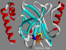
Sortase b from B. anthracis
2386
Structure of sortase b from the bacterium B. anthracis, which causes anthrax. Sortase b is an enzyme used to rob red blood cells of iron, which the bacteria need to survive. Midwest Center for Structural Genomics, PSI View Media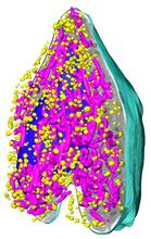
Soft X-ray tomography of a pancreatic beta cell
6605
A color-coded, 3D model of a rat pancreatic β cell. This type of cell produces insulin, a hormone that helps regulate blood sugar. Carolyn Larabell, University of California, San Francisco. View Media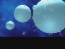
Diversity oriented synthesis: generating skeletal diversity using folding processes
3327
This 1 1/2-minute video animation was produced for chemical biologist Stuart Schreiber's lab page. The animation shows how diverse chemical structures can be produced in the lab. Eric Keller View Media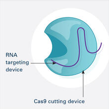
CRISPR Illustration Frame 1
6465
This illustration shows, in simplified terms, how the CRISPR-Cas9 system can be used as a gene-editing tool. This is the first frame in a series of four. National Institute of General Medical Sciences. View Media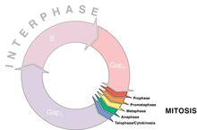
Cell cycle wheel
1310
A typical animal cell cycle lasts roughly 24 hours, but depending on the type of cell, it can vary in length from less than 8 hours to more than a year. Most of the variability occurs in Gap1. Judith Stoffer View Media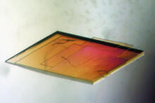
Pig trypsin crystal
2403
A crystal of pig trypsin protein created for X-ray crystallography, which can reveal detailed, three-dimensional protein structures. Alex McPherson, University of California, Irvine View Media
Life of an AIDS virus (with labels)
2514
HIV is a retrovirus, a type of virus that carries its genetic material not as DNA but as RNA. Crabtree + Company View Media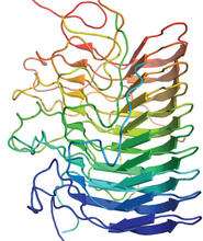
Structure of amyloid-forming prion protein
3542
This structure from an amyloid-forming prion protein shows one way beta sheets can stack. Douglas Fowler, University of Washington View Media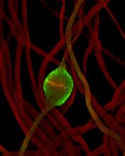
Fruit fly sperm cells
2433
Developing fruit fly spermatids require caspase activity (green) for the elimination of unwanted organelles and cytoplasm via apoptosis. Hermann Steller, Rockefeller University View Media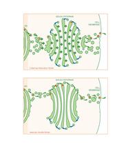
Golgi theories
1278
Two models for how material passes through the Golgi apparatus: the vesicular shuttle model and the cisternae maturation model. Judith Stoffer View Media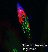
Proteasome
3451
This fruit fly spermatid recycles various molecules, including malformed or damaged proteins. Sigi Benjamin-Hong, Rockefeller University View Media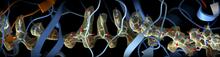
CRISPR
6351
RNA incorporated into the CRISPR surveillance complex is positioned to scan across foreign DNA. Cryo-EM density from a 3Å reconstruction is shown as a yellow mesh. NRAMM National Resource for Automated Molecular Microscopy http://nramm.nysbc.org/nramm-images/ Source: Bridget Carragher View Media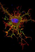
Abnormal, spiky fibroblast
3613
This is a fibroblast, a connective tissue cell that plays an important role in wound healing. Normal fibroblasts have smooth edges. Praveen Suraneni, Stowers Institute for Medical Research, Kansas City, Mo. View Media
Natcher Building 04
1084
NIGMS staff are located in the Natcher Building on the NIH campus. Alisa Machalek, National Institute of General Medical Sciences View Media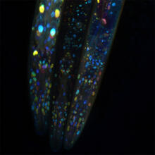
Mosaicism in C. elegans (Black Background)
6532
In the worm C. elegans, double-stranded RNA made in neurons can silence matching genes in a variety of cell types through the transport of RNA between cells. Snusha Ravikumar, Ph.D., University of Maryland, College Park, and Antony M. Jose, Ph.D., University of Maryland, College Park View Media
Neural tube development
2328
Proteins in the neural tissues of this zebrafish embryo direct cells to line up and form the neural tube, which will become the spinal cord and brain. Alexander Schier, Harvard University View Media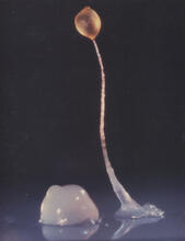
Dicty fruit
2684
Dictyostelium discoideum is a microscopic amoeba. A group of 100,000 form a mound as big as a grain of sand. Featured in The New Genetics. View Media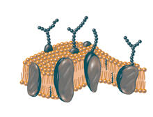
Plasma membrane
2523
The plasma membrane is a cell's protective barrier. See image 2524 for a labeled version of this illustration. Featured in The Chemistry of Health. Crabtree + Company View Media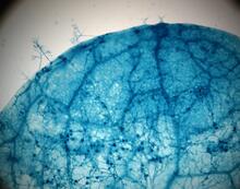
Disease-susceptible Arabidopsis leaf
2782
This is a magnified view of an Arabidopsis thaliana leaf after several days of infection with the pathogen Hyaloperonospora arabidopsidis. Jeff Dangl, University of North Carolina, Chapel Hill View Media
Thermotoga maritima and its metabolic network
2702
A combination of protein structures determined experimentally and computationally shows us the complete metabolic network of a heat-loving bacterium. View Media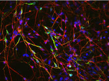
Neurons from human ES cells 02
3285
These neurons were derived from human embryonic stem cells. The neural cell bodies with axonal projections are visible in red, and the nuclei in blue. Xianmin Zeng lab, Buck Institute for Age Research, via CIRM View Media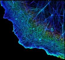
3D image of actin in a cell
3749
Actin is an essential protein in a cell's skeleton (cytoskeleton). It forms a dense network of thin filaments in the cell. Xiaowei Zhuang, Howard Hughes Medical Institute, Harvard University View Media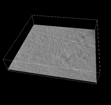
3D reconstruction of the Golgi apparatus in a pancreas cell
6609
Researchers used cryo-electron tomography (cryo-ET) to capture images of a rat pancreas cell that were then compiled and color-coded to produce a 3D reconstruction. Xianjun Zhang, University of Southern California. View Media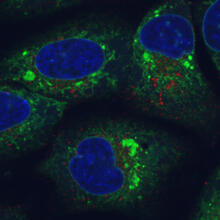
Endoplasmic reticulum abnormalities
6773
Human cells with the gene that codes for the protein FIT2 deleted. Green indicates an endoplasmic reticulum (ER) resident protein. Michel Becuwe, Harvard University. View Media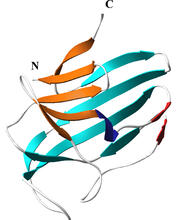
Secreted protein from Mycobacteria
2379
Model of a major secreted protein of unknown function, which is only found in mycobacteria, the class of bacteria that causes tuberculosis. Mycobacterium Tuberculosis Center, PSI View Media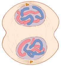
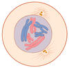
Mitosis - telophase
1332
Telophase during mitosis: Nuclear membranes form around each of the two sets of chromosomes, the chromosomes begin to spread out, and the spindle begins to break down. Judith Stoffer View Media