Switch to Gallery View
Image and Video Gallery
This is a searchable collection of scientific photos, illustrations, and videos. The images and videos in this gallery are licensed under Creative Commons Attribution Non-Commercial ShareAlike 3.0. This license lets you remix, tweak, and build upon this work non-commercially, as long as you credit and license your new creations under identical terms.
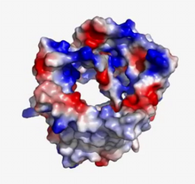
VDAC video 02
2571
This video shows the structure of the pore-forming protein VDAC-1 from humans. Gerhard Wagner, Harvard Medical School View Media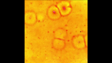
Cell-like compartments emerging from scrambled frog eggs 2
6588
Cell-like compartments spontaneously emerge from scrambled frog eggs, with nuclei (blue) from frog sperm. Endoplasmic reticulum (red) and microtubules (green) are also visible. Xianrui Cheng, Stanford University School of Medicine. View Media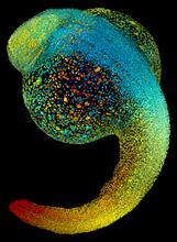
Zebrafish embryo
3644
Just 22 hours after fertilization, this zebrafish embryo is already taking shape. By 36 hours, all of the major organs will have started to form. Philipp Keller, Bill Lemon, Yinan Wan, and Kristin Branson, Janelia Farm Research Campus, Howard Hughes Medical Institute, Ashburn, Va. View Media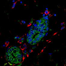
Mouse mammary cells lacking anti-cancer protein
3432
Shortly after a pregnant woman gives birth, her breasts start to secrete milk. This process is triggered by hormonal and genetic cues, including the protein Elf5. Nature Cell Biology, November 2012, Volume 14 No 11 pp1113-1231 View Media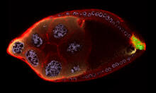
Cellular polarity
2309
As an egg cell develops, a process called polarization controls what parts ultimately become the embryo's head and tail. This picture shows an egg of the fruit fly Drosophila. Wu-Min Deng, Florida State University View Media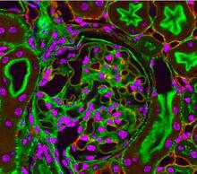
Fluorescent microscopy of kidney tissue--close-up
3725
This photograph of kidney tissue, taken using fluorescent light microscopy, shows a close-up view of part of image 3723. Tom Deerinck , National Center for Microscopy and Imaging Research View Media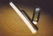
Bovine milk alpha-lactalbumin (2)
2404
Crystals of bovine milk alpha-lactalbumin protein created for X-ray crystallography, which can reveal detailed, three-dimensional protein structures. Alex McPherson, University of California, Irvine View Media
Draper, shown in the fatbody of a Drosophila melanogaster larva
2757
The fly fatbody is a nutrient storage and mobilization organ akin to the mammalian liver. The engulfment receptor Draper (green) is located at the cell surface of fatbody cells. Christina McPhee and Eric Baehrecke, University of Massachusetts Medical School View Media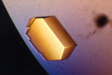
Pig trypsin (2)
2413
A crystal of porcine trypsin protein created for X-ray crystallography, which can reveal detailed, three-dimensional protein structures. Alex McPherson, University of California, Irvine View Media
Cross section of a Drosophila melanogaster pupa lacking Draper
2759
In the absence of the engulfment receptor Draper, salivary gland cells (light blue) persist in the thorax of a developing Drosophila melanogaster pupa. Christina McPhee and Eric Baehrecke, University of Massachusetts Medical School View Media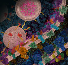
Molecular view of glutamatergic synapse
6992
This illustration highlights spherical pre-synaptic vesicles that carry the neurotransmitter glutamate. Amy Wu and Christine Zardecki, RCSB Protein Data Bank. View Media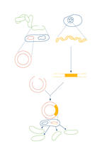
Recombinant DNA
2564
To splice a human gene into a plasmid, scientists take the plasmid out of an E. coli bacterium, cut the plasmid with a restriction enzyme, and splice in human DNA. Crabtree + Company View Media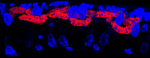
Microsporidia in roundworm 3
5779
Many disease-causing microbes manipulate their host’s metabolism and cells for their own ends. Keir Balla and Emily Troemel, University of California San Diego View Media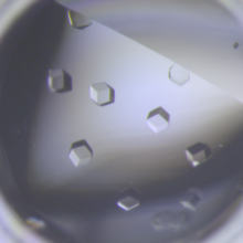
Crystals of CCD-1 in complex with cefotaxime
6764
CCD-1 is an enzyme produced by the bacterium Clostridioides difficile that helps it resist antibiotics. Keith Hodgson, Stanford University. View Media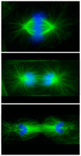
Cell division phases in Xenopus frog cells
3442
These images show three stages of cell division in Xenopus XL177 cells, which are derived from tadpole epithelial cells. They are (from top): metaphase, anaphase and telophase. Claire Walczak, who took them while working as a postdoc in the laboratory of Timothy Mitchison View Media
Yeast art depicting the New York City skyline
6521
This skyline of New York City was created by “printing” nanodroplets containing yeast (Saccharomyces cerevisiae) onto a large plate. Each dot is a separate yeast colony. Michael Shen, Ph.D., Jasmine Temple, Leslie Mitchell, Ph.D., and Jef Boeke, Ph.D., New York University School of Medicine; and Nick Phillips, James Chuang, Ph.D., and Jiarui Wang, Johns Hopkins University. View Media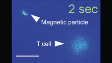
Magnetic Janus particle activating a T cell
6800
A Janus particle being used to activate a T cell, a type of immune cell. Yan Yu, Indiana University, Bloomington. View Media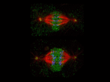
Katanin protein regulates anaphase
2594
The microtubule severing protein, katanin, localizes to chromosomes and regulates anaphase A in mitosis. David Sharp, Albert Einstein College of Medicine View Media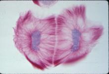
Lily mitosis 13
1019
A light microscope image of cells from the endosperm of an African globe lily (Scadoxus katherinae). This is one frame of a time-lapse sequence that shows cell division in action. Andrew S. Bajer, University of Oregon, Eugene View Media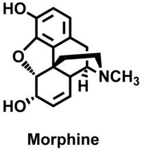
Morphine Structure
3438
The chemical structure of the morphine molecule Judy Coyle, Donald Danforth Plant Science Center View Media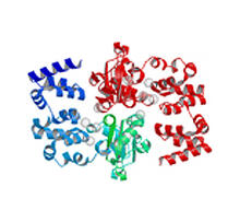
Magnesium transporter protein from E. faecalis
2345
Structure of a magnesium transporter protein from an antibiotic-resistant bacterium (Enterococcus faecalis) found in the human gut. New York Structural GenomiX Consortium View Media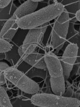
Flagellated bacterial cells
7014
Vibrio fischeri (2 mm in length) is the exclusive symbiotic partner of the Hawaiian bobtail squid, Euprymna scolopes. Margaret J. McFall-Ngai, Carnegie Institution for Science/California Institute of Technology, and Edward G. Ruby, California Institute of Technology. View Media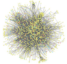
Molecular interactions
2743
This network map shows molecular interactions (yellow) associated with a congenital condition that causes heart arrhythmias and the targets for drugs that alter these interactions (red and blue). Ravi Iyengar, Mount Sinai School of Medicine View Media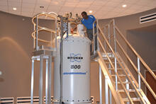
800 MHz NMR magnet
3526
Scientists use nuclear magnetic spectroscopy (NMR) to determine the detailed, 3D structures of molecules. Asokan Anbanandam, University of Kansas View Media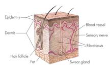
Skin cross-section
1056
Cross-section of skin anatomy shows layers and different tissue types. National Institutes of Health Medical Arts View Media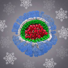
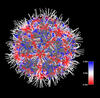
H1N1 Influenza Virus
6356
Related to image 6355. Dr. Rommie Amaro, University of California, San Diego View Media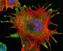
mDia1 antibody staining- 02
3331
Cells move forward with lamellipodia and filopodia supported by networks and bundles of actin filaments. Proper, controlled cell movement is a complex process. Rong Li and Praveen Suraneni, Stowers Institute for Medical Research View Media
Life of an AIDS virus (with labels)
2514
HIV is a retrovirus, a type of virus that carries its genetic material not as DNA but as RNA. Crabtree + Company View Media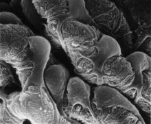
Podocytes from a chronically diseased kidney
3565
This scanning electron microscope (SEM) image shows podocytes--cells in the kidney that play a vital role in filtering waste from the bloodstream--from a patient with chronic kidney disease. Olga Troyanskaya, Princeton University and Matthias Kretzler, University of Michigan View Media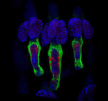
Wound healing in process
3497
Wound healing requires the action of stem cells. Hermann Steller, Rockefeller University View Media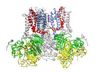
Ion channel
3487
A special "messy" region of a potassium ion channel is important in its function. Yu Zhoi, Christopher Lingle Laboratory, Washington University School of Medicine in St. Louis View Media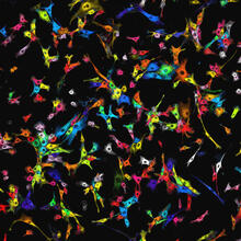
Single-cell “radios” image
7021
Individual cells are color-coded based on their identity and signaling activity using a protein circuit technology developed by the Coyle Lab. Scott Coyle, University of Wisconsin-Madison. View Media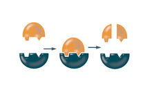
Enzymes convert subtrates into products
2521
Enzymes convert substrates into products very quickly. See image 2522 for a labeled version of this illustration. Featured in The Chemistry of Health. Crabtree + Company View Media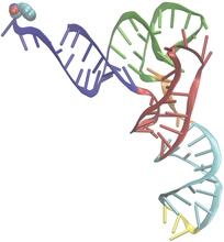
Phenylalanine tRNA molecule
3406
Phenylalanine tRNA showing the anticodon (yellow) and the amino acid, phenylalanine (blue and red spheres). Patrick O'Donoghue and Dieter Soll, Yale University View Media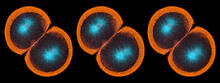
Sea urchin embryo 06
1052
Stereo triplet of a sea urchin embryo stained to reveal actin filaments (orange) and microtubules (blue). George von Dassow, University of Washington View Media
Natcher Building 05
1085
NIGMS staff are located in the Natcher Building on the NIH campus. Alisa Machalek, National Institute of General Medical Sciences View Media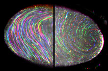
Fruit fly egg ooplasmic streaming
6809
Two fruit fly (Drosophila melanogaster) egg cells, one on each side of the central black line. Vladimir I. Gelfand, Feinberg School of Medicine, Northwestern University. View Media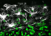
Vimentin in a quail embryo
2807
Confocal image showing high levels of the protein vimentin (white) at the edge zone of a quail embryo. Cell nuclei are labeled green. Andrés Garcia, Georgia Tech View Media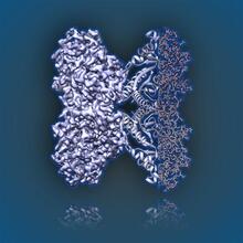
Aldolase
6350
2.5Å resolution reconstruction of rabbit muscle aldolase collected on a FEI/Thermo Fisher Titan Krios with energy filter and image corrector. National Resource for Automated Molecular Microscopy http://nramm.nysbc.org/nramm-images/ Source: Bridget Carragher View Media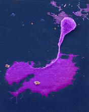
Crab nerve cell
1247
Neuron from a crab showing the cell body (bottom), axon (rope-like extension), and growth cone (top right). Tina Weatherby Carvalho, University of Hawaii at Manoa View Media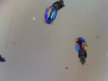
Young squids
6903
Real-time movie of young squids. Michael Shribak, Marine Biological Laboratory/University of Chicago. View Media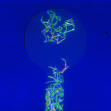
Genetically identical mycobacteria respond differently to antibiotic 1
5751
Antibiotic resistance in microbes is a serious health concern. So researchers have turned their attention to how bacteria undo the action of some antibiotics. Bree Aldridge, Tufts University View Media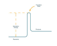
Activation energy (with labels)
2526
To become products, reactants must overcome an energy hill. See image 2525 for an unlabeled version of this illustration. Crabtree + Company View Media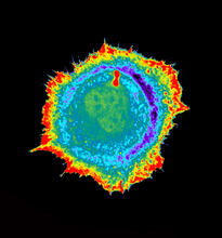
Seeing signaling protein activation in cells 01
2451
Cdc42, a member of the Rho family of small guanosine triphosphatase (GTPase) proteins, regulates multiple cell functions, including motility, proliferation, apoptosis, and cell morphology. Klaus Hahn, University of North Carolina, Chapel Hill Medical School View Media
Mitochondria from rat heart muscle cell_2
3664
These mitochondria (brown) are from the heart muscle cell of a rat. Mitochondria have an inner membrane that folds in many places (and that appears here as striations). National Center for Microscopy and Imaging Research View Media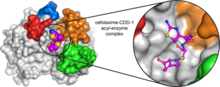
Space-filling model of a cefotaxime-CCD-1 complex
6767
CCD-1 is an enzyme produced by the bacterium Clostridioides difficile that helps it resist antibiotics. Keith Hodgson, Stanford University. View Media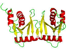
tRNA splicing enzyme endonuclease in humans
2351
An NMR solution structure model of the transfer RNA splicing enzyme endonuclease in humans (subunit Sen15). This represents the first structure of a eukaryotic tRNA splicing endonuclease subunit. Center for Eukaryotic Structural Genomics, PSI View Media
Prion protein fibrils 1
3460
Recombinant proteins such as the prion protein shown here are often used to model how proteins misfold and sometimes polymerize in neurodegenerative disorders. This prion protein was expressed in E. Ken Pekoc (public affairs officer) and Julie Marquardt, NIAID/ Rocky Mountain Laboratories View Media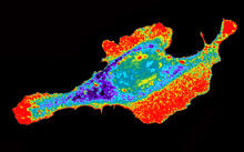
Seeing signaling protein activation in cells 03
2453
Cdc42, a member of the Rho family of small guanosine triphosphatase (GTPase) proteins, regulates multiple cell functions, including motility, proliferation, apoptosis, and cell morphology. Klaus Hahn, University of North Carolina, Chapel Hill Medical School View Media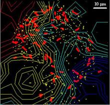
Cellular traffic
2310
Like tractor-trailers on a highway, small sacs called vesicles transport substances within cells. This image tracks the motion of vesicles in a living cell. Alexey Sharonov and Robin Hochstrasser, University of Pennsylvania View Media