
Image Gallery: Tracking embryonic zebrafish cells
ID
6775
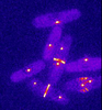
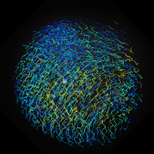
To better understand cell movements in developing embryos, researchers isolated cells from early zebrafish embryos and grew them as clusters. Provided with the right signals, the clusters replicated some cell movements seen in intact embryos. Each line in this image depicts the movement of a single cell. The image was created using time-lapse confocal microscopy. Related to video 6776.
Source
Liliana Solnica-Krezel, Washington University School of Medicine in St. Louis.
Topics