Switch to Gallery View
Image and Video Gallery
This is a searchable collection of scientific photos, illustrations, and videos. The images and videos in this gallery are licensed under Creative Commons Attribution Non-Commercial ShareAlike 3.0. This license lets you remix, tweak, and build upon this work non-commercially, as long as you credit and license your new creations under identical terms.
Central dogma, illustrated (with labels)
2548
DNA encodes RNA, which encodes protein. DNA is transcribed to make messenger RNA (mRNA). The mRNA sequence (dark red strand) is complementary to the DNA sequence (blue strand). Crabtree + Company View Media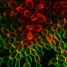
Snowflake yeast 1
6969
Multicellular yeast called snowflake yeast that researchers created through many generations of directed evolution from unicellular yeast. William Ratcliff, Georgia Institute of Technology. View Media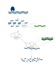
RNA interference
2558
RNA interference or RNAi is a gene-silencing process in which double-stranded RNAs trigger the destruction of specific RNAs. Crabtree + Company View Media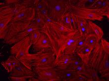
Mouse heart fibroblasts
3281
This image shows mouse fetal heart fibroblast cells. The muscle protein actin is stained red, and the cell nuclei are stained blue. Kara McCloskey lab, University of California, Merced, via CIRM View Media
Multivesicular bodies containing intralumenal vesicles assemble at the vacuole 2
5768
Collecting and transporting cellular waste and sorting it into recylable and nonrecylable pieces is a complex business in the cell. Matthew West and Greg Odorizzi, University of Colorado View Media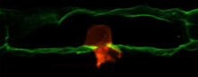
Anchor cell in basement membrane
2707
An anchor cell (red) pushes through the basement membrane (green) that surrounds it. Elliott Hagedorn, Duke University. View Media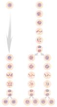
Mitosis and meiosis compared
1333
Meiosis is used to make sperm and egg cells. During meiosis, a cell's chromosomes are copied once, but the cell divides twice. Judith Stoffer View Media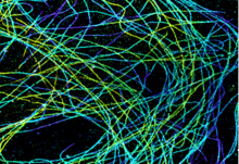
Microtubules in African green monkey cells
6891
Microtubules in African green monkey cells. Microtubules are strong, hollow fibers that provide cells with structural support. Melike Lakadamyali, Perelman School of Medicine at the University of Pennsylvania. View Media
Mitochondria from rat heart muscle cell_2
3664
These mitochondria (brown) are from the heart muscle cell of a rat. Mitochondria have an inner membrane that folds in many places (and that appears here as striations). National Center for Microscopy and Imaging Research View Media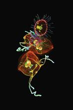
Jellyfish, viewed with ZEISS Lightsheet Z.1 microscope
3636
Jellyfish are especially good models for studying the evolution of embryonic tissue layers. Despite being primitive, jellyfish have a nervous system (stained green here) and musculature (red). Helena Parra, Pompeu Fabra University, Spain View Media
Alternative splicing
2552
Arranging exons in different patterns, called alternative splicing, enables cells to make different proteins from a single gene. Crabtree + Company View Media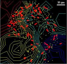
Cellular traffic
2310
Like tractor-trailers on a highway, small sacs called vesicles transport substances within cells. This image tracks the motion of vesicles in a living cell. Alexey Sharonov and Robin Hochstrasser, University of Pennsylvania View Media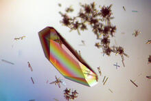
Hen egg lysozyme (1)
2396
Crystals of hen egg lysozyme protein created for X-ray crystallography, which can reveal detailed, three-dimensional protein structures. Alex McPherson, University of California, Irvine View Media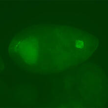
Fly cells
3594
If a picture is worth a thousand words, what's a movie worth? Denise Montell, Johns Hopkins University School of Medicine View Media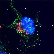
ARTS triggers apoptosis
2432
Cell showing overproduction of the ARTS protein (red). ARTS triggers apoptosis, as shown by the activation of caspase-3 (green) a key tool in the cell's destruction. The nucleus is shown in blue. Hermann Steller, Rockefeller University View Media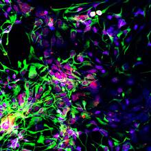
Motor neuron progenitors derived from human ES cells
3280
Motor neuron progenitors (green) were derived from human embryonic stem cells. Image and caption information courtesy of the California Institute for Regenerative Medicine. Hans Keirstead lab, University of California, Irvine, via CIRM View Media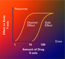
Dose response curves
2533
Dose-response curves determine how much of a drug (X-axis) causes a particular effect, or a side effect, in the body (Y-axis). Featured in Medicines By Design. Crabtree + Company View Media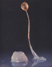
Dicty fruit
2684
Dictyostelium discoideum is a microscopic amoeba. A group of 100,000 form a mound as big as a grain of sand. Featured in The New Genetics. View Media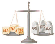
Life in balance
1336
Mitosis creates cells, and apoptosis kills them. The processes often work together to keep us healthy. Judith Stoffer View Media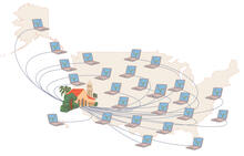
Folding@Home
1276
Stanford University scientist Vijay Pande decided to couple the power of computers with the help of the public. Judith Stoffer View Media
Blood clots show their flex
2450
Blood clots stop bleeding, but they also can cause heart attacks and strokes. Eric Lee, University of Illinois at Urbana-Champaign View Media
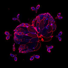
Wild-type and mutant fruit fly ovaries
6806
The two large, central, round shapes are ovaries from a typical fruit fly (Drosophila melanogaster). Vladimir I. Gelfand, Feinberg School of Medicine, Northwestern University. View Media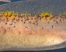
Pigment cells in fish skin
5756
Pigment cells are cells that give skin its color. David Parichy, University of Washington View Media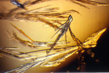
Pig trypsin (3)
2414
Crystals of porcine trypsin protein created for X-ray crystallography, which can reveal detailed, three-dimensional protein structures. Alex McPherson, University of California, Irvine View Media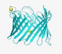
VDAC video 01
2570
This video shows the structure of the pore-forming protein VDAC-1 from humans. Gerhard Wagner, Harvard Medical School View Media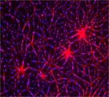
In vitro assembly of a cell-signaling pathway
3787
T cells are white blood cells that are important in defending the body against bacteria, viruses and other pathogens. Xiaolei Su, HHMI Whitman Center of the Marine Biological Laboratory View Media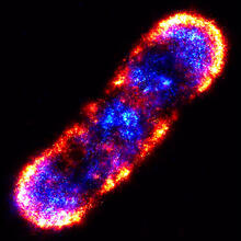
Self-organizing proteins
2771
Under the microscope, an E. coli cell lights up like a fireball. Each bright dot marks a surface protein that tells the bacteria to move toward or away from nearby food and toxins. View Media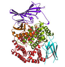
Aminopeptidase N from N. meningitidis
2341
Model of the enzyme aminopeptidase N from the human pathogen Neisseria meningitidis, which can cause meningitis epidemics. Midwest Center for Structural Genomics, PSI View Media
Zika virus
6998
Zika virus is shown in cross section at center left. On the outside, it includes envelope protein (red) and membrane protein (magenta) embedded in a lipid membrane (light purple). Amy Wu and Christine Zardecki, RCSB Protein Data Bank. View Media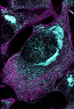
Microtubules and tau aggregates
6892
Microtubules (magenta) and tau protein (light blue) in a cell model of tauopathy. Melike Lakadamyali, Perelman School of Medicine at the University of Pennsylvania. View Media
Virtual snow world
2335
Glide across an icy canyon, where you see smiling snowmen and waddling penguins. Toss a snowball, hear it smash against an igloo, and then watch it explode in bright colors. David Patterson and Hunter Hoffmann, University of Washington View Media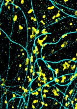
Lysosomes and microtubules
6889
Lysosomes (yellow) and detyrosinated microtubules (light blue). Lysosomes are bubblelike organelles that take in molecules and use enzymes to break them down. Melike Lakadamyali, Perelman School of Medicine at the University of Pennsylvania. View Media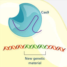
CRISPR Illustration Frame 4
6488
This illustration shows, in simplified terms, how the CRISPR-Cas9 system can be used as a gene-editing tool. National Institute of General Medical Sciences. View Media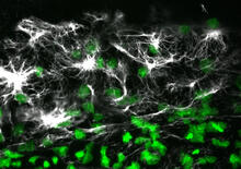
Vimentin in a quail embryo
2807
Confocal image showing high levels of the protein vimentin (white) at the edge zone of a quail embryo. Cell nuclei are labeled green. Andrés Garcia, Georgia Tech View Media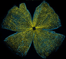
Mouse retina
5793
What looks like the gossamer wings of a butterfly is actually the retina of a mouse, delicately snipped to lay flat and sparkling with fluorescent molecules. Tom Deerinck and Keunyoung (“Christine”) Kim, NCMIR View Media
Blinking bacteria
2724
Like a pulsing blue shower, E. coli cells flash in synchrony. Genes inserted into each cell turn a fluorescent protein on and off at regular intervals. Jeff Hasty, University of California, San Diego View Media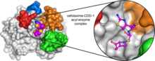
Space-filling model of a cefotaxime-CCD-1 complex
6767
CCD-1 is an enzyme produced by the bacterium Clostridioides difficile that helps it resist antibiotics. Keith Hodgson, Stanford University. View Media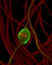
Fruit fly sperm cells
2433
Developing fruit fly spermatids require caspase activity (green) for the elimination of unwanted organelles and cytoplasm via apoptosis. Hermann Steller, Rockefeller University View Media
Cell-like compartments from frog eggs
6584
Cell-like compartments that spontaneously emerged from scrambled frog eggs, with nuclei (blue) from frog sperm. Endoplasmic reticulum (red) and microtubules (green) are also visible. Xianrui Cheng, Stanford University School of Medicine. View Media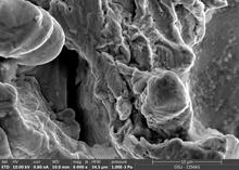
Staphylococcus aureus in the porous coating of a femoral hip stem
6804
Staphylococcus aureus bacteria (blue) on the porous coating of a femoral hip stem used in hip replacement surgery. Paul Stoodley, The Ohio State University. View Media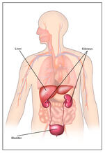
Body toxins (with labels)
2497
Body organs such as the liver and kidneys process chemicals and toxins. These "target" organs are susceptible to damage caused by these substances. Crabtree + Company View Media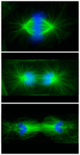
Cell division phases in Xenopus frog cells
3442
These images show three stages of cell division in Xenopus XL177 cells, which are derived from tadpole epithelial cells. They are (from top): metaphase, anaphase and telophase. Claire Walczak, who took them while working as a postdoc in the laboratory of Timothy Mitchison View Media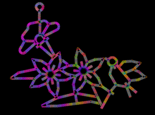
Computer sketch of bird-and-flower DNA origami
3689
A computer-generated sketch of a DNA origami folded into a flower-and-bird structure. See also related image 3690. Hao Yan, Arizona State University View Media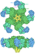
Plant resistosome
7002
The research organism Arabidopsis thaliana forms a large molecular machine called a resistosome to fight off infections. Amy Wu and Christine Zardecki, RCSB Protein Data Bank. View Media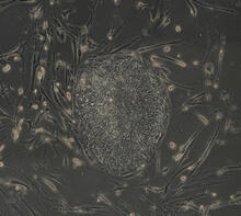
Induced stem cells from adult skin 03
2605
The human skin cells pictured contain genetic modifications that make them pluripotent, essentially equivalent to embryonic stem cells. James Thomson, University of Wisconsin-Madison View Media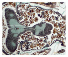
Cross section of a Drosophila melanogaster pupa
2758
This photograph shows a magnified view of a Drosophila melanogaster pupa in cross section. Compare this normal pupa to one that lacks an important receptor, shown in image 2759. Christina McPhee and Eric Baehrecke, University of Massachusetts Medical School View Media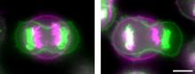
Symmetrically and asymmetrically elongating cells
3648
Merged fluorescent images of symmetrically (left) or asymmetrically (right) elongating HeLa cells at the end of early anaphase (magenta) and late anaphase (green). Tomomi Kiyomitsu and Iain M. Cheeseman, Whitehead Institute for Biomedical Research View Media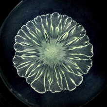
Floral pattern in a mixture of two bacterial species, Acinetobacter baylyi and Escherichia coli, grown on a semi-solid agar for 24 hours
6557
Floral pattern emerging as two bacterial species, motile Acinetobacter baylyi and non-motile Escherichia coli (green), are grown together for 24 hours on 0.75% agar surface from a small L. Xiong et al, eLife 2020;9: e48885 View Media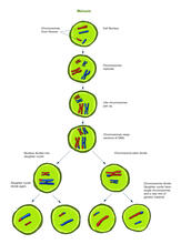
Meiosis illustration (with labels)
2546
Meiosis is the process whereby a cell reduces its chromosomes from diploid to haploid in creating eggs or sperm. Crabtree + Company View Media