Switch to Gallery View
Image and Video Gallery
This is a searchable collection of scientific photos, illustrations, and videos. The images and videos in this gallery are licensed under Creative Commons Attribution Non-Commercial ShareAlike 3.0. This license lets you remix, tweak, and build upon this work non-commercially, as long as you credit and license your new creations under identical terms.
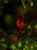
Fruit fly nurse cells transporting their contents during egg development
6754
In many animals, the egg cell develops alongside sister cells. Adam C. Martin, Massachusetts Institute of Technology. View Media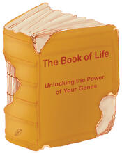
Aging book of life
1334
Damage to each person's genome, often called the "Book of Life," accumulates with time. Judith Stoffer View Media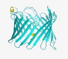
VDAC video 01
2570
This video shows the structure of the pore-forming protein VDAC-1 from humans. Gerhard Wagner, Harvard Medical School View Media
Fluorescence in situ hybridization (FISH) in mouse ES cells shows DNA interactions
3296
Researchers used fluorescence in situ hybridization (FISH) to confirm the presence of long range DNA-DNA interactions in mouse embryonic stem cells. Kathrin Plath, University of California, Los Angeles View Media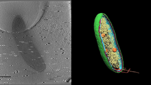
Cryo-electron tomography of a Caulobacter bacterium
6569
3D image of Caulobacter bacterium with various components highlighted: cell membranes (red and blue), protein shell (green), protein factories known as ribosomes (yellow), and storage granules Peter Dahlberg, Stanford University. View Media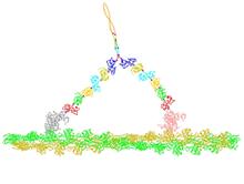
Myosin V binding to actin
2754
This simulation of myosin V binding to actin was created using the software tool Protein Mechanica. Simbios, NIH Center for Biomedical Computation at Stanford View Media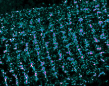
Single-Molecule Imaging
3339
This is a super-resolution light microscope image taken by Hiro Hakozaki and Masa Hoshijima of NCMIR. Tom Deerinck, NCMIR View Media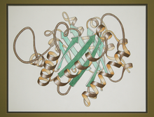
Early ribbon drawing of a protein
2748
This ribbon drawing of a protein hand drawn and colored by researcher Jane Richardson in 1981 helped originate the ribbon representation of proteins that is now ubiquitous in molecular graphics. Jane Richardson, Duke University Medical Center View Media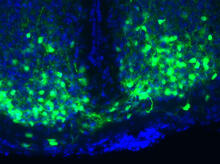
Master clock of the mouse brain
3547
An image of the area of the mouse brain that serves as the 'master clock,' which houses the brain's time-keeping neurons. The nuclei of the clock cells are shown in blue. Erik Herzog, Washington University in St. Louis View Media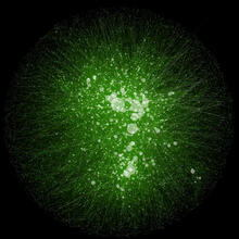
Cytoscape network wiring diagram 2
2749
This image integrates the thousands of known molecular and genetic interactions happening inside our bodies using a computer program called Cytoscape. Trey Ideker, University of California, San Diego View Media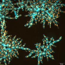
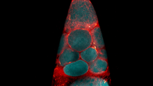
Snowflake yeast 3
6971
Multicellular yeast called snowflake yeast that researchers created through many generations of directed evolution from unicellular yeast. William Ratcliff, Georgia Institute of Technology. View Media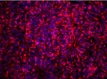
Neurons from human ES cells
3284
These neural precursor cells were derived from human embryonic stem cells. The neural cell bodies are stained red, and the nuclei are blue. Xianmin Zeng lab, Buck Institute for Age Research, via CIRM View Media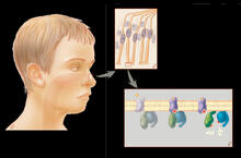
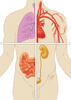
Olfactory system
1291
Sensory organs have cells equipped for detecting signals from the environment, such as odors. Judith Stoffer View Media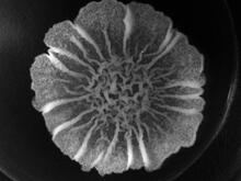
A Bacillus subtilis biofilm grown in a Petri dish
3718
Bacterial biofilms are tightly knit communities of bacterial cells growing on, for example, solid surfaces, such as in water pipes or on teeth. Gürol Süel, UCSD View Media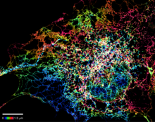
Dense tubular matrices in the peripheral endoplasmic reticulum (ER) 1
5855
Superresolution microscopy work on endoplasmic reticulum (ER) in the peripheral areas of the cell showing details of the structure and arrangement in a complex web of tubes. Jennifer Lippincott-Schwartz, Howard Hughes Medical Institute Janelia Research Campus, Virginia View Media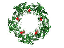
Cas4 nuclease protein structure
3720
This wreath represents the molecular structure of a protein, Cas4, which is part of a system, known as CRISPR, that bacteria use to protect themselves against viral invaders. Fred Dyda, NIDDK View Media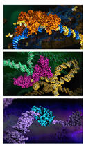
HIV enzyme
6999
These images model the molecular structures of three enzymes with critical roles in the life cycle of the human immunodeficiency virus (HIV). Amy Wu and Christine Zardecki, RCSB Protein Data Bank. View Media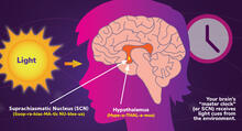
Circadian rhythms and the SCN
6613
Circadian rhythms are physical, mental, and behavioral changes that follow a 24-hour cycle. NIGMS View Media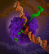
RNA polymerase
6993
RNA polymerase (purple) is a complex enzyme at the heart of transcription. Amy Wu and Christine Zardecki, RCSB Protein Data Bank. View Media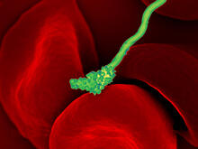
Human blood cells with Borrelia hermsii, a bacterium that causes relapsing fever
3586
Relapsing fever is caused by a bacterium and transmitted by certain soft-bodied ticks or body lice. The disease is seldom fatal in humans, but it can be very serious and prolonged. NIAID View Media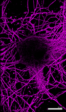
Microtubules in hippocampal neurons
6890
Microtubules (magenta) in neurons of the hippocampus, a part of the brain involved in learning and memory. Microtubules are strong, hollow fibers that provide structural support to cells. Melike Lakadamyali, Perelman School of Medicine at the University of Pennsylvania. View Media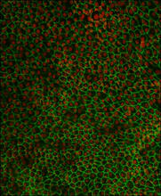
Retinal pigment epithelium derived from human ES cells 02
3287
This image shows a layer of retinal pigment epithelium cells derived from human embryonic stem cells, highlighting the nuclei (red) and cell surfaces (green). David Buckholz and Sherry Hikita, University of California, Santa Barbara, via CIRM View Media
Introduction to Genome Editing Using CRISPR/Cas9
5815
Genome editing using CRISPR/Cas9 is a rapidly expanding field of scientific research with emerging applications in disease treatment, medical therapeutics and bioenergy, just to name a few. Janet Iwasa View Media
Natcher Building 09
1089
NIGMS staff are located in the Natcher Building on the NIH campus. Alisa Machalek, National Institute of General Medical Sciences View Media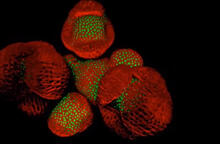
Arabidopsis Thaliana: Flowers Spring to Life
6503
This image capture shows how a single gene, STM, plays a starring role in plant development. Nathanaёl Prunet NIH Support: National Institute of General Medical Sciences View Media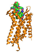
Chemokine CXCR4 receptor
3365
The receptor is shown bound to a small molecule peptide called CVX15. Raymond Stevens, The Scripps Research Institute View Media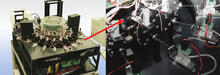
Capillary protein crystallization robot
2357
This ACAPELLA robot for capillary protein crystallization grows protein crystals, freezes them, and centers them without manual intervention. Structural Genomics of Pathogenic Protozoa Consortium View Media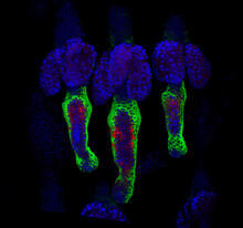
Wound healing in process
3497
Wound healing requires the action of stem cells. Hermann Steller, Rockefeller University View Media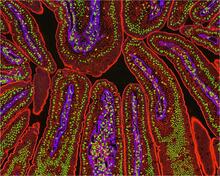
NCMIR Intestine-2
3390
The small intestine is where most of our nutrients from the food we eat are absorbed into the bloodstream. Tom Deerinck, National Center for Microscopy and Imaging Research (NCMIR) View Media
Natcher Building 05
1085
NIGMS staff are located in the Natcher Building on the NIH campus. Alisa Machalek, National Institute of General Medical Sciences View Media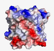
VDAC video 03
2572
This video shows the structure of the pore-forming protein VDAC-1 from humans. Gerhard Wagner, Harvard Medical School View Media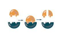
Enzymes convert subtrates into products
2521
Enzymes convert substrates into products very quickly. See image 2522 for a labeled version of this illustration. Featured in The Chemistry of Health. Crabtree + Company View Media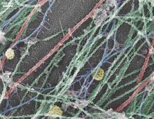
Cells use bubble-like structures called vesicles to transport cargo
3634
Cells use bubble-like structures called vesicles (yellow) to import, transport, and export cargo and in cellular communication. A single cell may be filled with thousands of moving vesicles.Tatyana Svitkina, University of Pennsylvania View Media
Cytoskeleton
1272
The three fibers of the cytoskeleton--microtubules in blue, intermediate filaments in red, and actin in green--play countless roles in the cell. Judith Stoffer View Media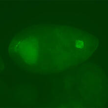
Fly cells
3594
If a picture is worth a thousand words, what's a movie worth? Denise Montell, Johns Hopkins University School of Medicine View Media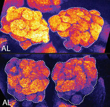
Sleep and the fly brain
2596
In the top snapshots, the brain of a sleep-deprived fruit fly glows orange, marking high concentrations of a synaptic protein called Bruchpilot (BRP) involved in communication between neurons. Chiara Cirelli, University of Wisconsin-Madison View Media
Yeast cells with nuclear envelopes and tubulin
6798
Yeast cells with nuclear envelopes shown in magenta and tubulin shown in light blue. The nuclear envelope defines the borders of the nucleus, which houses DNA. Alaina Willet, Kathy Gould’s lab, Vanderbilt University. View Media
Cell-like compartments from frog eggs 4
6591
Cell-like compartments that spontaneously emerged from scrambled frog eggs, with nuclei (blue) from frog sperm. Endoplasmic reticulum (red) and microtubules (green) are also visible. Xianrui Cheng, Stanford University School of Medicine. View Media
Mitochondria from rat heart muscle cell_2
3664
These mitochondria (brown) are from the heart muscle cell of a rat. Mitochondria have an inner membrane that folds in many places (and that appears here as striations). National Center for Microscopy and Imaging Research View Media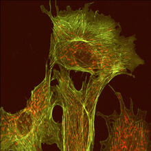
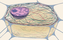
Endothelial cell
1102
This image shows two components of the cytoskeleton, microtubules (green) and actin filaments (red), in an endothelial cell derived from a cow lung. Tina Weatherby Carvalho, University of Hawaii at Manoa View Media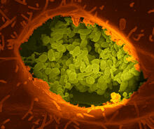
Q fever bacteria in an infected cell
3621
This image shows Q fever bacteria (yellow), which infect cows, sheep, and goats around the world and can infect humans, as well. When caught early, Q fever can be cured with antibiotics. Robert Heinzen, Elizabeth Fischer, and Anita Mora, National Institute of Allergy and Infectious Diseases, National Institutes of Health View Media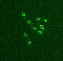
Dividing yeast cells with nuclear envelopes and spindle pole bodies
6795
Time-lapse video of yeast cells undergoing cell division. Nuclear envelopes are shown in green, and spindle pole bodies, which help pull apart copied genetic information, are shown in magenta. Alaina Willet, Kathy Gould’s lab, Vanderbilt University. View Media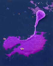
Crab nerve cell
1247
Neuron from a crab showing the cell body (bottom), axon (rope-like extension), and growth cone (top right). Tina Weatherby Carvalho, University of Hawaii at Manoa View Media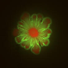
Floral pattern in a mixture of two bacterial species, Acinetobacter baylyi and Escherichia coli, grown on a semi-solid agar for 48 hours (photo 1)
6553
Floral pattern emerging as two bacterial species, motile Acinetobacter baylyi (red) and non-motile Escherichia coli (green), are grown together for 48 hours on 1% agar surface from a sma L. Xiong et al, eLife 2020;9: e48885 View Media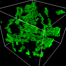
3D reconstruction of a tubular matrix in peripheral endoplasmic reticulum
5857
Detailed three-dimensional reconstruction of a tubular matrix in a thin section of the peripheral endoplasmic reticulum between the plasma membranes of the cell. Jennifer Lippincott-Schwartz, Howard Hughes Medical Institute Janelia Research Campus, Virginia View Media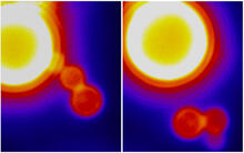
Dynamin Fission
3448
Time lapse series shows short dynamin assemblies (not visible) constricting a lipid tube to make a "beads on a string" appearance, then cutting off one of the beads i.e., catalyzing membrane fission). Ramachandran, Pucadyil et al. , The Scripps Research Institute View Media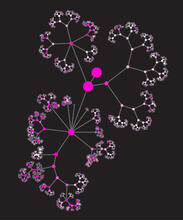
Network diagram of genes, cellular components and processes (unlabeled)
3436
This image shows the hierarchical ontology of genes, cellular components and processes derived from large genomic datasets. From Dutkowski et al. Janusz Dutkowski and Trey Ideker View Media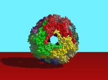
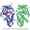
Heat shock protein complex from Methanococcus jannaschii
2385
Model based on X-ray crystallography of the structure of a small heat shock protein complex from the bacteria, Methanococcus jannaschii. Berkeley Structural Genomics Center, PSI-1 View Media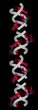
Zinc finger
2426
The structure of a gene-regulating zinc finger protein bound to DNA. Jeremy M. Berg, National Institute of General Medical Sciences View Media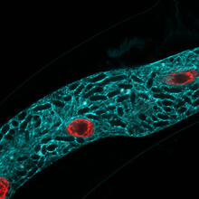
Microsporidia in roundworm 2
5778
Many disease-causing microbes manipulate their host’s metabolism and cells for their own ends. Keir Balla and Emily Troemel, University of California San Diego View Media