Switch to Gallery View
Image and Video Gallery
This is a searchable collection of scientific photos, illustrations, and videos. The images and videos in this gallery are licensed under Creative Commons Attribution Non-Commercial ShareAlike 3.0. This license lets you remix, tweak, and build upon this work non-commercially, as long as you credit and license your new creations under identical terms.
Stress Response in Cells
6570
Two highly stressed osteosarcoma cells are shown with a set of green droplet-like structures followed by a second set of magenta droplets. Julia F. Riley and Carlos A. Castañeda, Syracuse University View Media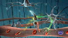
Motion in the brain
2323
Amid a network of blood vessels and star-shaped support cells, neurons in the brain signal each other. The mists of color show the flow of important molecules like glucose and oxygen. Kim Hager and Neal Prakash, University of California, Los Angeles View Media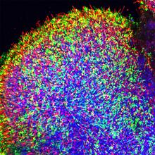
Human retinal organoid
6748
A replica of a human retina grown from stem cells. Kevin Eliceiri, University of Wisconsin-Madison. View Media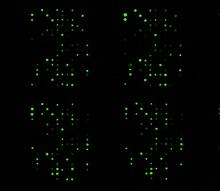
Glycan arrays
1265
The signal is obtained by allowing proteins in human serum to interact with glycan (polysaccharide) arrays. The arrays are shown in replicate so the pattern is clear. Ola Blixt, Scripps Research Institute View Media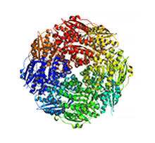
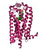
Mandelate racemase from B. subtilis
2350
Model of the mandelate racemase enzyme from Bacillus subtilis, a bacterium commonly found in soil. New York Structural GenomiX Research Consortium, PSI View Media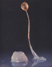
Dicty fruit
2684
Dictyostelium discoideum is a microscopic amoeba. A group of 100,000 form a mound as big as a grain of sand. Featured in The New Genetics. View Media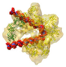
DNA replication origin recognition complex (ORC)
3597
A study published in March 2012 used cryo-electron microscopy to determine the structure of the DNA replication origin recognition complex (ORC), a semi-circular, protein complex (yellow) that recogni Huilin Li, Brookhaven National Laboratory View Media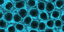
Movements of myosin
2324
Inside the fertilized egg cell of a fruit fly, we see a type of myosin (related to the protein that helps muscles contract) made to glow by attaching a fluorescent protein. Victoria Foe, University of Washington View Media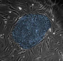
Human embryonic stem cells
2608
The center cluster of cells, colored blue, shows a colony of human embryonic stem cells. James Thomson, University of Wisconsin-Madison View Media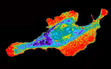
Seeing signaling protein activation in cells 03
2453
Cdc42, a member of the Rho family of small guanosine triphosphatase (GTPase) proteins, regulates multiple cell functions, including motility, proliferation, apoptosis, and cell morphology. Klaus Hahn, University of North Carolina, Chapel Hill Medical School View Media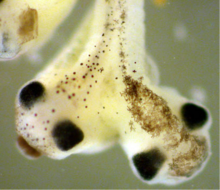
Two-headed Xenopus laevis tadpole
2755
Xenopus laevis, the African clawed frog, has long been used as a research organism for studying embryonic development. Michael Klymkowsky, University of Colorado, Boulder View Media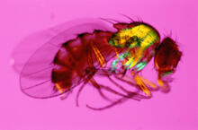
Fruit fly in the pink
2693
Fruit flies are a common model organism for basic medical research. Crabtree + Company View Media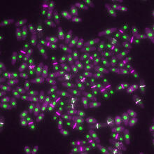
Yeast cells with nuclei and contractile rings
6792
Yeast cells with nuclei shown in green and contractile rings shown in magenta. Nuclei store DNA, and contractile rings help cells divide. Alaina Willet, Kathy Gould’s lab, Vanderbilt University. View Media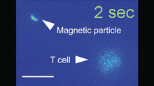
Magnetic Janus particle activating a T cell
6800
A Janus particle being used to activate a T cell, a type of immune cell. Yan Yu, Indiana University, Bloomington. View Media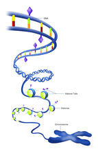
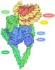
Epigenetic code (with labels)
2563
The "epigenetic code" controls gene activity with chemical tags that mark DNA (purple diamonds) and the "tails" of histone proteins (purple triangles). Crabtree + Company View Media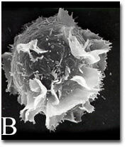
Activated mast cell surface
2637
A scanning electron microscope image of an activated mast cell. This image illustrates the interesting topography of the cell membrane, which is populated with receptors. Bridget Wilson, University of New Mexico View Media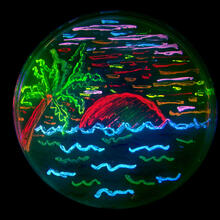
Glowing bacteria make a pretty postcard
3492
This tropical scene, reminiscent of a postcard from Key West, is actually a petri dish containing an artistic arrangement of genetically engineered bacteria. Nathan C. Shaner, The Scintillon Institute View Media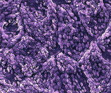
Retinal pigment epithelium derived from human ES cells
3286
This color-enhanced image is a scanning electron microscope image of retinal pigment epithelial (RPE) cells derived from human embryonic stem cells. David Hinton lab, University of Southern California, via CIRM View Media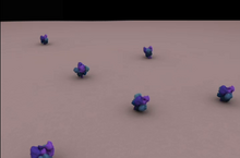
Clathrin-mediated endocytosis
5753
Endocytosis is the process by which cells are able to take up membrane and extracellular materials through the formation of a small intracellular bubble, called a vesicle. Janet Iwasa, University of Utah View Media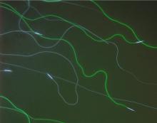
GFP sperm
2683
Fruit fly sperm cells glow bright green when they express the gene for green fluorescent protein (GFP). View Media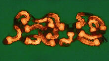
Group of Culex quinquefasciatus mosquito larvae
6770
Mosquito larvae with genes edited by CRISPR. Valentino Gantz, University of California, San Diego. View Media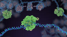
Cas9 protein involved in the CRISPR gene-editing technology
5816
In the gene-editing tool CRISPR, a small strand of RNA identifies a specific chunk of DNA. Janet Iwasa View Media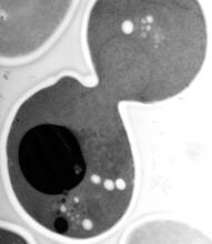
EM of yeast cell division
5770
Cell division is an incredibly coordinated process. Matthew West and Greg Odorizzi, University of Colorado View Media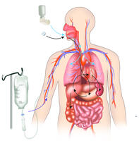
A drug's life in the body
2527
A drug's life in the body. Medicines taken by mouth pass through the liver before they are absorbed into the bloodstream. Crabtree + Company View Media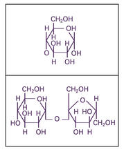
Glucose and sucrose
2500
Glucose (top) and sucrose (bottom) are sugars made of carbon, hydrogen, and oxygen atoms. Carbohydrates include simple sugars like these and are the main source of energy for the human body. Crabtree + Company View Media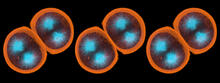
Sea urchin embryo 01
1047
Stereo triplet of a sea urchin embryo stained to reveal actin filaments (orange) and microtubules (blue). George von Dassow, University of Washington View Media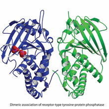
Dimeric association of receptor-type tyrosine-protein phosphatase
2349
Model of the catalytic portion of an enzyme, receptor-type tyrosine-protein phosphatase from humans. The enzyme consists of two identical protein subunits, shown in blue and green. New York Structural GenomiX Research Consortium, PSI View Media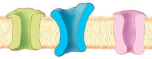
Ion channels
1284
The body uses a variety of ion channels to transport small molecules across cell membranes. Judith Stoffer View Media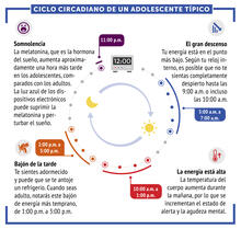
Ciclo circadiano de un adolescente típico
6612
Los ritmos circadianos son cambios físicos, mentales y conductuales que siguen un ciclo de 24 horas. NIGMS View Media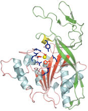
Human aspartoacylase
2352
Model of aspartoacylase, a human enzyme involved in brain metabolism. Center for Eukaryotic Structural Genomics, PSI View Media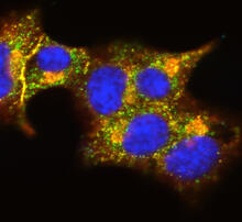
Insulin and protein interact in pancreatic beta cells
3546
A large number of proteins interact with the hormone insulin as it is produced in and secreted from the beta cells of the pancreas. William E. Balch, The Scripps Research Institute View Media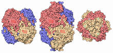
Catalase diversity
7003
Catalases are some of the most efficient enzymes found in cells. Amy Wu and Christine Zardecki, RCSB Protein Data Bank. View Media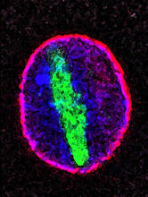
Cellular aging
2578
A protein called tubulin (green) accumulates in the center of a nucleus (outlined in pink) from an aging cell. Maximiliano D'Angelo and Martin Hetzer, Salk Institute View Media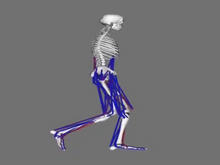
Simulation of leg muscles moving
6598
When we walk, muscles and nerves interact in intricate ways. This simulation, which is based on data from a six-foot-tall man, shows these interactions. Chand John and Eran Guendelman, Stanford University View Media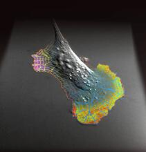
Biosensors illustration
2802
A rendering of an activity biosensor image overlaid with a cell-centered frame of reference used for image analysis of signal transduction. Gaudenz Danuser, Harvard Medical School View Media
A multicolored fish scale 1
3782
Each of the colored specs in this image is a cell on the surface of a fish scale. Chen-Hui Chen and Kenneth Poss, Duke University View Media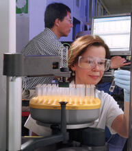
Protein purification robot
2375
Irina Dementieva, a biochemist, and Youngchang Kim, a biophysicist and crystallographer, work with the first robot of its type in the U.S. to automate protein purification. Midwest Center for Structural Genomics View Media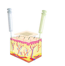
Drugs enter skin
2531
Drugs enter different layers of skin via intramuscular, subcutaneous, or transdermal delivery methods. See image 2532 for a labeled version of this illustration. Crabtree + Company View Media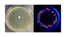
Antibiotic-surviving bacteria
6802
Colonies of bacteria growing despite high concentrations of antibiotics. These colonies are visible both by eye, as seen on the left, and by bioluminescence imaging, as seen on the right. Paul Stoodley, The Ohio State University. View Media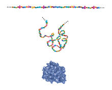
Building blocks and folding of proteins
2508
Proteins are made of amino acids hooked end-to-end like beads on a necklace. To become active, proteins must twist and fold into their final, or "native," conformation. Crabtree + Company View Media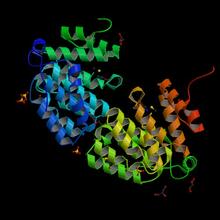
Protein from E. faecalis
2342
X-ray structure of a DNA repair enzyme superfamily representative from the human gastrointestinal bacterium Enterococcus faecalis. Midwest Center for Structural Genomics View Media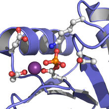
Active Site of E. coli response regulator PhoB
3412
Active site of E. coli response regulator PhoB. Ann Stock, Rutgers University View Media
Bacillus anthracis being killed
3525
Bacillus anthracis (anthrax) cells being killed by a fluorescent trans-translation inhibitor, which disrupts bacterial protein synthesis. Kenneth Keiler, Penn State University View Media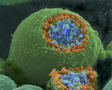
Nerve ending
1244
A scanning electron microscope picture of a nerve ending. It has been broken open to reveal vesicles (orange and blue) containing chemicals used to pass messages in the nervous system. Tina Weatherby Carvalho, University of Hawaii at Manoa View Media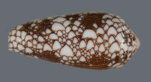
Cone snail shell
2576
A shell from the venomous cone snail Conus omaria, which lives in the Pacific and Indian oceans and eats other snails. Kerry Matz, University of Utah View Media
Mapping metabolic activity
2319
Like a map showing heavily traveled roads, this mathematical model of metabolic activity inside an E. coli cell shows the busiest pathway in white. Albert-László Barabási, University of Notre Dame View Media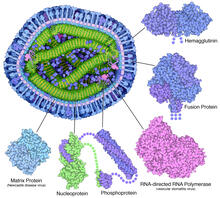
Measles virus proteins
6996
A cross section of the measles virus in which six proteins (enlarged on the outside of the virus) work together to infect cells. Amy Wu and Christine Zardecki, RCSB Protein Data Bank. View Media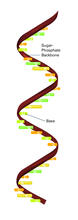
RNA strand (with labels)
2555
Ribonucleic acid (RNA) has a sugar-phosphate backbone and the bases adenine (A), cytosine (C), guanine (G), and uracil (U). Featured in The New Genetics. Crabtree + Company View Media
Mouse sperm sections
1191
This transmission electron micrograph shows sections of mouse sperm tails, or flagella. Tina Weatherby Carvalho, University of Hawaii at Manoa View Media
Glow-in-the-dark salamanders
2715
These six-month-old axolotls, a kind of salamander, glow green and blue under ultraviolet light. That's because they were genetically modified to make harmless green fluorescent protein, or GFP. View Media