Switch to Gallery View
Image and Video Gallery
This is a searchable collection of scientific photos, illustrations, and videos. The images and videos in this gallery are licensed under Creative Commons Attribution Non-Commercial ShareAlike 3.0. This license lets you remix, tweak, and build upon this work non-commercially, as long as you credit and license your new creations under identical terms.
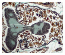
Cross section of a Drosophila melanogaster pupa
2758
This photograph shows a magnified view of a Drosophila melanogaster pupa in cross section. Compare this normal pupa to one that lacks an important receptor, shown in image 2759. Christina McPhee and Eric Baehrecke, University of Massachusetts Medical School View Media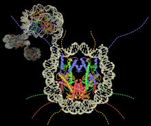
Nucleosome
2741
Like a strand of white pearls, DNA wraps around an assembly of special proteins called histones (colored) to form the nucleosome, a structure responsible for regulating genes and condensing DNA strand Karolin Luger, Colorado State University View Media
Pigment cells in the fin of pearl danio
5757
Pigment cells are cells that give skin its color. David Parichy, University of Washington View Media
Polarized cells- 02
3333
Cells move forward with lamellipodia and filopodia supported by networks and bundles of actin filaments. Proper, controlled cell movement is a complex process. Rong Li and Praveen Suraneni, Stowers Institute for Medical Research View Media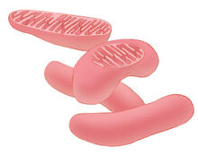
Mitochondria
1287
Bean-shaped mitochondria are cells' power plants. These organelles have their own DNA and replicate independently. The highly folded inner membranes are the site of energy generation. Judith Stoffer View Media
Skin cancer cells (squamous cell carcinoma)
3628
This image shows the uncontrolled growth of cells in squamous cell carcinoma, the second most common form of skin cancer. If caught early, squamous cell carcinoma is usually not life-threatening. Markus Schober and Elaine Fuchs, The Rockefeller University View Media
Video of Calling Cards in a mouse brain
6781
The green spots in this mouse brain are cells labeled with Calling Cards, a technology that records molecular events in brain cells as they mature. NIH Director's Blog View Media
Crane fly spermatocyte undergoing meiosis
6898
A crane fly spermatocyte during metaphase of meiosis-I, a step in the production of sperm. Michael Shribak, Marine Biological Laboratory/University of Chicago. View Media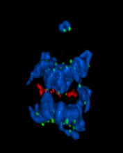
A chromosome goes missing in anaphase
5766
Anaphase is the critical step during mitosis when sister chromosomes are disjoined and directed to opposite spindle poles, ensuring equal distribution of the genome during cell division. View Media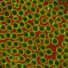
Fruit fly embryo
2431
Cells in an early-stage fruit fly embryo, showing the DIAP1 protein (pink), an inhibitor of apoptosis. Hermann Steller, Rockefeller University View Media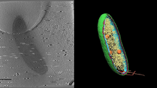
Cryo-electron tomography of a Caulobacter bacterium
6569
3D image of Caulobacter bacterium with various components highlighted: cell membranes (red and blue), protein shell (green), protein factories known as ribosomes (yellow), and storage granules Peter Dahlberg, Stanford University. View Media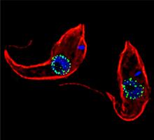
Trypanosoma brucei, the cause of sleeping sickness
3765
Trypanosoma brucei is a single-cell parasite that causes sleeping sickness in humans. Michael Rout, Rockefeller University View Media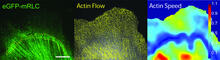
Actin flow
2798
Speckle microscopy analysis of actin cytoskeleton force. This is an example of NIH-supported research on single-cell analysis. Gaudenz Danuser, Harvard Medical School View Media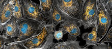
Panorama view of golden mitochondria
5762
Mitochondria are the powerhouses of the cells, generating the energy the cells need to do their tasks and to stay alive. Torsten Wittmann, University of California, San Francisco View Media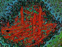
NCMIR Tongue 2
5811
Microscopy image of a tongue. One in a series of two, see image 5810 National Center for Microscopy and Imaging Research (NCMIR) View Media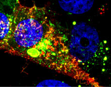
RSV-Infected Cell
3567
Viral RNA (red) in an RSV-infected cell. Eric Alonas and Philip Santangelo, Georgia Institute of Technology and Emory University View Media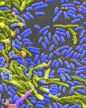
Vibrio bacteria
1160
Vibrio, a type (genus) of rod-shaped bacteria. Some Vibrio species cause cholera in humans. Tina Weatherby Carvalho, University of Hawaii at Manoa View Media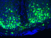
Master clock of the mouse brain
3547
An image of the area of the mouse brain that serves as the 'master clock,' which houses the brain's time-keeping neurons. The nuclei of the clock cells are shown in blue. Erik Herzog, Washington University in St. Louis View Media
Stretch detectors
2714
Muscles stretch and contract when we walk, and skin splits open and knits back together when we get a paper cut. Christopher Chen, University of Pennsylvania View Media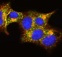
Insulin and protein interact in pancreatic beta cells
3546
A large number of proteins interact with the hormone insulin as it is produced in and secreted from the beta cells of the pancreas. William E. Balch, The Scripps Research Institute View MediaTranslation
1281
Ribosomes manufacture proteins based on mRNA instructions. Each ribosome reads mRNA, recruits tRNA molecules to fetch amino acids, and assembles the amino acids in the proper order. Judith Stoffer View Media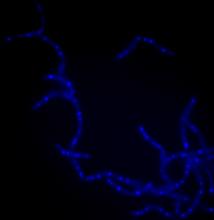
Bacillus anthracis being killed
3525
Bacillus anthracis (anthrax) cells being killed by a fluorescent trans-translation inhibitor, which disrupts bacterial protein synthesis. Kenneth Keiler, Penn State University View Media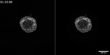
Breast cancer cells change migration phenotypes
6986
Cancer cells can change their migration phenotype, which includes their shape and the way that they move to invade different tissues. Bo Sun, Oregon State University. View Media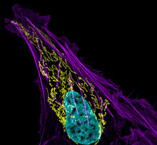
Bone cancer cell
3626
This image shows an osteosarcoma cell with DNA in blue, energy factories (mitochondria) in yellow, and actin filaments—part of the cellular skeleton—in purple. Dylan Burnette and Jennifer Lippincott-Schwartz, NICHD View Media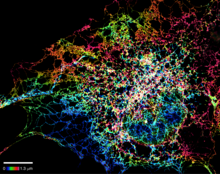
Dense tubular matrices in the peripheral endoplasmic reticulum (ER) 1
5855
Superresolution microscopy work on endoplasmic reticulum (ER) in the peripheral areas of the cell showing details of the structure and arrangement in a complex web of tubes. Jennifer Lippincott-Schwartz, Howard Hughes Medical Institute Janelia Research Campus, Virginia View MediaAssembly of the HIV capsid
5729
The HIV capsid is a pear-shaped structure that is made of proteins the virus needs to mature and become infective. John Grime and Gregory Voth, The University of Chicago View Media
TEM cross-section of C. elegans (roundworm)
5759
The worm Caenorhabditis elegans is a popular laboratory animal because its small size and fairly simple body make it easy to study. Piali Sengupta, Brandeis University View Media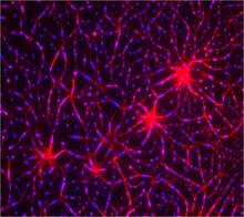
In vitro assembly of a cell-signaling pathway
3787
T cells are white blood cells that are important in defending the body against bacteria, viruses and other pathogens. Xiaolei Su, HHMI Whitman Center of the Marine Biological Laboratory View Media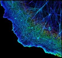
3D image of actin in a cell
3749
Actin is an essential protein in a cell's skeleton (cytoskeleton). It forms a dense network of thin filaments in the cell. Xiaowei Zhuang, Howard Hughes Medical Institute, Harvard University View Media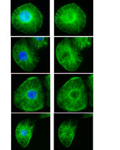
Interphase in Xenopus frog cells
3443
These images show frog cells in interphase. The cells are Xenopus XL177 cells, which are derived from tadpole epithelial cells. The microtubules are green and the chromosomes are blue. Claire Walczak, who took them while working as a postdoc in the laboratory of Timothy Mitchison. View Media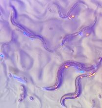
C. elegans with blue and yellow lights in the background
6750
These microscopic roundworms, called Caenorhabditis elegans, lack eyes and the opsin proteins used by visual systems to detect colors. H. Robert Horvitz and Dipon Ghosh, Massachusetts Institute of Technology. View Media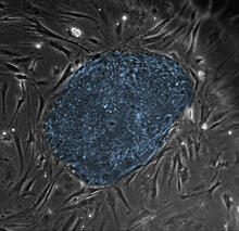
Human embryonic stem cells
2608
The center cluster of cells, colored blue, shows a colony of human embryonic stem cells. James Thomson, University of Wisconsin-Madison View Media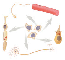
Stem cell differentiation
1294
Undifferentiated embryonic stem cells cease to exist a few days after conception. In this image, ES cells are shown to differentiate into sperm, muscle fiber, hair cells, nerve cells, and cone cells. Judith Stoffer View Media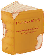
Aging book of life
1334
Damage to each person's genome, often called the "Book of Life," accumulates with time. Judith Stoffer View Media
3D reconstruction of a tubular matrix in peripheral endoplasmic reticulum
5857
Detailed three-dimensional reconstruction of a tubular matrix in a thin section of the peripheral endoplasmic reticulum between the plasma membranes of the cell. Jennifer Lippincott-Schwartz, Howard Hughes Medical Institute Janelia Research Campus, Virginia View Media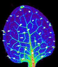
Zinc levels in a plant leaf
3727
Zinc is required for the function of more than 300 enzymes, including those that help regulate gene expression, in various organisms including humans. Suzana Car, Dartmouth College View Media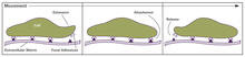
Focal adhesions (with labels)
2503
Cells walk along body surfaces via tiny "feet," called focal adhesions, that connect with the extracellular matrix. Crabtree + Company View Media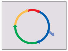
Cell cycle
2498
Cells progress through a cycle that consists of phases for growth (blue, green, yellow) and division (red). Cells become quiescent when they exit this cycle (purple). Crabtree + Company View Media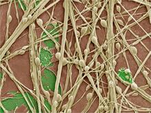
Synapses in culture
3399
Cultured hippocampal neurons grown on a substrate of glial cells (astrocytes). The glial cells form the pink/brown underlayment in this image. The tan threads are the neurons. National Center for Microscopy and Imaging Research View Media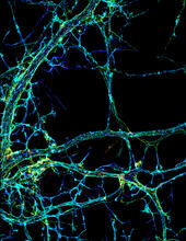
STORM image of axonal cytoskeleton
3678
This image shows the long, branched structures (axons) of nerve cells. Xiaowei Zhuang Laboratory, Howard Hughes Medical Institute, Harvard University View Media
Fruit fly egg ooplasmic streaming
6809
Two fruit fly (Drosophila melanogaster) egg cells, one on each side of the central black line. Vladimir I. Gelfand, Feinberg School of Medicine, Northwestern University. View Media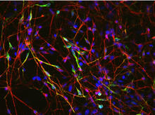
Neurons from human ES cells 02
3285
These neurons were derived from human embryonic stem cells. The neural cell bodies with axonal projections are visible in red, and the nuclei in blue. Xianmin Zeng lab, Buck Institute for Age Research, via CIRM View Media
A multicolored fish scale 2
3783
Each of the tiny colored specs in this image is a cell on the surface of a fish scale. Chen-Hui Chen and Kenneth Poss, Duke University View Media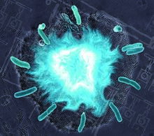
Supernova bacteria
2725
Bacteria engineered to act as genetic clocks flash in synchrony. Here, a "supernova" burst in a colony of coupled genetic clocks just after reaching critical cell density. Jeff Hasty, UCSD View Media
Biofilm formed by a pathogen
6518
A biofilm is a highly organized community of microorganisms that develops naturally on certain surfaces. Scott Chimileski, Ph.D., and Roberto Kolter, Ph.D., Harvard Medical School. View Media
“Two-faced” Janus particle activating a macrophage
6801
A macrophage—a type of immune cell that engulfs invaders—“eats” and is activated by a “two-faced” Janus particle. Yan Yu, Indiana University, Bloomington. View Media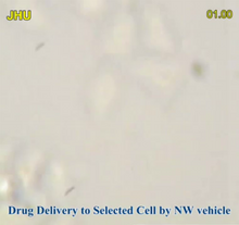
Precisely Delivering Chemical Cargo to Cells
3779
Moving protein or other molecules to specific cells to treat or examine them has been a major biological challenge. Nature Nanotechnology View Media
Vimentin in a quail embryo
2809
Video of high-resolution confocal images depicting vimentin immunofluorescence (green) and nuclei (blue) at the edge of a quail embryo yolk. Andrés Garcia, Georgia Tech View Media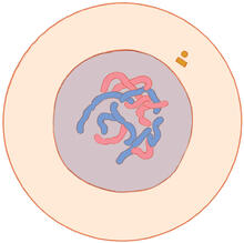
Mitosis - interphase
1316
A cell in interphase, at the start of mitosis: Chromosomes duplicate, and the copies remain attached to each other. Judith Stoffer View Media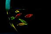
Glowing fish
2667
Professor Marc Zimmer's family pets, including these fish, glow in the dark in response to blue light. Featured in the September 2009 issue of Findings. View Media