Switch to Gallery View
Image and Video Gallery
This is a searchable collection of scientific photos, illustrations, and videos. The images and videos in this gallery are licensed under Creative Commons Attribution Non-Commercial ShareAlike 3.0. This license lets you remix, tweak, and build upon this work non-commercially, as long as you credit and license your new creations under identical terms.
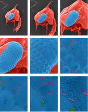
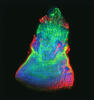
Crab larva eye
1251
Colorized scanning electron micrographs progressively zoom in on the eye of a crab larva. In the higher-resolution frames, bacteria are visible on the eye. Tina Weatherby Carvalho, University of Hawaii at Manoa View Media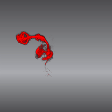
Color coding of the Drosophila brain - video
5843
This video results from a research project to visualize which regions of the adult fruit fly (Drosophila) brain derive from each neural stem cell. Yong Wan from Charles Hansen’s lab, University of Utah. Data preparation and visualization by Masayoshi Ito in the lab of Kei Ito, University of Tokyo. View Media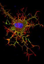
Abnormal, spiky fibroblast
3613
This is a fibroblast, a connective tissue cell that plays an important role in wound healing. Normal fibroblasts have smooth edges. Praveen Suraneni, Stowers Institute for Medical Research, Kansas City, Mo. View Media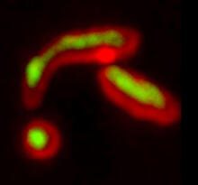
Misfolded proteins within in the mitochondria
5878
Misfolded proteins (green) within mitochondria (red). Related to video 5877. Rong Li rong@jhu.edu Department of Chemical and Biomolecular Engineering, Whiting School of Engineering, Johns Hopkins University, Baltimore, Maryland 21218, USA. View Media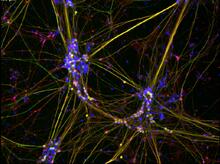
Peripheral nerve cells derived from ES cells
3263
Peripheral nerve cells made from human embryonic stem cell-derived neural crest stem cells. Stephen Dalton, University of Georgia View Media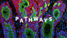
Pathways: What is It? | Why Scientists Study Cells
6540
Learn how curiosity about the world and our cells is key to scientific discoveries. National Institute of General Medical Sciences View Media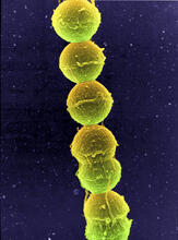
Streptococcus bacteria
1157
Image of Streptococcus, a type (genus) of spherical bacteria that can colonize the throat and back of the mouth. Stroptococci often occur in pairs or in chains, as shown here. Tina Weatherby Carvalho, University of Hawaii at Manoa View Media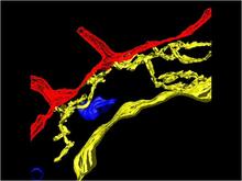
Computer model of cell membrane
2636
A computer model of the cell membrane, where the plasma membrane is red, endoplasmic reticulum is yellow, and mitochondria are blue. Bridget Wilson, University of New Mexico View Media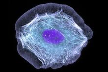
Skin cell (keratinocyte)
3599
This normal human skin cell was treated with a growth factor that triggered the formation of specialized protein structures that enable the cell to move. Torsten Wittmann, University of California, San Francisco View Media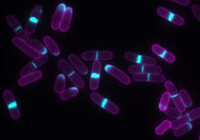
Yeast cells with accumulated cell wall material
6797
Yeast cells that abnormally accumulate cell wall material (blue) at their ends and, when preparing to divide, in their middles. This image was captured using wide-field microscopy with deconvolution. Alaina Willet, Kathy Gould’s lab, Vanderbilt University. View Media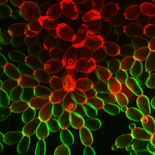
Snowflake yeast 1
6969
Multicellular yeast called snowflake yeast that researchers created through many generations of directed evolution from unicellular yeast. William Ratcliff, Georgia Institute of Technology. View Media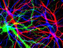
Brain cells in the hippocampus
3688
Hippocampal cells in culture with a neuron in green, showing hundreds of the small protrusions known as dendritic spines. Shelley Halpain, UC San Diego View Media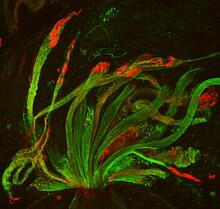
Fruit fly spermatids
3590
Developing spermatids (precursors of mature sperm cells) begin as small, round cells and mature into long-tailed, tadpole-shaped ones. Lacramioara Fabian, The Hospital for Sick Children, Toronto, Canada View Media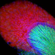
Developing zebrafish fin
3598
Originally from the waters of India, Nepal, and neighboring countries, zebrafish can now be found swimming in science labs (and home aquariums) throughout the world. Jessica Plavicki View Media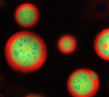
Nucleolus subcompartments spontaneously self-assemble 4
3793
What looks a little like distant planets with some mysterious surface features are actually assemblies of proteins normally found in the cell's nucleolus, a small but very important protein complex lo Nilesh Vaidya, Princeton University View Media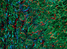
Optic nerve astrocytes
5852
Astrocytes in the cross section of a human optic nerve head Tom Deerinck and Keunyoung (“Christine”) Kim, NCMIR View Media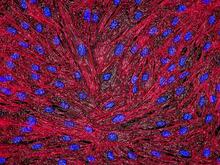
DNA and actin in cultured fibroblast cells
3670
DNA (blue) and actin (red) in cultured fibroblast cells. Tom Deerinck, National Center for Microscopy and Imaging Research (NCMIR) View Media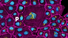
Pathways: The Fascinating Cells of Research Organisms
6538
Learn how research organisms, such as fruit flies and mice, can help us understand and treat human diseases. National Institute of General Medical Sciences View Media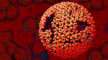
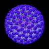
Molecular model of freshly made Rous sarcoma virus (RSV)
3771
Viruses have been the foes of animals and other organisms for time immemorial. Boon Chong Goh, University of Illinois at Urbana-Champaign View Media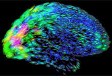
Mapping brain differences
2419
This image of the human brain uses colors and shapes to show neurological differences between two people. Arthur Toga, University of California, Los Angeles View Media
Smooth ER
1292
The endoplasmic reticulum comes in two types: Rough ER is covered with ribosomes and prepares newly made proteins; smooth ER specializes in making lipids and breaking down toxic molecules. Judith Stoffer View Media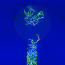
Genetically identical mycobacteria respond differently to antibiotic 1
5751
Antibiotic resistance in microbes is a serious health concern. So researchers have turned their attention to how bacteria undo the action of some antibiotics. Bree Aldridge, Tufts University View Media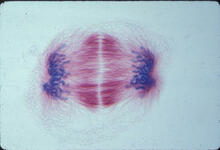
Lily mitosis 12
1018
A light microscope image of a cell from the endosperm of an African globe lily (Scadoxus katherinae). This is one frame of a time-lapse sequence that shows cell division in action. Andrew S. Bajer, University of Oregon, Eugene View Media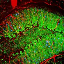
Brain showing hallmarks of Alzheimer's disease
3604
Along with blood vessels (red) and nerve cells (green), this mouse brain shows abnormal protein clumps known as plaques (blue). Alvin Gogineni, Genentech View Media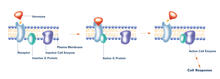
G switch (with labels)
2537
The G switch allows our bodies to respond rapidly to hormones. G proteins act like relay batons to pass messages from circulating hormones into cells. Crabtree + Company View Media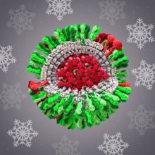
H1N1 Influenza Virus
6355
CellPack image of the H1N1 influenza virus, with hemagglutinin and neuraminidase glycoproteins in green and red, respectively, on the outer envelope (white); matrix protein in gray, and ribonucleoprot Dr. Rommie Amaro, University of California, San Diego View Media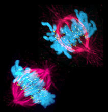
Cell in two stages of division
3541
This image shows a cell in two stages of division: prometaphase (top) and metaphase (bottom). Lilian Kabeche, Dartmouth View Media
Lily mitosis 06
1016
A light microscope image of a cell from the endosperm of an African globe lily (Scadoxus katherinae). This is one frame of a time-lapse sequence that shows cell division in action. Andrew S. Bajer, University of Oregon, Eugene View Media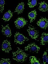
Calcium uptake during ATP production in mitochondria
3449
Living primary mouse embryonic fibroblasts. Mitochondria (green) stained with the mitochondrial membrane potential indicator, rhodamine 123. Nuclei (blue) are stained with DAPI. Lili Guo, Perelman School of Medicine, University of Pennsylvania View Media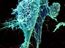
String-like Ebola virus peeling off an infected cell
3619
After multiplying inside a host cell, the stringlike Ebola virus is emerging to infect more cells. Heinz Feldmann, Peter Jahrling, Elizabeth Fischer and Anita Mora, National Institute of Allergy and Infectious Diseases, National Institutes of Health View MediaAssembly of the HIV capsid
5729
The HIV capsid is a pear-shaped structure that is made of proteins the virus needs to mature and become infective. John Grime and Gregory Voth, The University of Chicago View Media
Cytonemes in developing fruit fly cells
3574
Scientists have long known that multicellular organisms use biological molecules produced by one cell and sensed by another to transmit messages that, for instance, guide proper development of organs Sougata Roy, University of California, San Francisco View MediaTracking cells in a gastrulating zebrafish embryo
6776
During development, a zebrafish embryo is transformed from a ball of cells into a recognizable body plan by sweeping convergence and extension cell movements. This process is called gastrulation. Liliana Solnica-Krezel, Washington University School of Medicine in St. Louis. View Media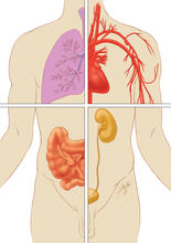
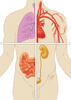
Quartered torso
1280
Cells function within organs and tissues, such as the lungs, heart, intestines, and kidney. Judith Stoffer View MediaNuclear Lamina – Three Views
6573
Three views of the entire nuclear lamina of a HeLa cell produced by tilted light sheet 3D single-molecule super-resolution imaging using a platform termed TILT3D. Anna-Karin Gustavsson, Ph.D. View Media
Dopaminergic neurons from ES cells
3270
Human embryonic stem cells differentiated into dopaminergic neurons, the type that degenerate in Parkinson's disease. Image courtesy of the California Institute for Regenerative Medicine. Jeannie Liu, Lab of Jan Nolta, University of California, Davis, via CIRM View Media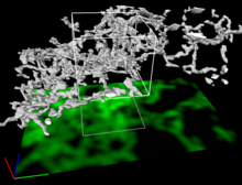
Dense tubular matrices in the peripheral endoplasmic reticulum (ER) 2
5856
Three-dimensional reconstruction of a tubular matrix in a thin section of the peripheral endoplasmic reticulum between the plasma membranes of the cell. Jennifer Lippincott-Schwartz, Howard Hughes Medical Institute Janelia Research Campus, Virginia View Media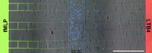
Neutrophil-like cells migrating in a microfluidic chip
6886
Neutrophil-like cells (blue) in a microfluidic chip preferentially migrating toward LTB4 over fMLP. Caroline Jones, University of Texas at Dallas. View Media
Centrioles anchor cilia in planaria
3292
Centrioles (green) anchor cilia (red), which project on the surface of pharynx cells of the freshwater planarian Schmidtea mediterranea. Juliette Azimzadeh, University of California, San Francisco View Media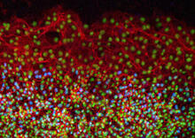
Cell proliferation in a quail embryo
2808
Image showing that the edge zone (top of image) of the quail embryo shows no proliferating cells (cyan), unlike the interior zone (bottom of image). Non-proliferating cell nuclei are labeled green. Andrés Garcia, Georgia Tech View Media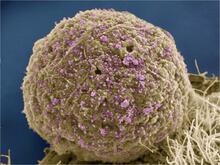
HIV Infected Cell
3386
The human immunodeficiency virus (HIV), shown here as tiny purple spheres, causes the disease known as AIDS (for acquired immunodeficiency syndrome). Tom Deerinck, National Center for Microscopy and Imaging Research (NCMIR) View Media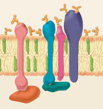
Animal cell membrane
1286
The membrane that surrounds a cell is made up of proteins and lipids. Judith Stoffer View Media
Yeast art depicting the New York City skyline
6521
This skyline of New York City was created by “printing” nanodroplets containing yeast (Saccharomyces cerevisiae) onto a large plate. Each dot is a separate yeast colony. Michael Shen, Ph.D., Jasmine Temple, Leslie Mitchell, Ph.D., and Jef Boeke, Ph.D., New York University School of Medicine; and Nick Phillips, James Chuang, Ph.D., and Jiarui Wang, Johns Hopkins University. View Media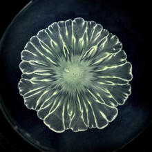
Floral pattern in a mixture of two bacterial species, Acinetobacter baylyi and Escherichia coli, grown on a semi-solid agar for 24 hours
6557
Floral pattern emerging as two bacterial species, motile Acinetobacter baylyi and non-motile Escherichia coli (green), are grown together for 24 hours on 0.75% agar surface from a small L. Xiong et al, eLife 2020;9: e48885 View Media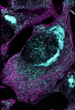
Microtubules and tau aggregates
6892
Microtubules (magenta) and tau protein (light blue) in a cell model of tauopathy. Melike Lakadamyali, Perelman School of Medicine at the University of Pennsylvania. View Media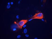
Smooth muscle from mouse stem cells
3289
These smooth muscle cells were derived from mouse neural crest stem cells. Red indicates smooth muscle proteins, blue indicates nuclei. Deepak Srivastava, Gladstone Institutes, via CIRM View Media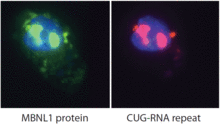
Proteins related to myotonic dystrophy
2727
Myotonic dystrophy is thought to be caused by the binding of a protein called Mbnl1 to abnormal RNA repeats. Manuel Ares, University of California, Santa Cruz View Media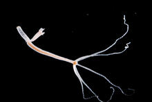
Hydra 06
2442
Hydra magnipapillata is an invertebrate animal used as a model organism to study developmental questions, for example the formation of the body axis. Hiroshi Shimizu, National Institute of Genetics in Mishima, Japan View Media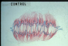
Lily mitosis 09
1022
A light microscope image of a cell from the endosperm of an African globe lily (Scadoxus katherinae). This is one frame of a time-lapse sequence that shows cell division in action. Andrew S. Bajer, University of Oregon, Eugene View Media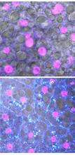
Fruit fly starvation leads to adipokine accumulation
6984
Adult Drosophila abdominal fat tissue showing cell nuclei labelled in magenta. Akhila Rajan, Fred Hutchinson Cancer Center View Media