Switch to Gallery View
Image and Video Gallery
This is a searchable collection of scientific photos, illustrations, and videos. The images and videos in this gallery are licensed under Creative Commons Attribution Non-Commercial ShareAlike 3.0. This license lets you remix, tweak, and build upon this work non-commercially, as long as you credit and license your new creations under identical terms.
G switch (with labels and stages)
2538
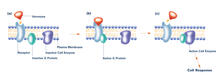
The G switch allows our bodies to respond rapidly to hormones. G proteins act like relay batons to pass messages from circulating hormones into cells. Crabtree + Company View Media
Repairing DNA
3493
Like a watch wrapped around a wrist, a special enzyme encircles the double helix to repair a broken strand of DNA. Tom Ellenberger, Washington University School of Medicine View Media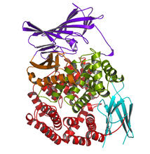
Aminopeptidase N from N. meningitidis
2341
Model of the enzyme aminopeptidase N from the human pathogen Neisseria meningitidis, which can cause meningitis epidemics. Midwest Center for Structural Genomics, PSI View Media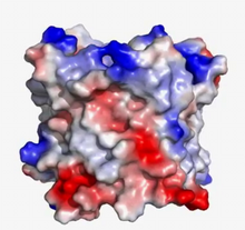
VDAC video 03
2572
This video shows the structure of the pore-forming protein VDAC-1 from humans. Gerhard Wagner, Harvard Medical School View Media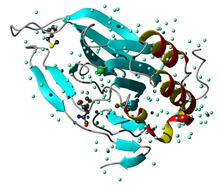
Cysteine dioxygenase from mouse
2347
Model of the mammalian iron enzyme cysteine dioxygenase from a mouse. Center for Eukaryotic Structural Genomics, PSI View Media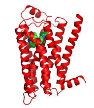
Beta 2-adrenergic receptor
3358
The receptor is shown bound to a partial inverse agonist, carazolol. Raymond Stevens, The Scripps Research Institute View Media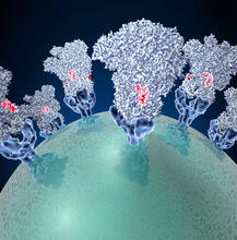
Coronavirus spike protein structure
3753
Coronaviruses are enveloped viruses responsible for 30 percent of mild respiratory infections and atypical deadly pneumonia in humans worldwide. Melody Campbell, UCSF View Media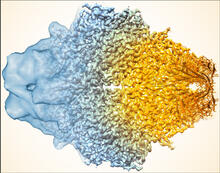
Beta-galactosidase montage showing cryo-EM improvement--gradient background
5883
Composite image of beta-galactosidase showing how cryo-EM’s resolution has improved dramatically in recent years. Older images to the left, more recent to the right. Veronica Falconieri, Sriram Subramaniam Lab, National Cancer Institute View Media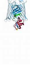
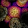
Bacteria working to eat
2304
Gram-negative bacteria perform molecular acrobatics just to eat. Because they're encased by two membranes, they must haul nutrients across both. Emad Tajkhorshid, University of Illinois at Urbana-Champaign View Media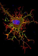
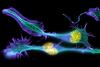
Abnormal, spiky fibroblast
3613
This is a fibroblast, a connective tissue cell that plays an important role in wound healing. Normal fibroblasts have smooth edges. Praveen Suraneni, Stowers Institute for Medical Research, Kansas City, Mo. View Media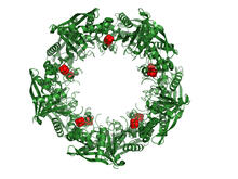
Cas4 nuclease protein structure
3720
This wreath represents the molecular structure of a protein, Cas4, which is part of a system, known as CRISPR, that bacteria use to protect themselves against viral invaders. Fred Dyda, NIDDK View Media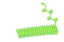
Histones in chromatin
2560
Histone proteins loop together with double-stranded DNA to form a structure that resembles beads on a string. Crabtree + Company View Media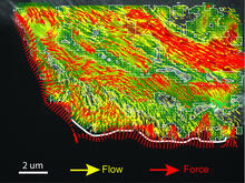
Intracellular forces
2799
Force vectors computed from actin cytoskeleton flow. This is an example of NIH-supported research on single-cell analysis. Gaudenz Danuser, Harvard Medical School View Media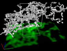
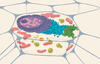
Dense tubular matrices in the peripheral endoplasmic reticulum (ER) 2
5856
Three-dimensional reconstruction of a tubular matrix in a thin section of the peripheral endoplasmic reticulum between the plasma membranes of the cell. Jennifer Lippincott-Schwartz, Howard Hughes Medical Institute Janelia Research Campus, Virginia View Media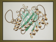
Early ribbon drawing of a protein
2748
This ribbon drawing of a protein hand drawn and colored by researcher Jane Richardson in 1981 helped originate the ribbon representation of proteins that is now ubiquitous in molecular graphics. Jane Richardson, Duke University Medical Center View Media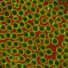
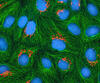
Fruit fly embryo
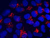
2431
Cells in an early-stage fruit fly embryo, showing the DIAP1 protein (pink), an inhibitor of apoptosis. Hermann Steller, Rockefeller University View Media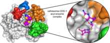
Space-filling model of a cefotaxime-CCD-1 complex
6767
CCD-1 is an enzyme produced by the bacterium Clostridioides difficile that helps it resist antibiotics. Keith Hodgson, Stanford University. View Media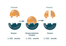
Enzymes convert subtrates into products (with labels)
2522
Enzymes convert substrates into products very quickly. See image 2521 for an unlabeled version of this illustration. Featured in The Chemistry of Health. Crabtree + Company View Media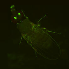
Fly by night
2417
This fruit fly expresses green fluorescent protein (GFP) in the same pattern as the period gene, a gene that regulates circadian rhythm and is expressed in all sensory neurons on the surface of the fl Jay Hirsh, University of Virginia View Media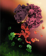
Shiga toxin
6997
E. coli bacteria normally live harmlessly in our intestines, but some cause disease by making toxins. Amy Wu and Christine Zardecki, RCSB Protein Data Bank. View Media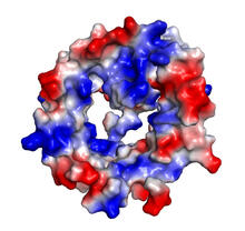
VDAC-1 (4)
2495
The structure of the pore-forming protein VDAC-1 from humans. Gerhard Wagner, Harvard Medical School View Media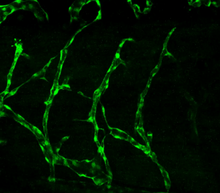
Disrupted vascular development in frog embryos
3403
Disassembly of vasculature in kdr:GFP frogs following addition of 250 µM TBZ. Related to images 3404 and 3505. Hye Ji Cha, University of Texas at Austin View Media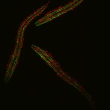
Group of fluorescent C. elegans showing muscle and ribosomal protein
6582
Three C. elegans, tiny roundworms, with a ribosomal protein glowing red and muscle fibers glowing green. Researchers used these worms to study a molecular pathway that affects aging. Jarod Rollins, Mount Desert Island Biological Laboratory. View Media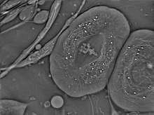
Crane fly spermatocyte undergoing meiosis
6898
A crane fly spermatocyte during metaphase of meiosis-I, a step in the production of sperm. Michael Shribak, Marine Biological Laboratory/University of Chicago. View Media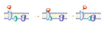
G switch
2536
The G switch allows our bodies to respond rapidly to hormones. See images 2537 and 2538 for labeled versions of this image. Crabtree + Company View Media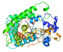
Cytochrome structure with anticancer drug
3326
This image shows the structure of the CYP17A1 enzyme (ribbons colored from blue N-terminus to red C-terminus), with the associated heme colored black. Emily Scott, University of Kansas View Media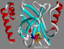
Sortase b from B. anthracis
2386
Structure of sortase b from the bacterium B. anthracis, which causes anthrax. Sortase b is an enzyme used to rob red blood cells of iron, which the bacteria need to survive. Midwest Center for Structural Genomics, PSI View Media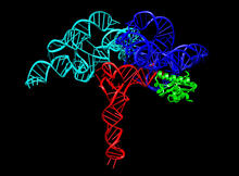
Ribonuclease P structure
3660
Ribbon diagram showing the structure of Ribonuclease P with tRNA. PDB entry 3Q1Q, molecular modeling by Fred Friedman, NIGMS View Media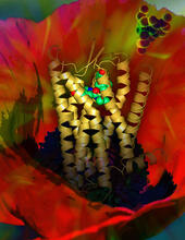
Human opioid receptor structure superimposed on poppy
3314
Opioid receptors on the surfaces of brain cells are involved in pleasure, pain, addiction, depression, psychosis, and other conditions. Raymond Stevens, The Scripps Research Institute View Media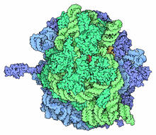
Ribosome illustration from PDB
5780
Ribosomes are complex machines made up of more than 50 proteins and three or four strands of genetic material called ribosomal RNA (rRNA). From PDB’s Molecule of the Month collection (direct link: http://pdb101.rcsb.org/motm/121) Molecule of the Month illustrations are available under a CC-BY-4.0 license. Attribution should be given to David S. Goodsell and the RCSB PDB. View Media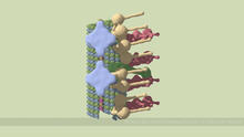
Partial Model of a Cilium’s Doublet Microtubule
6548
Cilia (cilium in singular) are complex molecular machines found on many of our cells. Brown Lab, Harvard Medical School and Veronica Falconieri Hays. View Media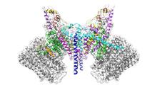
ATP Synthase
6353
Atomic model of the membrane region of the mitochondrial ATP synthase built into a cryo-EM map at 3.6 Å resolution. ATP synthase is the primary producer of ATP in aerobic cells. Bridget Carragher, <a href="http://nramm.nysbc.org/">NRAMM National Resource for Automated Molecular Microscopy</a> View Media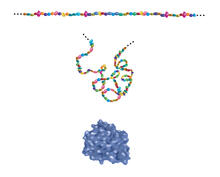
Building blocks and folding of proteins
2508
Proteins are made of amino acids hooked end-to-end like beads on a necklace. To become active, proteins must twist and fold into their final, or "native," conformation. Crabtree + Company View Media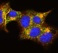
Insulin and protein interact in pancreatic beta cells
3546
A large number of proteins interact with the hormone insulin as it is produced in and secreted from the beta cells of the pancreas. William E. Balch, The Scripps Research Institute View Media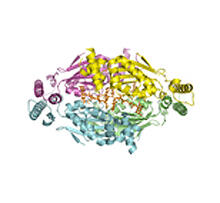
Thymidylate synthase complementing protein from Thermotoga maritime
2387
A model of thymidylate synthase complementing protein from Thermotoga maritime. Joint Center for Structural Genomics, PSI View Media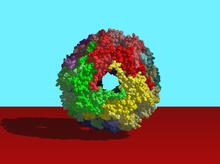
Heat shock protein complex from Methanococcus jannaschii
2385
Model based on X-ray crystallography of the structure of a small heat shock protein complex from the bacteria, Methanococcus jannaschii. Berkeley Structural Genomics Center, PSI-1 View Media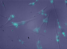
Molecules blocking Huntington's protein production
2600
The molecules that glow blue in these cultured cells prevent the expression of the mutant proteins that cause Huntington's disease. Jiaxin Hu, David W. Dodd and Robert H. E. Hudson, UT Southwestern Medical Center View Media
The Structure of Cilia’s Doublet Microtubules
6549
Cilia (cilium in singular) are complex molecular machines found on many of our cells. Brown Lab, Harvard Medical School and Veronica Falconieri Hays View Media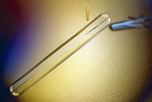
Bovine milk alpha-lactalbumin (1)
2397
A crystal of bovine milk alpha-lactalbumin protein created for X-ray crystallography, which can reveal detailed, three-dimensional protein structures. Alex McPherson, University of California, Irvine View Media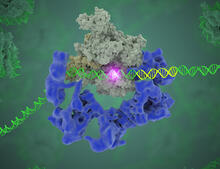
TFIID complex binds DNA to start gene transcription
3766
Gene transcription is a process by which the genetic information encoded in DNA is transcribed into RNA. Eva Nogales, Berkeley Lab View Media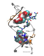
Myotonic dystrophy type 2 genetic defect
3573
Scientists revealed a detailed image of the genetic defect that causes myotonic dystrophy type 2 and used that information to design drug candidates to counteract the disease. Matthew Disney, Scripps Research Institute and Ilyas Yildirim, Northwestern University View Media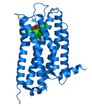
Dopamine D3 receptor
3363
The receptor is shown bound to an antagonist, eticlopride Raymond Stevens, The Scripps Research Institute View Media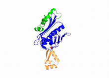
Hsp33 Heat Shock Protein Inactive to Active
3402
When the heat shock protein hsp33 is folded, it is inactive and contains a zinc ion, stabilizing the redox sensitive domain (orange). Dana Reichmann, University of Michigan View Media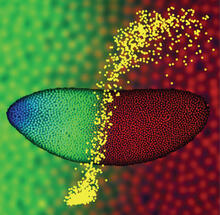
Precise development in the fruit fly embryo
2593
This 2-hour-old fly embryo already has a blueprint for its formation, and the process for following it is so precise that the difference of just a few key molecules can change the plans. Thomas Gregor, Princeton University View Media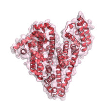
Serum albumin structure 1
3744
Serum albumin (SA) is the most abundant protein in the blood plasma of mammals. SA has a characteristic heart-shape structure and is a highly versatile protein. Wladek Minor, University of Virginia View Media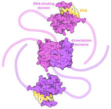
SARS-CoV-2 nucleocapsid dimer
6991
In SARS-CoV-2, the virus that causes COVID-19, nucleocapsid is a complex molecule with many functional parts. Amy Wu and Christine Zardecki, RCSB Protein Data Bank. View Media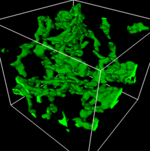
3D reconstruction of a tubular matrix in peripheral endoplasmic reticulum
5857
Detailed three-dimensional reconstruction of a tubular matrix in a thin section of the peripheral endoplasmic reticulum between the plasma membranes of the cell. Jennifer Lippincott-Schwartz, Howard Hughes Medical Institute Janelia Research Campus, Virginia View Media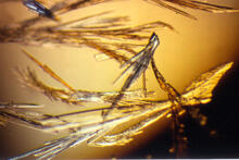
Pig trypsin (3)
2414
Crystals of porcine trypsin protein created for X-ray crystallography, which can reveal detailed, three-dimensional protein structures. Alex McPherson, University of California, Irvine View Media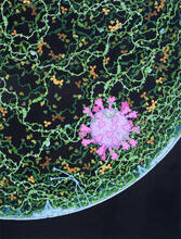
Respiratory droplet
6994
This painting shows a cross section of a small respiratory droplet, like the ones that are thought to transmit SARS-CoV-2, the virus that causes COVID-19. Amy Wu and Christine Zardecki, RCSB Protein Data Bank. View Media
Zinc finger
2426
The structure of a gene-regulating zinc finger protein bound to DNA. Jeremy M. Berg, National Institute of General Medical Sciences View Media