Switch to Gallery View
Image and Video Gallery
This is a searchable collection of scientific photos, illustrations, and videos. The images and videos in this gallery are licensed under Creative Commons Attribution Non-Commercial ShareAlike 3.0. This license lets you remix, tweak, and build upon this work non-commercially, as long as you credit and license your new creations under identical terms.
Human endoplasmic reticulum membrane protein complex
6777
A 3D model of the human endoplasmic reticulum membrane protein complex (EMC) that identifies its nine essential subunits. Rebecca Voorhees, California Institute of Technology. View Media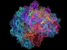
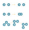
RNA Polymerase II
2484
NIGMS-funded researchers led by Roger Kornberg solved the structure of RNA polymerase II. David Bushnell, Ken Westover and Roger Kornberg, Stanford University View Media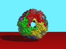
Heat shock protein complex from Methanococcus jannaschii
2385
Model based on X-ray crystallography of the structure of a small heat shock protein complex from the bacteria, Methanococcus jannaschii. Berkeley Structural Genomics Center, PSI-1 View Media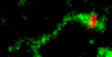
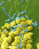
Shiga toxin being sorted inside a cell
3488
Shiga toxin (green) is sorted from the endosome into membrane tubules (red), which then pinch off and move to the Golgi apparatus. Somshuvra Mukhopadhyay, The University of Texas at Austin, and Adam D. Linstedt, Carnegie Mellon University View Media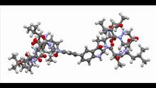
Himastatin, 360-degree view
6851
A 360-degree view of the molecule himastatin, which was first isolated from the bacterium Streptomyces himastatinicus. Himastatin shows antibiotic activity. Mohammad Movassaghi, Massachusetts Institute of Technology. View Media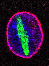
Cellular aging
2578
A protein called tubulin (green) accumulates in the center of a nucleus (outlined in pink) from an aging cell. Maximiliano D'Angelo and Martin Hetzer, Salk Institute View Media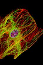
Cells keep their shape with actin filaments and microtubules
3617
This image shows a normal fibroblast, a type of cell that is common in connective tissue and frequently studied in research labs. James J. Faust and David G. Capco, Arizona State University View MediaNuclear Lamina
6572
The 3D single-molecule super-resolution reconstruction of the entire nuclear lamina in a HeLa cell was acquired using the TILT3D platform. Anna-Karin Gustavsson, Ph.D. View Media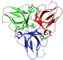
dUTP pyrophosphatase from M. tuberculosis
2381
Model of an enzyme, dUTP pyrophosphatase, from Mycobacterium tuberculosis. Drugs targeted to this enzyme might inhibit the replication of the bacterium that causes most cases of tuberculosis. Mycobacterium Tuberculosis Center, PSI View Media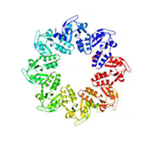
Protein involved in cell division from Mycoplasma pneumoniae
2377
Model of a protein involved in cell division from Mycoplasma pneumoniae. This model, based on X-ray crystallography, revealed a structural domain not seen before. Berkeley Structural Genomics Center, PSI View Media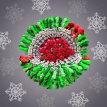
H1N1 Influenza Virus
6355
CellPack image of the H1N1 influenza virus, with hemagglutinin and neuraminidase glycoproteins in green and red, respectively, on the outer envelope (white); matrix protein in gray, and ribonucleoprot Dr. Rommie Amaro, University of California, San Diego View Media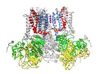
Ion channel
3487
A special "messy" region of a potassium ion channel is important in its function. Yu Zhoi, Christopher Lingle Laboratory, Washington University School of Medicine in St. Louis View Media
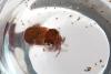
Blood clots show their flex
2450
Blood clots stop bleeding, but they also can cause heart attacks and strokes. Eric Lee, University of Illinois at Urbana-Champaign View Media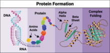
Protein formation
6603
Proteins are 3D structures made up of smaller units. DNA is transcribed to RNA, which in turn is translated into amino acids. NIGMS, with the folded protein illustration adapted from Jane Richardson, Duke University Medical Center View Media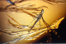
Pig trypsin (3)
2414
Crystals of porcine trypsin protein created for X-ray crystallography, which can reveal detailed, three-dimensional protein structures. Alex McPherson, University of California, Irvine View Media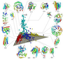
Map of protein structures 02
2367
A global "map of the protein structure universe" indicating the positions of specific proteins. Berkeley Structural Genomics Center, PSI View Media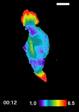
RAC1 activation in motile fibroblast
2457
Novel biosensor system maps the timing and location of Rac protein activation in a living mouse embryo fibroblast. Klaus Hahn, University of North Carolina, Chapel Hill Medical School View Media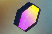
Hen egg lysozyme (2)
2406
A crystal of hen egg lysozyme protein created for X-ray crystallography, which can reveal detailed, three-dimensional protein structures. Alex McPherson, University of California, Irvine View Media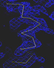
Section of an electron density map
2354
Electron density maps such as this one are generated from the diffraction patterns of X-rays passing through protein crystals. The Southeast Collaboratory for Structural Genomics View Media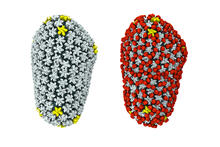
Cryo-EM reveals how the HIV capsid attaches to a human protein to evade immune detection
3755
The illustration shows the capsid of human immunodeficiency virus (HIV) whose molecular features were resolved with cryo-electron microscopy (cryo-EM). Juan R. Perilla, University of Illinois at Urbana-Champaign View Media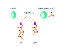
Kinases (with labels)
2535
Kinases are enzymes that add phosphate groups (red-yellow structures) to proteins (green), assigning the proteins a code. Crabtree + Company View Media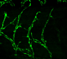
Disrupted vascular development in frog embryos
3403
Disassembly of vasculature in kdr:GFP frogs following addition of 250 µM TBZ. Related to images 3404 and 3505. Hye Ji Cha, University of Texas at Austin View Media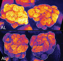
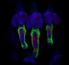
Sleep and the fly brain
2596
In the top snapshots, the brain of a sleep-deprived fruit fly glows orange, marking high concentrations of a synaptic protein called Bruchpilot (BRP) involved in communication between neurons. Chiara Cirelli, University of Wisconsin-Madison View Media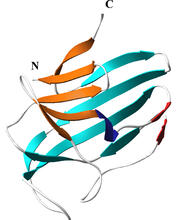
Secreted protein from Mycobacteria
2379
Model of a major secreted protein of unknown function, which is only found in mycobacteria, the class of bacteria that causes tuberculosis. Mycobacterium Tuberculosis Center, PSI View Media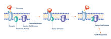
G switch (with labels)
2537
The G switch allows our bodies to respond rapidly to hormones. G proteins act like relay batons to pass messages from circulating hormones into cells. Crabtree + Company View Media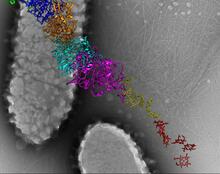
Bacterial nanowire model
6580
A model of a Geobacter sulfurreducens nanowire created from cryo-electron microscopy images. Edward Egelman, University of Virginia. View Media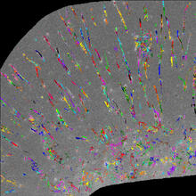
Trajectories of labeled cell receptors
2801
Trajectories of single molecule labeled cell surface receptors. This is an example of NIH-supported research on single-cell analysis. Gaudenz Danuser, Harvard Medical School View Media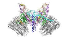
ATP Synthase
6353
Atomic model of the membrane region of the mitochondrial ATP synthase built into a cryo-EM map at 3.6 Å resolution. ATP synthase is the primary producer of ATP in aerobic cells. Bridget Carragher, <a href="http://nramm.nysbc.org/">NRAMM National Resource for Automated Molecular Microscopy</a> View MediaAssembly of the HIV capsid
5729
The HIV capsid is a pear-shaped structure that is made of proteins the virus needs to mature and become infective. John Grime and Gregory Voth, The University of Chicago View Media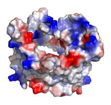
VDAC-1 (1)
2488
The structure of the pore-forming protein VDAC-1 from humans. Gerhard Wagner, Harvard Medical School View Media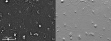
Epithelial cell migration
6899
High-resolution time lapse of epithelial (skin) cell migration and wound healing. It shows an image taken every 13 seconds over the course of almost 14 minutes. Michael Shribak, Marine Biological Laboratory/University of Chicago. View Media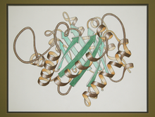
Early ribbon drawing of a protein
2748
This ribbon drawing of a protein hand drawn and colored by researcher Jane Richardson in 1981 helped originate the ribbon representation of proteins that is now ubiquitous in molecular graphics. Jane Richardson, Duke University Medical Center View Media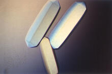
Fungal lipase (2)
2411
Crystals of fungal lipase protein created for X-ray crystallography, which can reveal detailed, three-dimensional protein structures. Alex McPherson, University of California, Irvine View Media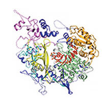
Kluyveromyces polysporus Argonaute bound to guide RNA
3408
A segment of siRNA, shown in red, guides a "slicer" protein called Argonaute (multi-colored twists and corkscrews) to the target RNA molecules. Kotaro Nakanishi and David Weinberg, Massachusetts Institute of Technology View Media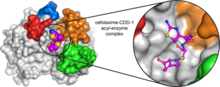
Space-filling model of a cefotaxime-CCD-1 complex
6767
CCD-1 is an enzyme produced by the bacterium Clostridioides difficile that helps it resist antibiotics. Keith Hodgson, Stanford University. View Media
Drosophila (fruit fly) myosin 1D motility assay
6562
Actin gliding powered by myosin 1D. Note the counterclockwise motion of the gliding actin filaments. Serapion Pyrpassopoulos and E. Michael Ostap, University of Pennsylvania View Media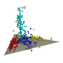
Map of protein structures 01
2365
A global "map of the protein structure universe." The Berkeley Structural Genomics Center has developed a method to visualize the vast universe of protein structures in which proteins of similar struc Berkeley Structural Genomics Center, PSI View Media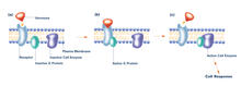
G switch (with labels and stages)
2538
The G switch allows our bodies to respond rapidly to hormones. G proteins act like relay batons to pass messages from circulating hormones into cells. Crabtree + Company View Media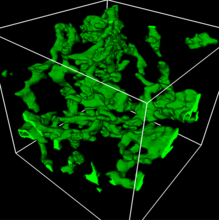
3D reconstruction of a tubular matrix in peripheral endoplasmic reticulum
5857
Detailed three-dimensional reconstruction of a tubular matrix in a thin section of the peripheral endoplasmic reticulum between the plasma membranes of the cell. Jennifer Lippincott-Schwartz, Howard Hughes Medical Institute Janelia Research Campus, Virginia View Media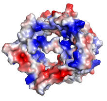
VDAC-1 (2)
2491
The structure of the pore-forming protein VDAC-1 from humans. Gerhard Wagner, Harvard Medical School View Media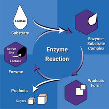
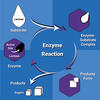
Enzyme reaction
6604
Enzymes speed up chemical reactions by reducing the amount of energy needed for the reactions. NIGMS View Media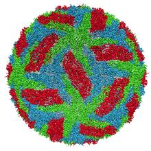
Protective membrane and membrane proteins of the dengue virus visualized with cryo-EM
3756
Dengue virus is a mosquito-borne illness that infects millions of people in the tropics and subtropics each year. Like many viruses, dengue is enclosed by a protective membrane. Hong Zhou, UCLA View Media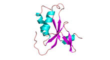
Antitoxin GhoS (Illustration 1)
3427
Structure of the bacterial antitoxin protein GhoS. GhoS inhibits the production of a bacterial toxin, GhoT, which can contribute to antibiotic resistance. Rebecca Page and Wolfgang Peti, Brown University and Thomas K. Wood, Pennsylvania State University View Media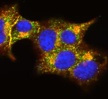
Insulin and protein interact in pancreatic beta cells
3546
A large number of proteins interact with the hormone insulin as it is produced in and secreted from the beta cells of the pancreas. William E. Balch, The Scripps Research Institute View Media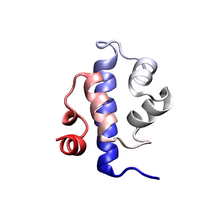
Protein folding video
3391
Proteins are long chains of amino acids. Each protein has a unique amino acid sequence. It is still a mystery how a protein folds into the proper shape based on its sequence. Theoretical and Computational Biophysics Group View Media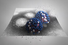
Stetten Lecture 2017poster image
5896
This image is featured on the poster for Dr. Rommie Amaro's 2017 Stetten Lecture. Dr. Rommie Amaro, University of California, San Diego View Media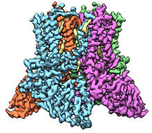
Transient receptor potential channel TRPV5
6577
A 3D reconstruction of a transient receptor potential channel called TRPV5 that was created based on cryo-electron microscopy images. Vera Moiseenkova-Bell, University of Pennsylvania. View Media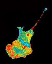
Seeing signaling protein activation in cells 04
2454
Cdc42, a member of the Rho family of small guanosine triphosphatase (GTPase) proteins, regulates multiple cell functions, including motility, proliferation, apoptosis, and cell morphology. Klaus Hahn, University of North Carolina, Chapel Hill Medical School View Media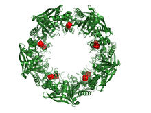
Cas4 nuclease protein structure
3720
This wreath represents the molecular structure of a protein, Cas4, which is part of a system, known as CRISPR, that bacteria use to protect themselves against viral invaders. Fred Dyda, NIDDK View Media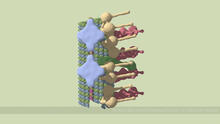
Partial Model of a Cilium’s Doublet Microtubule
6548
Cilia (cilium in singular) are complex molecular machines found on many of our cells. Brown Lab, Harvard Medical School and Veronica Falconieri Hays. View Media