Switch to Gallery View
Image and Video Gallery
This is a searchable collection of scientific photos, illustrations, and videos. The images and videos in this gallery are licensed under Creative Commons Attribution Non-Commercial ShareAlike 3.0. This license lets you remix, tweak, and build upon this work non-commercially, as long as you credit and license your new creations under identical terms.
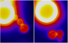
Dynamin Fission
3448
Time lapse series shows short dynamin assemblies (not visible) constricting a lipid tube to make a "beads on a string" appearance, then cutting off one of the beads i.e., catalyzing membrane fission). Ramachandran, Pucadyil et al. , The Scripps Research Institute View MediaNuclear Lamina
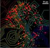
6572
The 3D single-molecule super-resolution reconstruction of the entire nuclear lamina in a HeLa cell was acquired using the TILT3D platform. Anna-Karin Gustavsson, Ph.D. View Media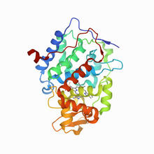
CCP enzyme
6762
The enzyme CCP is found in the mitochondria of baker’s yeast. Scientists study the chemical reactions that CCP triggers, which involve a water molecule, iron, and oxygen. Protein Data Bank. View Media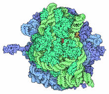
Ribosome illustration from PDB
5780
Ribosomes are complex machines made up of more than 50 proteins and three or four strands of genetic material called ribosomal RNA (rRNA). From PDB’s Molecule of the Month collection (direct link: http://pdb101.rcsb.org/motm/121) Molecule of the Month illustrations are available under a CC-BY-4.0 license. Attribution should be given to David S. Goodsell and the RCSB PDB. View Media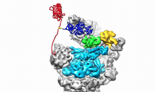
Movie of the 19S proteasome subunit processing a protein substrate
3764
The proteasome is a critical multiprotein complex in the cell that breaks down and recycles proteins that have become damaged or are no longer needed. Andreas Martin, HHMI View Media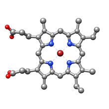
Structure of heme, top view
3539
Molecular model of the struture of heme. Heme is a small, flat molecule with an iron ion (dark red) at its center. Rachel Kramer Green, RCSB Protein Data Bank View Media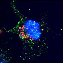
ARTS triggers apoptosis
2432
Cell showing overproduction of the ARTS protein (red). ARTS triggers apoptosis, as shown by the activation of caspase-3 (green) a key tool in the cell's destruction. The nucleus is shown in blue. Hermann Steller, Rockefeller University View Media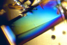
Bovine trypsin
2408
A crystal of bovine trypsin protein created for X-ray crystallography, which can reveal detailed, three-dimensional protein structures. Alex McPherson, University of California, Irvine View Media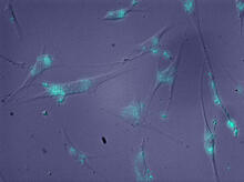
Molecules blocking Huntington's protein production
2600
The molecules that glow blue in these cultured cells prevent the expression of the mutant proteins that cause Huntington's disease. Jiaxin Hu, David W. Dodd and Robert H. E. Hudson, UT Southwestern Medical Center View Media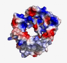
VDAC video 02
2571
This video shows the structure of the pore-forming protein VDAC-1 from humans. Gerhard Wagner, Harvard Medical School View Media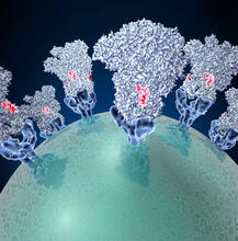
Coronavirus spike protein structure
3753
Coronaviruses are enveloped viruses responsible for 30 percent of mild respiratory infections and atypical deadly pneumonia in humans worldwide. Melody Campbell, UCSF View Media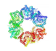
Nicotinic acid phosphoribosyltransferase
2355
Model of the enzyme nicotinic acid phosphoribosyltransferase. Berkeley Structural Genomics Center, PSI View Media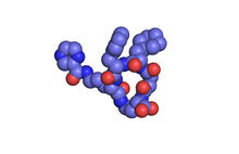
X-ray co-crystal structure of Src kinase bound to a DNA-templated macrocycle inhibitor 1
3413
X-ray co-crystal structure of Src kinase bound to a DNA-templated macrocycle inhibitor. Markus A. Seeliger, Stony Brook University Medical School and David R. Liu, Harvard University View Media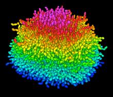
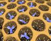
A Growing Bacterial Biofilm
5825
A growing Vibrio cholerae (cholera) biofilm. Cholera bacteria form colonies called biofilms that enable them to resist antibiotic therapy within the body and other challenges to their growth. Jing Yan, Ph.D., and Bonnie Bassler, Ph.D., Department of Molecular Biology, Princeton University, Princeton, NJ. View Media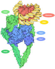
Histone deacetylases
7001
The human genome contains much of the information needed for every cell in the body to function. However, different types of cells often need different types of information. Amy Wu and Christine Zardecki, RCSB Protein Data Bank. View Media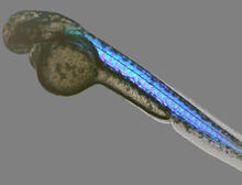
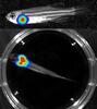
Zebrafish embryo
6897
A zebrafish embryo showing its natural colors. Zebrafish have see-through eggs and embryos, making them ideal research organisms for studying the earliest stages of development. Michael Shribak, Marine Biological Laboratory/University of Chicago. View Media
Early life of a protein
2740
This illustration represents the early life of a protein—specifically, apomyoglobin—as it is synthesized by a ribosome and emerges from the ribosomal tunnel, which contains the newly formed protein's Silvia Cavagnero, University of Wisconsin, Madison View Media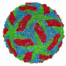
Cryo-electron microscopy of the dengue virus showing protective membrane and membrane proteins
3748
Dengue virus is a mosquito-borne illness that infects millions of people in the tropics and subtropics each year. Like many viruses, dengue is enclosed by a protective membrane. Hong Zhou, UCLA View Media