Switch to Gallery View
Image and Video Gallery
This is a searchable collection of scientific photos, illustrations, and videos. The images and videos in this gallery are licensed under Creative Commons Attribution Non-Commercial ShareAlike 3.0. This license lets you remix, tweak, and build upon this work non-commercially, as long as you credit and license your new creations under identical terms.
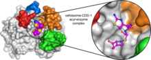
Space-filling model of a cefotaxime-CCD-1 complex
6767
CCD-1 is an enzyme produced by the bacterium Clostridioides difficile that helps it resist antibiotics. Keith Hodgson, Stanford University. View Media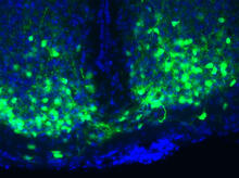
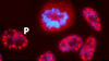
Master clock of the mouse brain
3547
An image of the area of the mouse brain that serves as the 'master clock,' which houses the brain's time-keeping neurons. The nuclei of the clock cells are shown in blue. Erik Herzog, Washington University in St. Louis View Media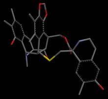
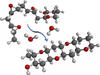
Anti-tumor drug ecteinascidin 743 (ET-743), structure without hydrogens 04
2797
Ecteinascidin 743 (ET-743, brand name Yondelis), was discovered and isolated from a sea squirt, Ecteinascidia turbinata, by NIGMS grantee Kenneth Rinehart at the University of Illinois. Timothy Jamison, Massachusetts Institute of Technology View Media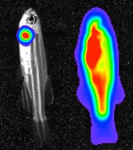
Bioluminescent imaging in adult zebrafish 04
3559
Luciferase-based imaging enables visualization and quantification of internal organs and transplanted cells in live adult zebrafish. View Media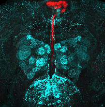
Fruit fly brain responds to adipokines
6985
Drosophila adult brain showing that an adipokine (fat hormone) generates a response from neurons (aqua) and regulates insulin-producing neurons (red).Akhila Rajan, Fred Hutchinson Cancer Center View Media
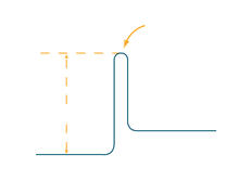
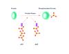
Activation energy
2525
To become products, reactants must overcome an energy hill. See image 2526 for a labeled version of this illustration. Featured in The Chemistry of Health. Crabtree + Company View Media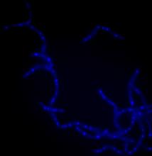
Bacillus anthracis being killed
3525
Bacillus anthracis (anthrax) cells being killed by a fluorescent trans-translation inhibitor, which disrupts bacterial protein synthesis. Kenneth Keiler, Penn State University View Media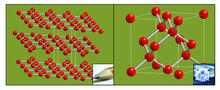
Carbon building blocks (with examples)
2507
The arrangement of identical molecular components can make a dramatic difference. For example, carbon atoms can be arranged into dull graphite (left) or sparkly diamonds (right). Crabtree + Company View Media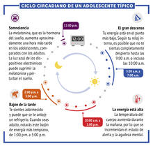
Ciclo circadiano de un adolescente típico
6612
Los ritmos circadianos son cambios físicos, mentales y conductuales que siguen un ciclo de 24 horas. NIGMS View Media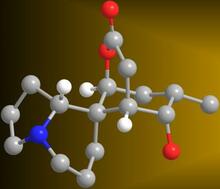
Serratezomine A
2687
A 3-D model of the alkaloid serratezomine A shows the molecule's complex ring structure. View Media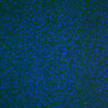
Movie of in vitro assembly of a cell-signaling pathway
3786
T cells are white blood cells that are important in defending the body against bacteria, viruses and other pathogens. Xiaolei Su, HHMI Whitman Center of the Marine Biological Laboratory View Media
Independence Day
5888
This graphic that resembles a firework was created from a picture of a fruit fly spermatid. Sigi Benjamin-Hong, Rockefeller University View Media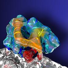
Kinesin moves cellular cargo
3491
A protein called kinesin (blue) is in charge of moving cargo around inside cells and helping them divide. Charles Sindelar, Yale University View Media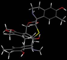
Anti-tumor drug ecteinascidin 743 (ET-743) with hydrogens 02
2791
Ecteinascidin 743 (ET-743, brand name Yondelis), was discovered and isolated from a sea squirt, Ecteinascidia turbinata, by NIGMS grantee Kenneth Rinehart at the University of Illinois. Timothy Jamison, Massachusetts Institute of Technology View Media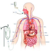
A drug's life in the body (with labels)
2528
A drug's life in the body. Medicines taken by mouth (oral) pass through the liver before they are absorbed into the bloodstream. Crabtree + Company View Media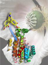
Atomic Structure of Poppy Enzyme
3422
The atomic structure of the morphine biosynthetic enzyme salutaridine reductase bound to the cofactor NADPH. The substrate salutaridine is shown entering the active site. Judy Coyle, Donald Danforth Plant Science Center View Media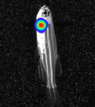
Bioluminescent imaging in adult zebrafish - lateral view
3558
Luciferase-based imaging enables visualization and quantification of internal organs and transplanted cells in live adult zebrafish. Kenneth Poss, Duke University View Media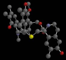
Anti-tumor drug ecteinascidin 743 (ET-743), structure without hydrogens 03
2796
Ecteinascidin 743 (ET-743, brand name Yondelis), was discovered and isolated from a sea squirt, Ecteinascidia turbinata, by NIGMS grantee Kenneth Rinehart at the University of Illinois. Timothy Jamison, Massachusetts Institute of Technology View Media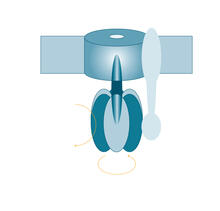
ATP synthase
2517
The world's smallest motor, ATP synthase, generates energy for the cell. See image 2518 for a labeled version of this illustration. Crabtree + Company View Media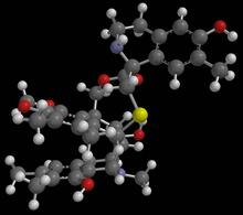
Anti-tumor drug ecteinascidin 743 (ET-743) with hydrogens 01
2790
Ecteinascidin 743 (ET-743, brand name Yondelis), was discovered and isolated from a sea squirt, Ecteinascidia turbinata, by NIGMS grantee Kenneth Rinehart at the University of Illinois. Timothy Jamison, Massachusetts Institute of Technology View Media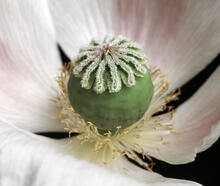
White Poppy (cropped)
3423
A cropped image of a white poppy. View poppy uncropped here 3424. Judy Coyle, Donald Danforth Plant Science Center View Media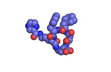
X-ray co-crystal structure of Src kinase bound to a DNA-templated macrocycle inhibitor 1
3413
X-ray co-crystal structure of Src kinase bound to a DNA-templated macrocycle inhibitor. Markus A. Seeliger, Stony Brook University Medical School and David R. Liu, Harvard University View Media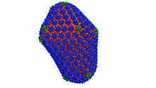
Atomic-level structure of the HIV capsid
6601
This animation shows atoms of the HIV capsid, the shell that encloses the virus's genetic material. Juan R. Perilla and the Theoretical and Computational Biophysics Group, University of Illinois at Urbana-Champaign View Media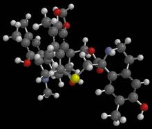
Anti-tumor drug ecteinascidin 743 (ET-743) with hydrogens 03
2792
Ecteinascidin 743 (ET-743, brand name Yondelis), was discovered and isolated from a sea squirt, Ecteinascidia turbinata, by NIGMS grantee Kenneth Rinehart at the University of Illinois. Timothy Jamison, Massachusetts Institute of Technology View Media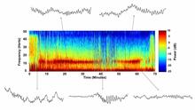
Brain waves of a patient anesthetized with propofol
6779
A representation of a patient’s brain waves after receiving the anesthetic propofol. Emery N. Brown, M.D., Ph.D., Massachusetts General Hospital/Harvard Medical School, Picower Institute for Learning and Memory, and Massachusetts Institute of Technology. View Media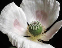
White Poppy
3424
A white poppy. View cropped image of a poppy here 3423. Judy Coyle, Donald Danforth Plant Science Center View Media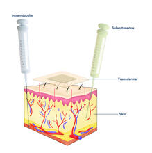
Drugs enter skin (with labels)
2532
Drugs enter different layers of skin via intramuscular, subcutaneous, or transdermal delivery methods. See image 2531 for an unlabeled version of this illustration. Crabtree + Company View Media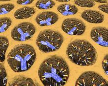
Antibodies in silica honeycomb
2750
Antibodies are among the most promising therapies for certain forms of cancer, but patients must take them intravenously, exposing healthy tissues to the drug and increasing the risk of side effects. Chenghong Lei, Pacific Northwest National Laboratory & Karl Erik Hellstrom, University of Washington View Media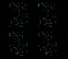
Glycan arrays
1265
The signal is obtained by allowing proteins in human serum to interact with glycan (polysaccharide) arrays. The arrays are shown in replicate so the pattern is clear. Ola Blixt, Scripps Research Institute View Media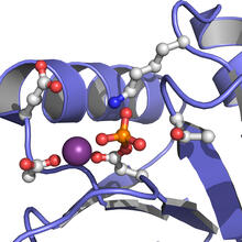
Active Site of E. coli response regulator PhoB
3412
Active site of E. coli response regulator PhoB. Ann Stock, Rutgers University View Media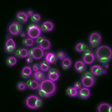
Yeast cells responding to a glucose shortage
6772
These yeast cells were exposed to a glucose (sugar) shortage. Mike Henne, University of Texas Southwestern Medical Center. View Media
Genetically identical mycobacteria respond differently to antibiotic 2
5752
Antibiotic resistance in microbes is a serious health concern. So researchers have turned their attention to how bacteria undo the action of some antibiotics. Bree Aldridge, Tufts University View Media
See how immune cell acid destroys bacterial proteins
6602
This animation shows the effect of exposure to hypochlorous acid, which is found in certain types of immune cells, on bacterial proteins. American Chemistry Council View Media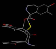
Anti-tumor drug ecteinascidin 743 (ET-743), structure without hydrogens 02
2795
Ecteinascidin 743 (ET-743, brand name Yondelis), was discovered and isolated from a sea squirt, Ecteinascidia turbinata, by NIGMS grantee Kenneth Rinehart at the University of Illinois. Timothy Jamison, Massachusetts Institute of Technology View Media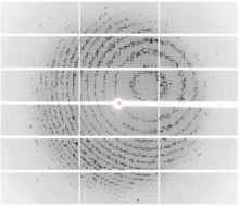
X-ray diffraction pattern from a crystallized cefotaxime-CCD-1 complex
6765
CCD-1 is an enzyme produced by the bacterium Clostridioides difficile that helps it resist antibiotics. Keith Hodgson, Stanford University. View Media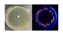
Antibiotic-surviving bacteria
6802
Colonies of bacteria growing despite high concentrations of antibiotics. These colonies are visible both by eye, as seen on the left, and by bioluminescence imaging, as seen on the right. Paul Stoodley, The Ohio State University. View Media
A drug's life in the body
2527
A drug's life in the body. Medicines taken by mouth pass through the liver before they are absorbed into the bloodstream. Crabtree + Company View Media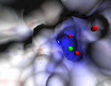
Active site of sulfite oxidase
2746
Sulfite oxidase is an enzyme that is essential for normal neurological development in children. John Enemark, University of Arizona View Media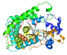
Cytochrome structure with anticancer drug
3326
This image shows the structure of the CYP17A1 enzyme (ribbons colored from blue N-terminus to red C-terminus), with the associated heme colored black. Emily Scott, University of Kansas View Media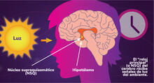
Los ritmos circadianos y el núcleo supraquiasmático
6614
Los ritmos circadianos son cambios físicos, mentales y de comportamiento que siguen un ciclo de 24 horas. NIGMS View Media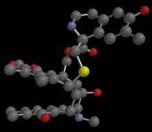
Anti-tumor drug ecteinascidin 743 (ET-743), structure without hydrogens 01
2794
Ecteinascidin 743 (ET-743, brand name Yondelis), was discovered and isolated from a sea squirt, Ecteinascidia turbinata, by NIGMS grantee Kenneth Rinehart at the University of Illinois. Timothy Jamison, Massachusetts Institute of Technology View Media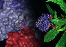
Chang Shan
3483
For thousands of years, Chinese herbalists have treated malaria using Chang Shan, a root extract from a type of hydrangea that grows in Tibet and Nepal. Paul Schimmel Lab, Scripps Research Institute View Media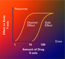
Dose response curves
2533
Dose-response curves determine how much of a drug (X-axis) causes a particular effect, or a side effect, in the body (Y-axis). Featured in Medicines By Design. Crabtree + Company View Media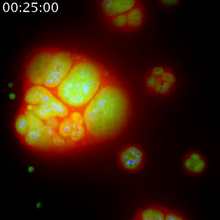
Nucleolus subcompartments spontaneously self-assemble 1
3789
The nucleolus is a small but very important protein complex located in the cell's nucleus. Nilesh Vaidya, Princeton University View Media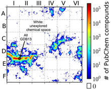
Computer algorithm
3458
This computer algorithm plots all feasible small carbon-based molecules as though they were cities on a map and identifies huge, unexplored spaces that may help fuel research into new drug therapies. Aaron Virshup, Julia Contreras-Garcia, Peter Wipf, Weitao Yang and David Beratan, University of Pittsburgh Center for Chemical Methodologies and Library Development View Media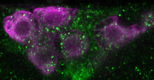
Insulin production and fat sensing in fruit flies
6982
Fourteen neurons (magenta) in the adult Drosophila brain produce insulin, and fat tissue sends packets of lipids to the brain via the lipoprotein carriers (green). Akhila Rajan, Fred Hutchinson Cancer Center View Media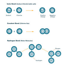
Bond types (with labels)
2520
Ionic and covalent bonds hold molecules, like sodium chloride and chlorine gas, together. Hydrogen bonds among molecules, notably involving water, also play an important role in biology. Crabtree + Company View Media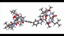
Himastatin, 360-degree view
6851
A 360-degree view of the molecule himastatin, which was first isolated from the bacterium Streptomyces himastatinicus. Himastatin shows antibiotic activity. Mohammad Movassaghi, Massachusetts Institute of Technology. View Media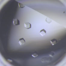
Crystals of CCD-1 in complex with cefotaxime
6764
CCD-1 is an enzyme produced by the bacterium Clostridioides difficile that helps it resist antibiotics. Keith Hodgson, Stanford University. View Media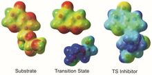
Enzyme transition states
3429
The molecule on the left is an electrostatic potential map of the van der Waals surface of the transition state for human purine nucleoside phosphorylase. Vern Schramm, Albert Einstein College of Medicine of Yeshiva University View Media