Switch to Gallery View
Image and Video Gallery
This is a searchable collection of scientific photos, illustrations, and videos. The images and videos in this gallery are licensed under Creative Commons Attribution Non-Commercial ShareAlike 3.0. This license lets you remix, tweak, and build upon this work non-commercially, as long as you credit and license your new creations under identical terms.
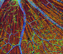
Mouse Retina
3309
A genetic disorder of the nervous system, neurofibromatosis causes tumors to form on nerves throughout the body, including a type of tumor called an optic nerve glioma that can result in childhood bli Tom Deerinck, NCMIR View Media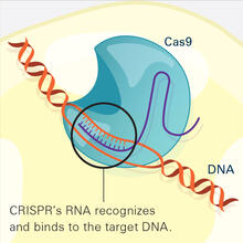
CRISPR Illustration Frame 2
6486
This illustration shows, in simplified terms, how the CRISPR-Cas9 system can be used as a gene-editing tool. National Institute of General Medical Sciences. View Media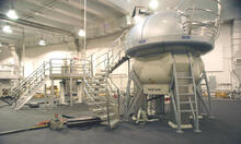
NMR spectrometer
2371
This photo shows a Varian Unity Inova 900 MHz, 21.1 T standard bore magnet Nuclear Magnetic Resonnance (NMR) spectrometer. Center for Eukaryotic Structural Genomics View Media
“Two-faced” Janus particle activating a macrophage
6801
A macrophage—a type of immune cell that engulfs invaders—“eats” and is activated by a “two-faced” Janus particle. Yan Yu, Indiana University, Bloomington. View Media
Cell proliferation in a quail embryo
2808
Image showing that the edge zone (top of image) of the quail embryo shows no proliferating cells (cyan), unlike the interior zone (bottom of image). Non-proliferating cell nuclei are labeled green. Andrés Garcia, Georgia Tech View Media
Zinc levels in a plant leaf
3727
Zinc is required for the function of more than 300 enzymes, including those that help regulate gene expression, in various organisms including humans. Suzana Car, Dartmouth College View Media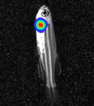
Bioluminescent imaging in adult zebrafish - lateral view
3558
Luciferase-based imaging enables visualization and quantification of internal organs and transplanted cells in live adult zebrafish. Kenneth Poss, Duke University View Media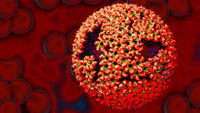
Molecular model of freshly made Rous sarcoma virus (RSV)
3771
Viruses have been the foes of animals and other organisms for time immemorial. Boon Chong Goh, University of Illinois at Urbana-Champaign View Media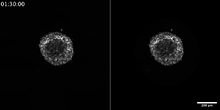
Breast cancer cells change migration phenotypes
6986
Cancer cells can change their migration phenotype, which includes their shape and the way that they move to invade different tissues. Bo Sun, Oregon State University. View Media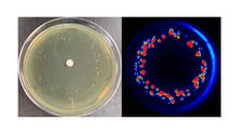
Antibiotic-surviving bacteria
6802
Colonies of bacteria growing despite high concentrations of antibiotics. These colonies are visible both by eye, as seen on the left, and by bioluminescence imaging, as seen on the right. Paul Stoodley, The Ohio State University. View Media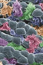
Cells lining the trachea
3646
In this image, viewed with a ZEISS ORION NanoFab microscope, the community of cells lining a mouse airway is magnified more than 10,000 times. Eva Mutunga and Kate Klein, University of the District of Columbia and National Institute of Standards and Technology View Media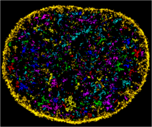
Chromatin in human fibroblast
6888
The nucleus of a human fibroblast cell with chromatin—a substance made up of DNA and proteins—shown in various colors. Melike Lakadamyali, Perelman School of Medicine at the University of Pennsylvania. View Media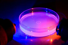
Petri dish containing C. elegans
6751
This Petri dish contains microscopic roundworms called Caenorhabditis elegans. Researchers used these particular worms to study how C. H. Robert Horvitz and Dipon Ghosh, Massachusetts Institute of Technology. View Media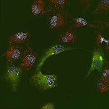
Endoplasmic reticulum abnormalities 2
6774
Human cells with the gene that codes for the protein FIT2 deleted. After an experimental intervention, they are expressing a nonfunctional version of FIT2, shown in green. Michel Becuwe, Harvard University. View Media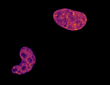
Cell division and cell death
6790
Two cells over a 2-hour period. The one on the bottom left goes through programmed cell death, also known as apoptosis. The one on the top right goes through cell division, also called mitosis. Dylan T. Burnette, Vanderbilt University School of Medicine. View MediaTracking embryonic zebrafish cells
6775
To better understand cell movements in developing embryos, researchers isolated cells from early zebrafish embryos and grew them as clusters. Liliana Solnica-Krezel, Washington University School of Medicine in St. Louis. View Media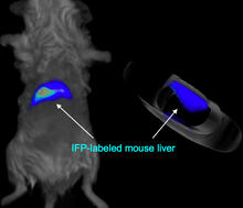
Mouse liver labeled with fluorescent probe
2601
A mouse liver glows after being tagged with specially designed infrared-fluorescent protein (IFP). Xiaokun Shu, University of California, San Diego View Media
C. elegans showing internal structures
6961
An image of Caenorhabditis elegans, a tiny roundworm, showing internal structures including the intestine, pharynx, and body wall muscle. C. Michael Shribak, Marine Biological Laboratory/University of Chicago. View Media
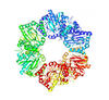
X-ray crystallography
2511
X-ray crystallography allows researchers to see structures too small to be seen by even the most powerful microscopes. Crabtree + Company View Media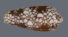
Cone snail shell
2576
A shell from the venomous cone snail Conus omaria, which lives in the Pacific and Indian oceans and eats other snails. Kerry Matz, University of Utah View Media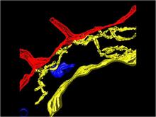
Computer model of cell membrane
2636
A computer model of the cell membrane, where the plasma membrane is red, endoplasmic reticulum is yellow, and mitochondria are blue. Bridget Wilson, University of New Mexico View Media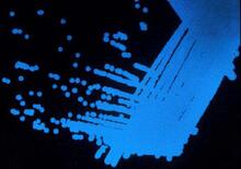
Colorful communication
2313
The marine bacterium Vibrio harveyi glows when near its kind. Bonnie Bassler, Princeton University View Media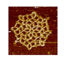
Snowflake DNA origami
3724
An atomic force microscopy image shows DNA folded into an intricate, computer-designed structure. The image is featured on Biomedical Beat blog post Cool Images: A Holiday-Themed Collection. Hao Yan, Arizona State University View Media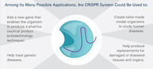
CRISPR Illustration Frame 5
6489
This illustration shows, in simplified terms, how the CRISPR-Cas9 system can be used as a gene-editing tool. This is the fifthframe in a series of five. View Media
Math from the heart
3592
Watch a cell ripple toward a beam of light that turns on a movement-related protein. View Media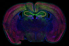
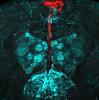
Calling Cards in a mouse brain
6780
The green spots in this mouse brain are cells labeled with Calling Cards, a technology that records molecular events in brain cells as they mature. Allen Yen, Lab of Joseph Dougherty, Washington University School of Medicine in St. Louis. View Media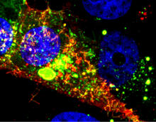
RSV-Infected Cell
3567
Viral RNA (red) in an RSV-infected cell. Eric Alonas and Philip Santangelo, Georgia Institute of Technology and Emory University View Media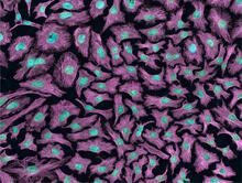
HeLa cells
3520
Multiphoton fluorescence image of HeLa cells with cytoskeletal microtubules (magenta) and DNA (cyan). Nikon RTS2000MP custom laser scanning microscope. National Center for Microscopy and Imaging Research (NCMIR) View Media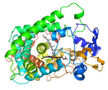
Cytochrome structure with anticancer drug
3326
This image shows the structure of the CYP17A1 enzyme (ribbons colored from blue N-terminus to red C-terminus), with the associated heme colored black. Emily Scott, University of Kansas View Media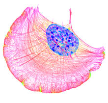
Crawling cell
6964
A crawling cell with DNA shown in blue and actin filaments, which are a major component of the cytoskeleton, visible in pink. Actin filaments help enable cells to crawl. Dylan T. Burnette, Vanderbilt University School of Medicine. View Media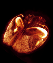
Mouse brain 2
6930
A mouse brain that was genetically modified so that subpopulations of its neurons glow. Prayag Murawala, MDI Biological Laboratory and Hannover Medical School. View Media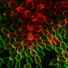
Snowflake yeast 1
6969
Multicellular yeast called snowflake yeast that researchers created through many generations of directed evolution from unicellular yeast. William Ratcliff, Georgia Institute of Technology. View Media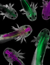
Axolotls showing nervous system components
6928
Axolotls—a type of salamander—that have been genetically modified so that various parts of their nervous systems glow purple and green. Prayag Murawala, MDI Biological Laboratory and Hannover Medical School. View Media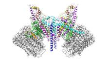
ATP Synthase
6353
Atomic model of the membrane region of the mitochondrial ATP synthase built into a cryo-EM map at 3.6 Å resolution. ATP synthase is the primary producer of ATP in aerobic cells. Bridget Carragher, <a href="http://nramm.nysbc.org/">NRAMM National Resource for Automated Molecular Microscopy</a> View Media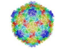
Bacteriophage P22 capsid
5874
Cryo-electron microscopy (cryo-EM) has the power to capture details of proteins and other small biological structures at the molecular level. This image shows proteins in the capsid, or outer co Dr. Wah Chiu, Baylor College of Medicine View Media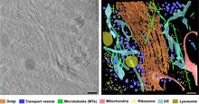
Cryo-ET cross-section of the Golgi apparatus
6606
On the left, a cross-section slice of a rat pancreas cell captured using cryo-electron tomography (cryo-ET). On the right, a 3D, color-coded version of the image highlighting cell structures. Xianjun Zhang, University of Southern California. View Media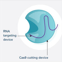
CRISPR Illustration Frame 1
6465
This illustration shows, in simplified terms, how the CRISPR-Cas9 system can be used as a gene-editing tool. This is the first frame in a series of four. National Institute of General Medical Sciences. View Media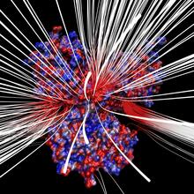
Electrostatic map of human spermine synthase
3658
From PDB entry 3c6k, Crystal structure of human spermine synthase in complex with spermidine and 5-methylthioadenosine. Emil Alexov, Clemson University View Media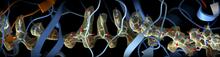
CRISPR
6351
RNA incorporated into the CRISPR surveillance complex is positioned to scan across foreign DNA. Cryo-EM density from a 3Å reconstruction is shown as a yellow mesh. NRAMM National Resource for Automated Molecular Microscopy http://nramm.nysbc.org/nramm-images/ Source: Bridget Carragher View Media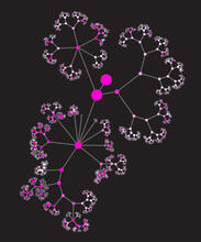
Network diagram of genes, cellular components and processes (unlabeled)
3436
This image shows the hierarchical ontology of genes, cellular components and processes derived from large genomic datasets. From Dutkowski et al. Janusz Dutkowski and Trey Ideker View Media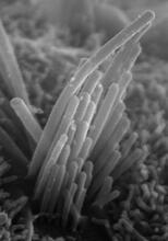
Ear hair cells derived from embryonic stem cells
3272
Mouse embryonic stem cells matured into this bundle of hair cells similar to the ones that transmit sound in the ear. Stefen Heller, Stanford University, via CIRM View Media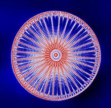
Arachnoidiscus diatom
6902
An Arachnoidiscus diatom with a diameter of 190µm. Michael Shribak, Marine Biological Laboratory/University of Chicago. View Media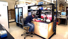
Protein purification facility
2376
The Center for Eukaryotic Structural Genomics protein purification facility is responsible for purifying all recombinant proteins produced by the center. Center for Eukaryotic Structural Genomics View Media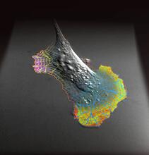
Biosensors illustration
2802
A rendering of an activity biosensor image overlaid with a cell-centered frame of reference used for image analysis of signal transduction. Gaudenz Danuser, Harvard Medical School View Media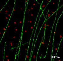
Multicolor STORM
2325
In 2006, scientists developed an optical microscopy technique enabling them to clearly see individual molecules within cells. In 2007, they took the technique, abbreviated STORM, a step further. Xiaowei Zhuang, Harvard University View Media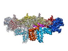
Dengue virus membrane protein structure
3758
Dengue virus is a mosquito-borne illness that infects millions of people in the tropics and subtropics each year. Like many viruses, dengue is enclosed by a protective membrane. Hong Zhou, UCLA View Media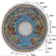
Annotated TEM cross-section of C. elegans (roundworm)
5760
The worm Caenorhabditis elegans is a popular laboratory animal because its small size and fairly simple body make it easy to study. Piali Sengupta, Brandeis University View MediaBeta-galactosidase montage showing cryo-EM improvement--transparent background
5882
Composite image of beta-galactosidase showing how cryo-EM’s resolution has improved dramatically in recent years. Older images to the left, more recent to the right. Veronica Falconieri, Sriram Subramaniam Lab, National Cancer Institute View Media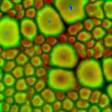
Cell-like compartments from frog eggs 2
6585
Cell-like compartments that spontaneously emerged from scrambled frog eggs, with nuclei (blue) from frog sperm. Endoplasmic reticulum (red) and microtubules (green) are also visible. Xianrui Cheng, Stanford University School of Medicine. View Media
Bovine milk alpha-lactalbumin (1)
2397
A crystal of bovine milk alpha-lactalbumin protein created for X-ray crystallography, which can reveal detailed, three-dimensional protein structures. Alex McPherson, University of California, Irvine View Media