Switch to Gallery View
Image and Video Gallery
This is a searchable collection of scientific photos, illustrations, and videos. The images and videos in this gallery are licensed under Creative Commons Attribution Non-Commercial ShareAlike 3.0. This license lets you remix, tweak, and build upon this work non-commercially, as long as you credit and license your new creations under identical terms.
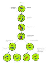
Meiosis illustration (with labels)
2546
Meiosis is the process whereby a cell reduces its chromosomes from diploid to haploid in creating eggs or sperm. Crabtree + Company View Media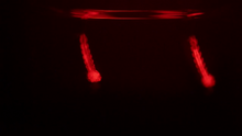
Culex quinquefasciatus mosquito larvae
6771
Mosquito larvae with genes edited by CRISPR swimming in water. Valentino Gantz, University of California, San Diego. View Media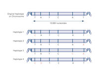
Haplotypes (with labels)
2567
Haplotypes are combinations of gene variants that are likely to be inherited together within the same chromosomal region. Crabtree + Company View Media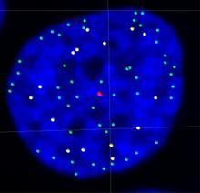
Telomeres on outer edge of nucleus during cell division
3484
New research shows telomeres moving to the outer edge of the nucleus after cell division, suggesting these caps that protect chromosomes also may play a role in organizing DNA. Laure Crabbe, Jamie Kasuboski and James Fitzpatrick, Salk Institute for Biological Studies View Media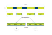
Alternative splicing (with labels)
2553
Arranging exons in different patterns, called alternative splicing, enables cells to make different proteins from a single gene. Featured in The New Genetics. Crabtree + Company View Media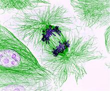
Dividing cells showing chromosomes and cell skeleton
3631
This pig cell is in the process of dividing. The chromosomes (purple) have already replicated and the duplicates are being pulled apart by fibers of the cell skeleton known as microtubules (green). Nasser Rusan, National Heart, Lung, and Blood Institute, National Institutes of Health View Media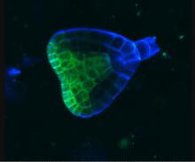
Early development in Arabidopsis
2733
Early on, this Arabidopsis plant embryo picks sides: While one end will form the shoot, the other will take root underground. Zachery R. Smith, Jeff Long lab at the Salk Institute for Biological Studies View Media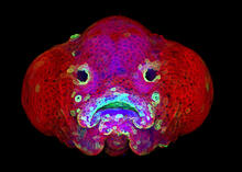
Zebrafish larva
5881
You are face to face with a 6-day-old zebrafish larva. What look like eyes will become nostrils, and the bulges on either side will become eyes. Oscar Ruiz and George Eisenhoffer, University of Texas MD Anderson Cancer Center, Houston View Media
CRISPR illustration
3719
This illustration shows, in simplified terms, how the CRISPR-Cas9 system can be used as a gene-editing tool. National Institute of General Medical Sciences. View Media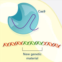
CRISPR Illustration Frame 4
6488
This illustration shows, in simplified terms, how the CRISPR-Cas9 system can be used as a gene-editing tool. National Institute of General Medical Sciences. View Media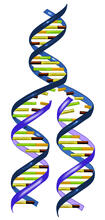
DNA replication illustration
2543
During DNA replication, each strand of the original molecule acts as a template for the synthesis of a new, complementary DNA strand. Crabtree + Company View Media
Lily mitosis 06
1016
A light microscope image of a cell from the endosperm of an African globe lily (Scadoxus katherinae). This is one frame of a time-lapse sequence that shows cell division in action. Andrew S. Bajer, University of Oregon, Eugene View Media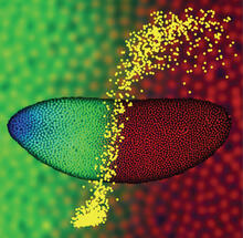
Precise development in the fruit fly embryo
2593
This 2-hour-old fly embryo already has a blueprint for its formation, and the process for following it is so precise that the difference of just a few key molecules can change the plans. Thomas Gregor, Princeton University View Media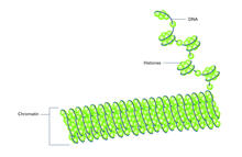
Histones in chromatin (with labels)
2561
Histone proteins loop together with double-stranded DNA to form a structure that resembles beads on a string. Crabtree + Company View Media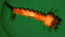
Culex quinquefasciatus mosquito larva
6769
A mosquito larva with genes edited by CRISPR. The red-orange glow is a fluorescent protein used to track the edits. Valentino Gantz, University of California, San Diego. View Media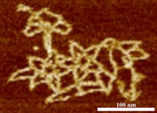
Microscopy image of bird-and-flower DNA origami
3690
An atomic force microscopy image shows DNA folded into an intricate, computer-designed structure. Hao Yan, Arizona State University View Media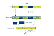
Introns (with labels)
2551
Genes are often interrupted by stretches of DNA (introns, blue) that do not contain instructions for making a protein. Crabtree + Company View Media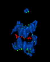
A chromosome goes missing in anaphase
5766
Anaphase is the critical step during mitosis when sister chromosomes are disjoined and directed to opposite spindle poles, ensuring equal distribution of the genome during cell division. View Media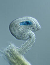
Genetic imprinting in Arabidopsis
2418
This delicate, birdlike projection is an immature seed of the Arabidopsis plant. The part in blue shows the cell that gives rise to the endosperm, the tissue that nourishes the embryo. Robert Fischer, University of California, Berkeley View Media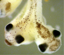
Two-headed Xenopus laevis tadpole
2755
Xenopus laevis, the African clawed frog, has long been used as a research organism for studying embryonic development. Michael Klymkowsky, University of Colorado, Boulder View Media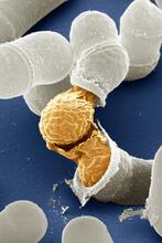
Birth of a yeast cell
3614
Yeast make bread, beer, and wine. And like us, yeast can reproduce sexually. A mother and father cell fuse and create one large cell that contains four offspring. Juergen Berger, Max Planck Institute for Developmental Biology, and Maria Langegger, Friedrich Miescher Laboratory of the Max Planck Society, Germany View Media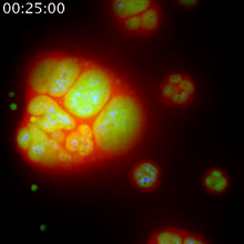
Nucleolus subcompartments spontaneously self-assemble 1
3789
The nucleolus is a small but very important protein complex located in the cell's nucleus. Nilesh Vaidya, Princeton University View Media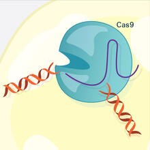
CRISPR Illustration Frame 3
6487
This illustration shows, in simplified terms, how the CRISPR-Cas9 system can be used as a gene-editing tool. National Institute of General Medical Sciences. View Media