Beginning in late July, NIH will start introducing a new website experience designed to make it easier to find health information, research, funding opportunities, and other resources. During the transition, you may notice changes to navigation, page layouts, and where some information is located.
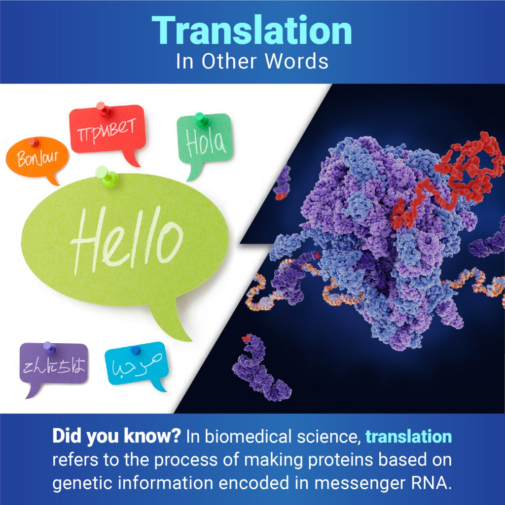
In everyday use, most people understand translation to mean converting words from one language to another. But when biologists talk about translation, they mean the process of making proteins based on the genetic information encoded in messenger RNA (mRNA). Proteins are essential for virtually every process in our bodies, from transporting oxygen to defending against infection, so translation is vital for keeping us alive and healthy.
Apoptosis is the process by which cells in the body die in a controlled and predictable way because they have DNA damage or are no longer needed. The term comes from a Greek word meaning “falling off,” as in leaves falling from a tree.
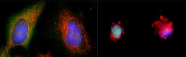
When a cell undergoes apoptosis, it shrinks and pulls away from its neighbors. As the cytoskeleton that gives it shape and structure collapses, the envelope around the cell’s nucleus breaks down, and its DNA breaks into pieces. Its surface changes, signaling its death to other cells and leading a healthy cell to engulf the dying one and recycle its components.
Two cells in a healthy state (left) and entering apoptosis (right). Credit: Hogan Tang of the Denise Montell Lab, Johns Hopkins School of Medicine.
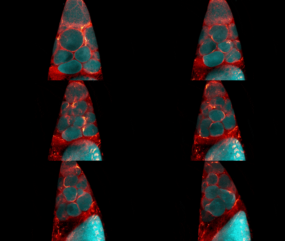
Credit: Jasmin Imran Alsous and Jonathan Jackson, Martin Lab, Massachusetts Institute of Technology.
What looks like a bubbling lava lamp is actually part of an egg cell’s maturation process. In many animals, the egg cell develops alongside sister cells. These sister cells are called nurse cells in the fruit fly (Drosophila melanogaster), and their job is to “nurse” an immature egg cell, or oocyte. Toward the end of oocyte development, the nurse cells transfer all their contents into the oocyte in a process called nurse cell dumping. This video captures this transfer, showing significant shape changes on the part of the nurse cells (blue), which are powered by wavelike activity of the protein myosin (red).
This post is a great supplement to Pathways: The Imaging Issue.
The video was taken using a confocal laser scanning microscopy (sometimes shortened to just “confocal microscopy”), one of the techniques mentioned in the Pathways timeline (1970s).
 National Institute of