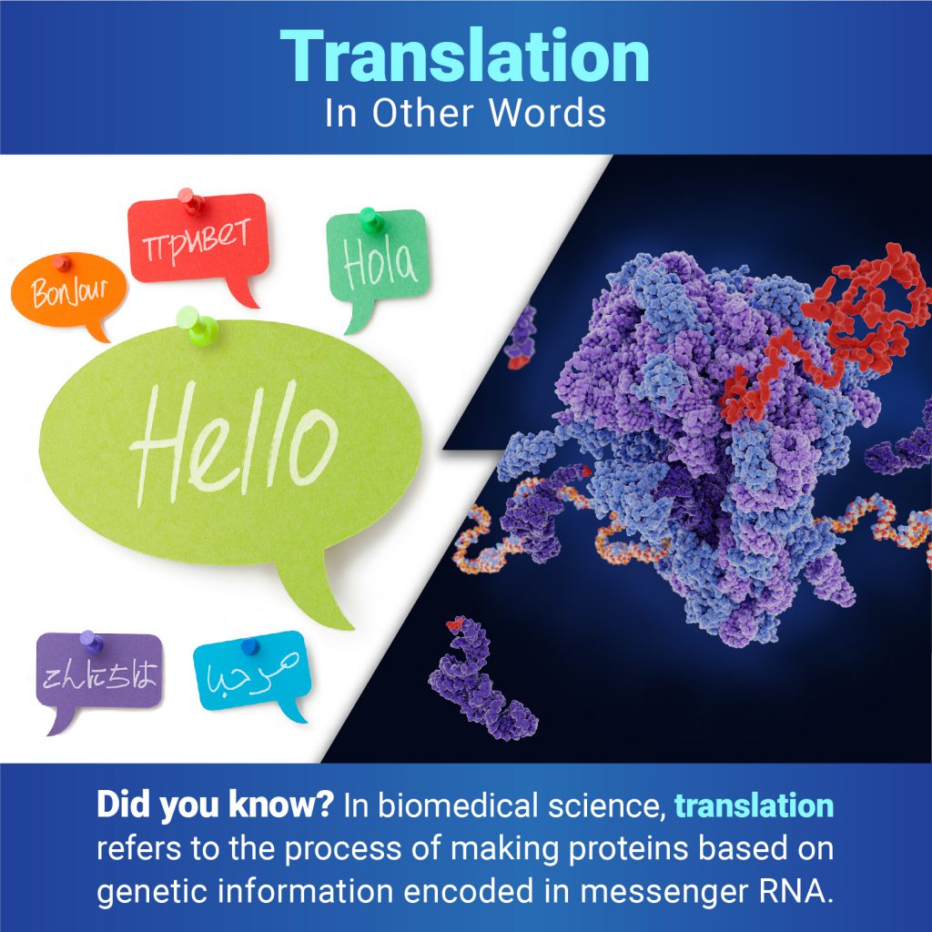
In everyday use, most people understand translation to mean converting words from one language to another. But when biologists talk about translation, they mean the process of making proteins based on the genetic information encoded in messenger RNA (mRNA). Proteins are essential for virtually every process in our bodies, from transporting oxygen to defending against infection, so translation is vital for keeping us alive and healthy.
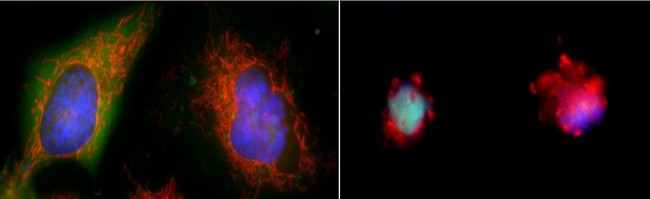
Apoptosis is the process by which cells in the body die in a controlled and predictable way because they have DNA damage or are no longer needed. The term comes from a Greek word meaning “falling off,” as in leaves falling from a tree.
When a cell undergoes apoptosis, it shrinks and pulls away from its neighbors. As the cytoskeleton that gives it shape and structure collapses, the envelope around the cell’s nucleus breaks down, and its DNA breaks into pieces. Its surface changes, signaling its death to other cells and leading a healthy cell to engulf the dying one and recycle its components.
Two cells in a healthy state (left) and entering apoptosis (right). Credit: Hogan Tang of the Denise Montell Lab, Johns Hopkins School of Medicine.
Credit: Jasmin Imran Alsous and Jonathan Jackson, Martin Lab, Massachusetts Institute of Technology.
What looks like a bubbling lava lamp is actually part of an egg cell’s maturation process. In many animals, the egg cell develops alongside sister cells. These sister cells are called nurse cells in the fruit fly (Drosophila melanogaster), and their job is to “nurse” an immature egg cell, or oocyte. Toward the end of oocyte development, the nurse cells transfer all their contents into the oocyte in a process called nurse cell dumping. This video captures this transfer, showing significant shape changes on the part of the nurse cells (blue), which are powered by wavelike activity of the protein myosin (red).
This post is a great supplement to Pathways: The Imaging Issue.
The video was taken using a confocal laser scanning microscopy (sometimes shortened to just “confocal microscopy”), one of the techniques mentioned in the Pathways timeline (1970s).
Proteins (such as hemoglobin, actin, and amylase) are workhorse molecules that contribute to virtually every activity in the body. Some of proteins’ many jobs include carrying oxygen from your lungs to the rest of your body (hemoglobin), allowing your muscles to move (actin and myosin), and digesting your food (amylase, pepsin, and lactase). All proteins are made up of chains of amino acids that fold into specific 3D structures, and each protein’s structure allows it to perform its distinct job. Proteins that are misfolded or misshapen can cause diseases such as Parkinson’s or cataracts.
While it’s straightforward to use the genetic code to predict amino acid sequences of proteins from gene sequences, the vast diversity of protein shapes and many factors that influence a protein’s 3D structure make it much more complicated to create simple folding rules that could be used to predict proteins’ structures from these sequences. Scientists have worked on this problem for nearly 50 years, and NIGMS has supported many of their efforts, including the Critical Assessment of Structure Prediction (CASP) program.
Dr. Hong Liu in the lab. Credit: Courtesy of Dr. Hong Liu.
“A scientific career is really worth it,” says Hong Liu, Ph.D., an assistant professor of biochemistry and molecular biology at Tulane University School of Medicine in New Orleans, Louisiana. Check out the highlights of our interview with Dr. Liu below to learn about his journey as a scientist and his advice for students.
Q: What makes a career in science exciting?
A: I think there are at least two things that make a science career very exciting. The first is that doing science means you have freedom to explore a lot of new ideas. The second thing is it’s rewarding. The "rewarding" I’m talking about here is not like how much money you can make. It’s rewarding in the answers you find and the new knowledge you reveal.
Have you ever wondered what creates striking images of cells and other tiny
structures? Most often, the answer is microscopes. Many of us have encountered
basic light microscopes in science classes, but those are just one of many
types that scientists use. Check out the slideshow to see images researchers
have captured using different kinds of microscopes. For even more images of
the microscopic world, visit the
NIGMS Image and Video Gallery.
Students, teachers, and other curious minds can step into a scientific imaging lab with a free online interactive developed by NIGMS and Scholastic. Imaging tools help scientists unlock the mysteries of our cells and molecules. A better understanding of this tiny world can help researchers learn about the body’s normal and abnormal processes and lead to more effective, targeted treatments for illnesses.