Switch to Gallery View
Image and Video Gallery
This is a searchable collection of scientific photos, illustrations, and videos. The images and videos in this gallery are licensed under Creative Commons Attribution Non-Commercial ShareAlike 3.0. This license lets you remix, tweak, and build upon this work non-commercially, as long as you credit and license your new creations under identical terms.
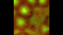
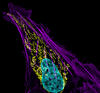
Cell-like compartments emerging from scrambled frog eggs 4
6590
Cell-like compartments that spontaneously emerged from scrambled frog eggs, with nuclei (blue) from frog sperm. Endoplasmic reticulum (red) and microtubules (green) are also visible. Xianrui Cheng, Stanford University School of Medicine. View Media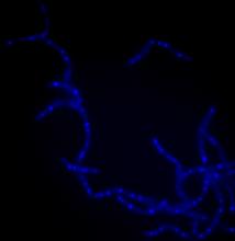
Bacillus anthracis being killed
3525
Bacillus anthracis (anthrax) cells being killed by a fluorescent trans-translation inhibitor, which disrupts bacterial protein synthesis. Kenneth Keiler, Penn State University View Media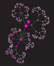
Network diagram of genes, cellular components and processes (unlabeled)
3436
This image shows the hierarchical ontology of genes, cellular components and processes derived from large genomic datasets. From Dutkowski et al. Janusz Dutkowski and Trey Ideker View MediaGenetic mosaicism in fruit flies
6983
Fat tissue from the abdomen of a genetically mosaic adult fruit fly. Genetic mosaicism means that the fly has cells with different genotypes even though it formed from a single zygote. Akhila Rajan, Fred Hutchinson Cancer Center View Media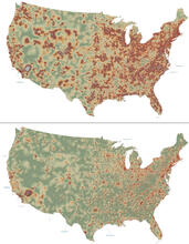
Mapping disease spread
2320
How far and fast an infectious disease spreads across a community depends on many factors, including transportation. These U.S. David Chrest, RTI International View Media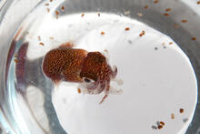
Adult and juvenile Hawaiian bobtail squids
7010
An adult Hawaiian bobtail squid, Euprymna scolopes, (~4 cm) surrounded by newly hatched juveniles (~2 mm) in a bowl of seawater.Margaret J. McFall-Ngai, Carnegie Institution for Science/California Institute of Technology, and Edward G. Ruby, California Institute of Technology. View Media
Rabbit GPDA
2405
A crystal of rabbit GPDA protein created for X-ray crystallography, which can reveal detailed, three-dimensional protein structures. Alex McPherson, University of California, Irvine View Media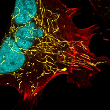
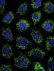
Multinucleated cancer cell
6967
A cancer cell with three nuclei, shown in turquoise. The abnormal number of nuclei indicates that the cell failed to go through cell division, probably more than once. Dylan T. Burnette, Vanderbilt University School of Medicine. View Media
C. elegans showing internal structures
6961
An image of Caenorhabditis elegans, a tiny roundworm, showing internal structures including the intestine, pharynx, and body wall muscle. C. Michael Shribak, Marine Biological Laboratory/University of Chicago. View Media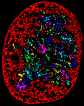
Chromatin in human fibroblast
6887
The nucleus of a human fibroblast cell with chromatin—a substance made up of DNA and proteins—shown in various colors. Melike Lakadamyali, Perelman School of Medicine at the University of Pennsylvania. View Media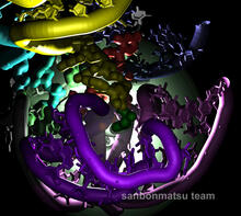
Natural nanomachine in action
2336
Using a supercomputer to simulate the movement of atoms in a ribosome, researchers looked into the core of this protein-making nanomachine and took snapshots. Kevin Sanbonmatsu, Los Alamos National Laboratory View Media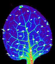
Zinc levels in a plant leaf
3727
Zinc is required for the function of more than 300 enzymes, including those that help regulate gene expression, in various organisms including humans. Suzana Car, Dartmouth College View Media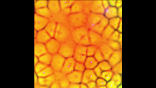
Cell-like compartments emerging from scrambled frog eggs
6587
Cell-like compartments spontaneously emerge from scrambled frog eggs, with nuclei (blue) from frog sperm. Endoplasmic reticulum (red) and microtubules (green) are also visible. Xianrui Cheng, Stanford University School of Medicine. View Media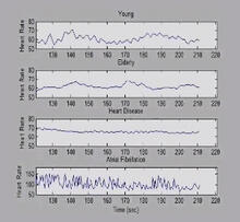
Heart rates time series image
3596
These time series show the heart rates of four different individuals. Madalena Costa and Ary Goldberger, Beth Israel Deaconess Medical Center View Media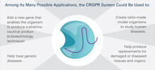
CRISPR Illustration Frame 5
6489
This illustration shows, in simplified terms, how the CRISPR-Cas9 system can be used as a gene-editing tool. This is the fifthframe in a series of five. View Media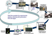
High-throughput protein structure determination pipeline
2364
This slide shows the technologies that the Joint Center for Structural Genomics developed for going from gene to structure and how the technologies have been integrated into a high-throughput pipeline Joint Center for Structural Genomics View Media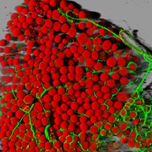
Fat cells (red) and blood vessels (green)
3600
A mouse's fat cells (red) are shown surrounded by a network of blood vessels (green). Daniela Malide, National Heart, Lung, and Blood Institute, National Institutes of Health View Media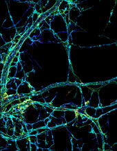
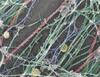
STORM image of axonal cytoskeleton
3678
This image shows the long, branched structures (axons) of nerve cells. Xiaowei Zhuang Laboratory, Howard Hughes Medical Institute, Harvard University View Media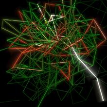
Mapping metabolic activity
2319
Like a map showing heavily traveled roads, this mathematical model of metabolic activity inside an E. coli cell shows the busiest pathway in white. Albert-László Barabási, University of Notre Dame View Media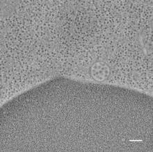
Multivesicular bodies containing intralumenal vesicles assemble at the vacuole 2
5768
Collecting and transporting cellular waste and sorting it into recylable and nonrecylable pieces is a complex business in the cell. Matthew West and Greg Odorizzi, University of Colorado View Media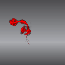
Color coding of the Drosophila brain - video
5843
This video results from a research project to visualize which regions of the adult fruit fly (Drosophila) brain derive from each neural stem cell. Yong Wan from Charles Hansen’s lab, University of Utah. Data preparation and visualization by Masayoshi Ito in the lab of Kei Ito, University of Tokyo. View Media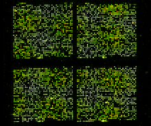
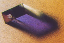
Microarray 01
1070
Microarrays, also called gene chips, are tools that let scientists track the activity of hundreds or thousands of genes simultaneously. Maggie Werner-Washburne, University of New Mexico, Albuquerque View Media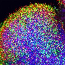
Human retinal organoid
6748
A replica of a human retina grown from stem cells. Kevin Eliceiri, University of Wisconsin-Madison. View Media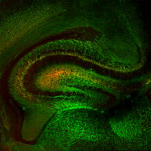
Mouse Brain Cross Section
5886
The brain sections are treated with fluorescent antibodies specific to a particular protein and visualized using serial electron microscopy (SEM). Anton Maximov, The Scripps Research Institute, La Jolla, CA View Media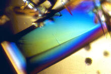
Bovine trypsin
2408
A crystal of bovine trypsin protein created for X-ray crystallography, which can reveal detailed, three-dimensional protein structures. Alex McPherson, University of California, Irvine View Media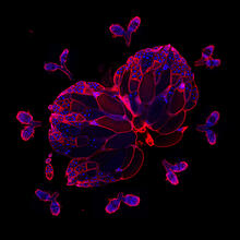
Wild-type and mutant fruit fly ovaries
6806
The two large, central, round shapes are ovaries from a typical fruit fly (Drosophila melanogaster). Vladimir I. Gelfand, Feinberg School of Medicine, Northwestern University. View Media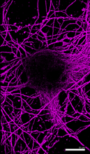
Microtubules in hippocampal neurons
6890
Microtubules (magenta) in neurons of the hippocampus, a part of the brain involved in learning and memory. Microtubules are strong, hollow fibers that provide structural support to cells. Melike Lakadamyali, Perelman School of Medicine at the University of Pennsylvania. View Media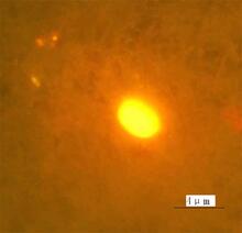
Finding one bug
2314
A nanometer-sized biosensor can detect a single deadly bacterium in tainted ground beef. How? Weihong Tan, University of Florida in Gainesville View Media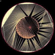
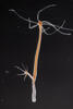
Modeling disease spread
2322
What looks like a Native American dream catcher is really a network of social interactions within a community. Stephen Eubank, University of Virginia Biocomplexity Institute (formerly Virginia Bioinformatics Institute) View Media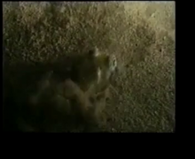
Adult Hawaiian bobtail squid burying in the sand
7012
Each morning, the nocturnal Hawaiian bobtail squid, Euprymna scolopes, hides from predators by digging into the sand. At dusk, it leaves the sand again to hunt. Margaret J. McFall-Ngai, Carnegie Institution for Science/California Institute of Technology, and Edward G. Ruby, California Institute of Technology. View Media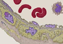
Transmission electron microscopy of coronary artery wall with elastin-rich ECM pseudocolored in light brown
3738
Elastin is a fibrous protein in the extracellular matrix (ECM). It is abundant in artery walls like the one shown here. As its name indicates, elastin confers elasticity. Tom Deerinck, National Center for Microscopy and Imaging Research (NCMIR) View Media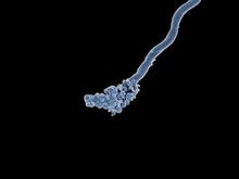
Relapsing fever bacterium (gray) and red blood cells
3585
Relapsing fever is caused by a bacterium and transmitted by certain soft-bodied ticks or body lice. The disease is seldom fatal in humans, but it can be very serious and prolonged. NIAID View Media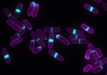
Yeast cells with accumulated cell wall material
6797
Yeast cells that abnormally accumulate cell wall material (blue) at their ends and, when preparing to divide, in their middles. This image was captured using wide-field microscopy with deconvolution. Alaina Willet, Kathy Gould’s lab, Vanderbilt University. View Media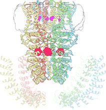
Cryo-electron microscopy revealing the "wasabi receptor"
3747
The TRPA1 protein is responsible for the burn you feel when you taste a bite of sushi topped with wasabi. Jean-Paul Armache, UCSF View Media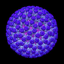
Rotavirus structure
3584
This image shows a computer-generated, three-dimensional map of the rotavirus structure. This virus infects humans and other animals and causes severe diarrhea in infants and young children. Bridget Carragher, The Scripps Research Institute, La Jolla, CA View Media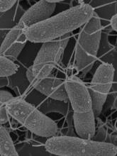
Flagellated bacterial cells
7014
Vibrio fischeri (2 mm in length) is the exclusive symbiotic partner of the Hawaiian bobtail squid, Euprymna scolopes. Margaret J. McFall-Ngai, Carnegie Institution for Science/California Institute of Technology, and Edward G. Ruby, California Institute of Technology. View Media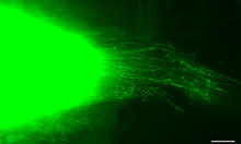
Cytonemes in developing fruit fly cells
3574
Scientists have long known that multicellular organisms use biological molecules produced by one cell and sensed by another to transmit messages that, for instance, guide proper development of organs Sougata Roy, University of California, San Francisco View Media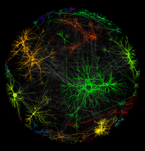
A molecular interaction network in yeast 1
3730
The image visualizes a part of the yeast molecular interaction network. Keiichiro Ono, UCSD View Media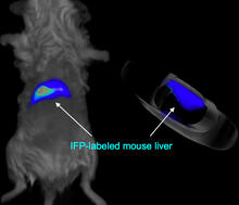
Mouse liver labeled with fluorescent probe
2601
A mouse liver glows after being tagged with specially designed infrared-fluorescent protein (IFP). Xiaokun Shu, University of California, San Diego View Media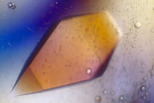
Pig trypsin (1)
2400
A crystal of porcine trypsin protein created for X-ray crystallography, which can reveal detailed, three-dimensional protein structures. Alex McPherson, University of California, Irvine View Media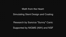
Math from the heart
3592
Watch a cell ripple toward a beam of light that turns on a movement-related protein. View Media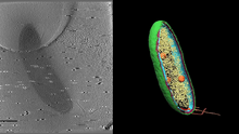
Cryo-electron tomography of a Caulobacter bacterium
6569
3D image of Caulobacter bacterium with various components highlighted: cell membranes (red and blue), protein shell (green), protein factories known as ribosomes (yellow), and storage granules Peter Dahlberg, Stanford University. View Media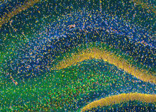
Rat Hippocampus
3308
This image of the hippocampus was taken with an ultra-widefield high-speed multiphoton laser microscope. Tom Deerinck, NCMIR View Media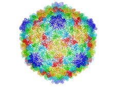
Bacteriophage P22 capsid
5874
Cryo-electron microscopy (cryo-EM) has the power to capture details of proteins and other small biological structures at the molecular level. This image shows proteins in the capsid, or outer co Dr. Wah Chiu, Baylor College of Medicine View Media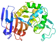
Ribbon diagram of a cefotaxime-CCD-1 complex
6766
CCD-1 is an enzyme produced by the bacterium Clostridioides difficile that helps it resist antibiotics. Keith Hodgson, Stanford University. View Media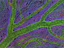
Small blood vessels in a mouse retina
3400
Blood vessels at the back of the eye (retina) are used to diagnose glaucoma and diabetic eye disease. They also display characteristic changes in people with high blood pressure. National Center for Microscopy and Imaging Research View Media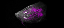
Zebrafish head vasculature video
6933
Various views of a zebrafish head with blood vessels shown in purple. Prayag Murawala, MDI Biological Laboratory and Hannover Medical School. View Media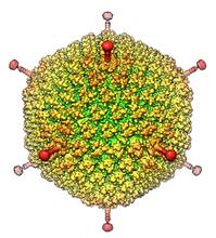
Human Adenovirus
6347
The cryo-EM structure of human adenovirus D26 (HAdV-D26) at near atomic resolution (3.7 Å), determined in collaboration with the NRAMM facility*. National Resource for Automated Molecular Microscopy http://nramm.nysbc.org/nramm-images/ Source: Bridget Carragher View Media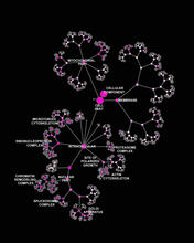
Network diagram of genes, cellular components and processes (labeled)
3437
This image shows the hierarchical ontology of genes, cellular components and processes derived from large genomic datasets. From Dutkowski et al. Janusz Dutkowski and Trey Ideker, University of California, San Diego View Media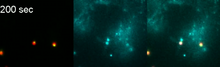
Phagosome in macrophage cell
6799
A sensor particle being engulfed by a macrophage—an immune cell—and encapsuled in a compartment called a phagosome. The phagosome then fuses with lysosomes—another type of compartment. Yan Yu, Indiana University, Bloomington. View Media