Switch to Gallery View
Image and Video Gallery
This is a searchable collection of scientific photos, illustrations, and videos. The images and videos in this gallery are licensed under Creative Commons Attribution Non-Commercial ShareAlike 3.0. This license lets you remix, tweak, and build upon this work non-commercially, as long as you credit and license your new creations under identical terms.
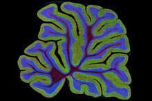
Cerebellum: the brain's locomotion control center
3639
The cerebellum of a mouse is shown here in cross-section. The cerebellum is the brain's locomotion control center. Thomas Deerinck, National Center for Microscopy and Imaging Research, University of California, San Diego View Media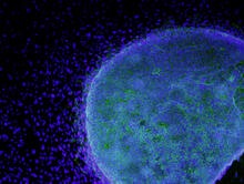
Colony of human ES cells
3269
A colony of human embryonic stem cells (light blue) grows on fibroblasts (dark blue). California Institute for Regenerative Medicine View Media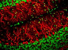
Purkinje cells are one of the main cell types in the brain
3637
This image captures Purkinje cells (red), one of the main types of nerve cell found in the brain. Yinghua Ma and Timothy Vartanian, Cornell University, Ithaca, N.Y. View Media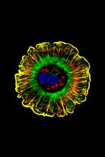
Human liver cell (hepatocyte)
3610
Hepatocytes, like the one shown here, are the most abundant type of cell in the human liver. Donna Beer Stolz, University of Pittsburgh View Media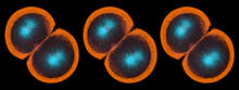
Sea urchin embryo 06
1052
Stereo triplet of a sea urchin embryo stained to reveal actin filaments (orange) and microtubules (blue). George von Dassow, University of Washington View Media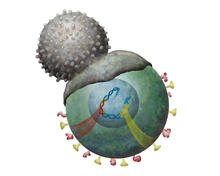
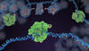
Immune cell attacks cell infected with a retrovirus
2489
T cells engulf and digest cells displaying markers (or antigens) for retroviruses, such as HIV. Kristy Whitehouse, science illustrator View Media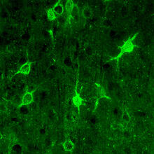
Confocal microscopy of perineuronal nets in the brain 1
3741
The photo shows a confocal microscopy image of perineuronal nets (PNNs), which are specialized extracellular matrix (ECM) structures in the brain. Tom Deerinck, National Center for Microscopy and Imaging Research (NCMIR) View Media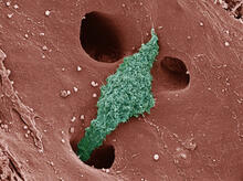
Kupffer cell residing in the liver
6535
Kupffer cells appear in the liver during the early stages of mammalian development and stay put throughout life to protect liver cells, clean up old red blood cells, and regulate iron levels. Thomas Deerinck, National Center for Microscopy and Imaging Research, University of California, San Diego. View Media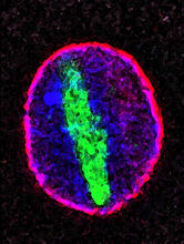
Cellular aging
2578
A protein called tubulin (green) accumulates in the center of a nucleus (outlined in pink) from an aging cell. Maximiliano D'Angelo and Martin Hetzer, Salk Institute View Media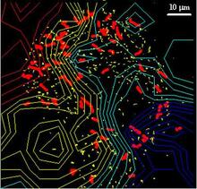
Cellular traffic
2310
Like tractor-trailers on a highway, small sacs called vesicles transport substances within cells. This image tracks the motion of vesicles in a living cell. Alexey Sharonov and Robin Hochstrasser, University of Pennsylvania View Media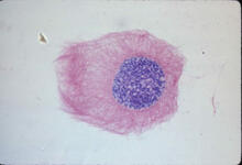
Lily mitosis 03
1013
A light microscope image of a cell from the endosperm of an African globe lily (Scadoxus katherinae). This is one frame of a time-lapse sequence that shows cell division in action. Andrew S. Bajer, University of Oregon, Eugene View Media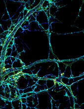
STORM image of axonal cytoskeleton
3678
This image shows the long, branched structures (axons) of nerve cells. Xiaowei Zhuang Laboratory, Howard Hughes Medical Institute, Harvard University View Media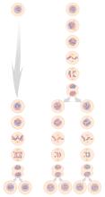
Mitosis and meiosis compared
1333
Meiosis is used to make sperm and egg cells. During meiosis, a cell's chromosomes are copied once, but the cell divides twice. Judith Stoffer View Media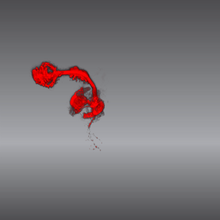
Color coding of the Drosophila brain - video
5843
This video results from a research project to visualize which regions of the adult fruit fly (Drosophila) brain derive from each neural stem cell. Yong Wan from Charles Hansen’s lab, University of Utah. Data preparation and visualization by Masayoshi Ito in the lab of Kei Ito, University of Tokyo. View Media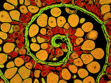
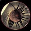
Anglerfish ovary cross-section
3620
This image captures the spiral-shaped ovary of an anglerfish in cross-section. Once matured, these eggs will be released in a gelatinous, floating mass. James E. Hayden, The Wistar Institute, Philadelphia, Pa. View Media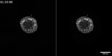
Breast cancer cells change migration phenotypes
6986
Cancer cells can change their migration phenotype, which includes their shape and the way that they move to invade different tissues. Bo Sun, Oregon State University. View Media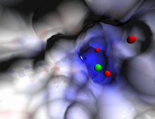
Active site of sulfite oxidase
2746
Sulfite oxidase is an enzyme that is essential for normal neurological development in children. John Enemark, University of Arizona View Media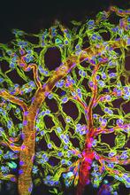
Weblike sheath covering developing egg chambers in a giant grasshopper
3616
The lubber grasshopper, found throughout the southern United States, is frequently used in biology classes to teach students about the respiratory system of insects. Kevin Edwards, Johny Shajahan, and Doug Whitman, Illinois State University. View Media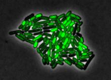
Pulsating response to stress in bacteria
3253
By attaching fluorescent proteins to the genetic circuit responsible for B. subtilis's stress response, researchers can observe the cells' pulses as green flashes. Michael Elowitz, Caltech University View Media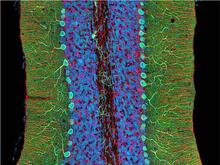
Mouse cerebellum close-up
3371
The cerebellum is the brain's locomotion control center. Every time you shoot a basketball, tie your shoe or chop an onion, your cerebellum fires into action. National Center for Microscopy and Imaging Research (NCMIR) View MediaAssembly of the HIV capsid
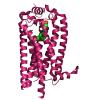
5729
The HIV capsid is a pear-shaped structure that is made of proteins the virus needs to mature and become infective. John Grime and Gregory Voth, The University of Chicago View Media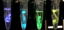
Bioluminescence in a Tube
5895
Details about the basic biology and chemistry of the ingredients that produce bioluminescence are allowing scientists to harness it as an imaging tool. Credit: Nathan Shaner, Scintillon Institute. Nathan Shaner, Scintillon Institute View Media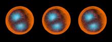
Sea urchin embryo 02
1048
Stereo triplet of a sea urchin embryo stained to reveal actin filaments (orange) and microtubules (blue). George von Dassow, University of Washington View Media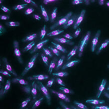
Yeast cells with nuclear envelopes and tubulin
6798
Yeast cells with nuclear envelopes shown in magenta and tubulin shown in light blue. The nuclear envelope defines the borders of the nucleus, which houses DNA. Alaina Willet, Kathy Gould’s lab, Vanderbilt University. View Media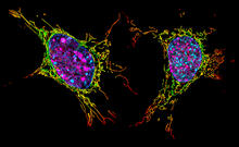
Two mouse fibroblast cells
6789
Two mouse fibroblasts, one of the most common types of cells in mammalian connective tissue. They play a key role in wound healing and tissue repair. Dylan T. Burnette, Vanderbilt University School of Medicine. View Media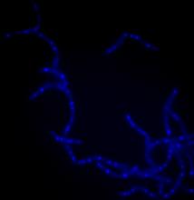
Bacillus anthracis being killed
3525
Bacillus anthracis (anthrax) cells being killed by a fluorescent trans-translation inhibitor, which disrupts bacterial protein synthesis. Kenneth Keiler, Penn State University View Media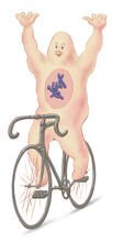
Bicycling cell
1337
A humorous treatment of the concept of a cycling cell. Judith Stoffer View Media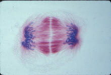
Lily mitosis 12
1018
A light microscope image of a cell from the endosperm of an African globe lily (Scadoxus katherinae). This is one frame of a time-lapse sequence that shows cell division in action. Andrew S. Bajer, University of Oregon, Eugene View Media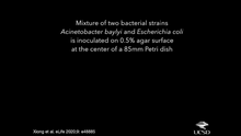
Time-lapse video of floral pattern in a mixture of two bacterial species, Acinetobacter baylyi and Escherichia coli, grown on a semi-solid agar for 24 hours
6550
This time-lapse video shows the emergence of a flower-like pattern in a mixture of two bacterial species, motile Acinetobacter baylyi and non-motile Escherichia coli (green), that are gr L. Xiong et al, eLife 2020;9: e48885 View Media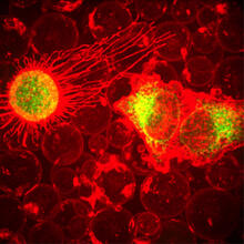
Plasma-Derived Membrane Vesicles
5887
This fiery image doesn’t come from inside a bubbling volcano. Instead, it shows animal cells caught in the act of making bubbles, or blebbing. Jeanne Stachowiak, University of Texas at Austin View Media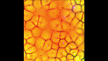
Cell-like compartments emerging from scrambled frog eggs
6587
Cell-like compartments spontaneously emerge from scrambled frog eggs, with nuclei (blue) from frog sperm. Endoplasmic reticulum (red) and microtubules (green) are also visible. Xianrui Cheng, Stanford University School of Medicine. View Media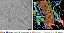
Cryo-ET cross-section of the Golgi apparatus
6606
On the left, a cross-section slice of a rat pancreas cell captured using cryo-electron tomography (cryo-ET). On the right, a 3D, color-coded version of the image highlighting cell structures. Xianjun Zhang, University of Southern California. View Media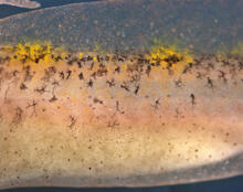
Migrating pigment cells
5758
Pigment cells are cells that give skin its color. David Parichy, University of Washington View Media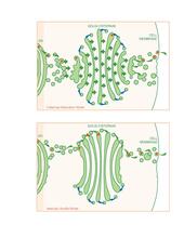
Golgi theories
1278
Two models for how material passes through the Golgi apparatus: the vesicular shuttle model and the cisternae maturation model. Judith Stoffer View Media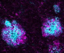
Lysosome clusters around amyloid plaques
5771
It's probably most people's least favorite activity, but we still need to do it--take out our trash. Otherwise our homes will get cluttered and smelly, and eventually, we'll get sick. Swetha Gowrishankar and Shawn Ferguson, Yale School of Medicine View Media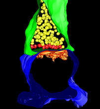
3-D Architecture of a Synapse
5885
This image shows the structure of a synapse, or junction between two nerve cells in three dimensions. From the brain of a mouse. Anton Maximov, The Scripps Research Institute, La Jolla, CA View Media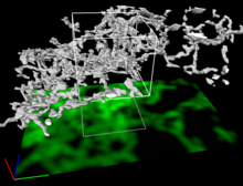
Dense tubular matrices in the peripheral endoplasmic reticulum (ER) 2
5856
Three-dimensional reconstruction of a tubular matrix in a thin section of the peripheral endoplasmic reticulum between the plasma membranes of the cell. Jennifer Lippincott-Schwartz, Howard Hughes Medical Institute Janelia Research Campus, Virginia View Media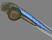
Zebrafish embryo
6897
A zebrafish embryo showing its natural colors. Zebrafish have see-through eggs and embryos, making them ideal research organisms for studying the earliest stages of development. Michael Shribak, Marine Biological Laboratory/University of Chicago. View Media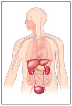
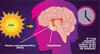
Body toxins
2496
Body organs such as the liver and kidneys process chemicals and toxins. These "target" organs are susceptible to damage caused by these substances. Crabtree + Company View Media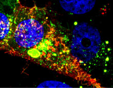
RSV-Infected Cell
3567
Viral RNA (red) in an RSV-infected cell. Eric Alonas and Philip Santangelo, Georgia Institute of Technology and Emory University View Media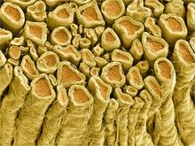
Myelinated axons 1
3396
Myelinated axons in a rat spinal root. Tom Deerinck, National Center for Microscopy and Imaging Research (NCMIR) View Media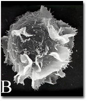
Activated mast cell surface
2637
A scanning electron microscope image of an activated mast cell. This image illustrates the interesting topography of the cell membrane, which is populated with receptors. Bridget Wilson, University of New Mexico View Media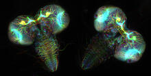
Fruit fly larvae brains showing tubulin
6808
Two fruit fly (Drosophila melanogaster) larvae brains with neurons expressing fluorescently tagged tubulin protein. Vladimir I. Gelfand, Feinberg School of Medicine, Northwestern University. View Media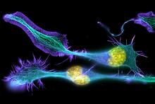
Developing nerve cells
3632
These developing mouse nerve cells have a nucleus (yellow) surrounded by a cell body, with long extensions called axons and thin branching structures called dendrites. Torsten Wittmann, University of California, San Francisco View Media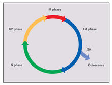
Cell cycle (with labels)
2499
Cells progress through a cycle that consists of phases for growth (G1, S, and G2) and division (M). Cells become quiescent when they exit this cycle (G0). Crabtree + Company View Media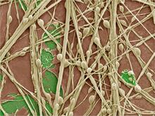
Synapses in culture
3399
Cultured hippocampal neurons grown on a substrate of glial cells (astrocytes). The glial cells form the pink/brown underlayment in this image. The tan threads are the neurons. National Center for Microscopy and Imaging Research View Media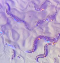
C. elegans with blue and yellow lights in the background
6750
These microscopic roundworms, called Caenorhabditis elegans, lack eyes and the opsin proteins used by visual systems to detect colors. H. Robert Horvitz and Dipon Ghosh, Massachusetts Institute of Technology. View Media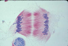
Lily mitosis 11
1011
A light microscope image of cells from the endosperm of an African globe lily (Scadoxus katherinae). This is one frame of a time-lapse sequence that shows cell division in action. Andrew S. Bajer, University of Oregon, Eugene View Media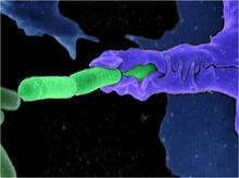
Anthrax bacteria (green) being swallowed by an immune system cell
3612
Multiple anthrax bacteria (green) being enveloped by an immune system cell (purple). Anthrax bacteria live in soil and form dormant spores that can survive for decades. Camenzind G. Robinson, Sarah Guilman, and Arthur Friedlander, United States Army Medical Research Institute of Infectious Diseases View Media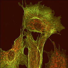
Endothelial cell
1102
This image shows two components of the cytoskeleton, microtubules (green) and actin filaments (red), in an endothelial cell derived from a cow lung. Tina Weatherby Carvalho, University of Hawaii at Manoa View Media