Beginning in late July, NIH will start introducing a new website experience designed to make it easier to find health information, research, funding opportunities, and other resources. During the transition, you may notice changes to navigation, page layouts, and where some information is located.
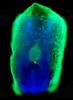
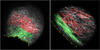
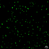
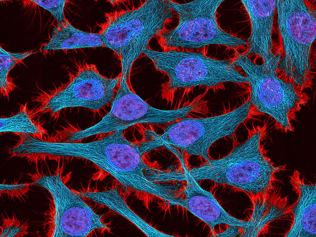
Multiphoton fluorescence image of HeLa cells stained with the actin binding toxin phalloidin (red), microtubules (cyan) and cell nuclei (blue). Nikon RTS2000MP custom laser scanning microscope. See related images 3518, 3519, 3520, 3522.
Source
National Center for Microscopy and Imaging Research (NCMIR)
