Switch to Gallery View
Image and Video Gallery
This is a searchable collection of scientific photos, illustrations, and videos. The images and videos in this gallery are licensed under Creative Commons Attribution Non-Commercial ShareAlike 3.0. This license lets you remix, tweak, and build upon this work non-commercially, as long as you credit and license your new creations under identical terms.
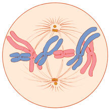
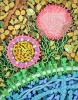
Mitosis - metaphase
1329
A cell in metaphase during mitosis: The copied chromosomes align in the middle of the spindle. Judith Stoffer View Media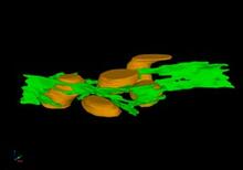
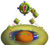
Mitochondria and endoplasmic reticulum
2635
A computer model shows how the endoplasmic reticulum is close to and almost wraps around mitochondria in the cell. The endoplasmic reticulum is lime green and the mitochondria are yellow. Bridget Wilson, University of New Mexico View Media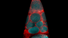
Fruit fly nurse cells transporting their contents during egg development
6754
In many animals, the egg cell develops alongside sister cells. Adam C. Martin, Massachusetts Institute of Technology. View Media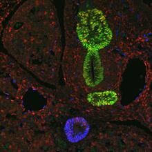
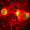
Transcription factor Sox17 controls embryonic development of certain internal organs
3440
During embryonic development, transcription factors (proteins that regulate gene expression) govern the differentiation of cells into separate tissues and organs. James M. Wells, Cincinnati Children's Hospital Medical Center View Media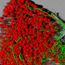
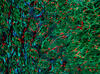
Fat cells (red) and blood vessels (green)
3600
A mouse's fat cells (red) are shown surrounded by a network of blood vessels (green). Daniela Malide, National Heart, Lung, and Blood Institute, National Institutes of Health View Media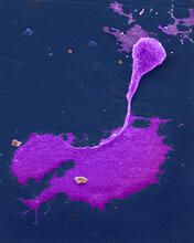
Crab nerve cell
1247
Neuron from a crab showing the cell body (bottom), axon (rope-like extension), and growth cone (top right). Tina Weatherby Carvalho, University of Hawaii at Manoa View Media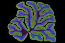
Cerebellum: the brain's locomotion control center
3639
The cerebellum of a mouse is shown here in cross-section. The cerebellum is the brain's locomotion control center. Thomas Deerinck, National Center for Microscopy and Imaging Research, University of California, San Diego View Media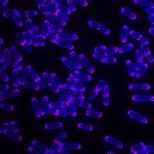
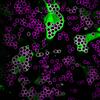
Yeast cells with Fimbrin Fim1
6794
Yeast cells with the protein Fimbrin Fim1 shown in magenta. This protein plays a role in cell division. This image was captured using wide-field microscopy with deconvolution.Alaina Willet, Kathy Gould’s lab, Vanderbilt University. View Media
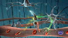
Motion in the brain
2323
Amid a network of blood vessels and star-shaped support cells, neurons in the brain signal each other. The mists of color show the flow of important molecules like glucose and oxygen. Kim Hager and Neal Prakash, University of California, Los Angeles View MediaTranslation
1281
Ribosomes manufacture proteins based on mRNA instructions. Each ribosome reads mRNA, recruits tRNA molecules to fetch amino acids, and assembles the amino acids in the proper order. Judith Stoffer View Media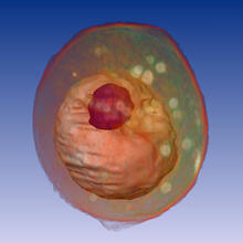
Yeast cell
1092
A whole yeast (Saccharomyces cerevisiae) cell viewed by X-ray microscopy. Inside, the nucleus and a large vacuole (red) are visible. Carolyn Larabell, University of California, San Francisco and the Lawrence Berkeley National Laboratory View Media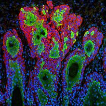
Skin cancer cells (squamous cell carcinoma)
3628
This image shows the uncontrolled growth of cells in squamous cell carcinoma, the second most common form of skin cancer. If caught early, squamous cell carcinoma is usually not life-threatening. Markus Schober and Elaine Fuchs, The Rockefeller University View Media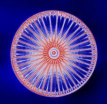
Arachnoidiscus diatom
6902
An Arachnoidiscus diatom with a diameter of 190µm. Michael Shribak, Marine Biological Laboratory/University of Chicago. View Media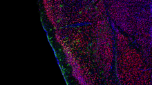
Video of Calling Cards in a mouse brain
6781
The green spots in this mouse brain are cells labeled with Calling Cards, a technology that records molecular events in brain cells as they mature. NIH Director's Blog View Media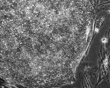
Induced stem cells from adult skin 01
2603
These cells are induced stem cells made from human adult skin cells that were genetically reprogrammed to mimic embryonic stem cells. James Thomson, University of Wisconsin-Madison View Media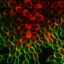
Snowflake yeast 1
6969
Multicellular yeast called snowflake yeast that researchers created through many generations of directed evolution from unicellular yeast. William Ratcliff, Georgia Institute of Technology. View Media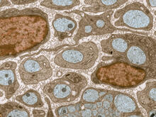
Transmission electron microscopy of myelinated axons with ECM between the axons
3736
The extracellular matrix (ECM) is most prevalent in connective tissues but also is present between the stems (axons) of nerve cells, as shown here. Tom Deerinck, National Center for Microscopy and Imaging Research (NCMIR) View Media
Cell division with late aligning chromosomes
2747
This video shows an instance of abnormal mitosis where chromosomes are late to align. Gary Gorbsky, Oklahoma Medical Research Foundation View Media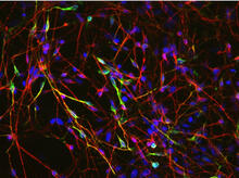
Neurons from human ES cells 02
3285
These neurons were derived from human embryonic stem cells. The neural cell bodies with axonal projections are visible in red, and the nuclei in blue. Xianmin Zeng lab, Buck Institute for Age Research, via CIRM View Media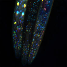
Mosaicism in C. elegans (Black Background)
6532
In the worm C. elegans, double-stranded RNA made in neurons can silence matching genes in a variety of cell types through the transport of RNA between cells. Snusha Ravikumar, Ph.D., University of Maryland, College Park, and Antony M. Jose, Ph.D., University of Maryland, College Park View Media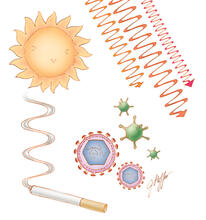
Cell toxins
1312
A number of environmental factors cause DNA mutations that can lead to cancer: toxins in cigarette smoke, sunlight and other radiation, and some viruses. Judith Stoffer View Media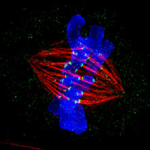
Dividing cell in metaphase
3445
This image of a mammalian epithelial cell, captured in metaphase, was the winning image in the high- and super-resolution microscopy category of the 2012 GE Healthcare Life Sciences Cell Imaging Compe Jane Stout in the laboratory of Claire Walczak, Indiana University, GE Healthcare 2012 Cell Imaging Competition View Media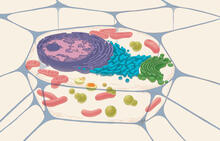
Animal cell
1274
A typical animal cell, sliced open to reveal a cross-section of organelles. Judith Stoffer View Media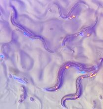
C. elegans with blue and yellow lights in the background
6750
These microscopic roundworms, called Caenorhabditis elegans, lack eyes and the opsin proteins used by visual systems to detect colors. H. Robert Horvitz and Dipon Ghosh, Massachusetts Institute of Technology. View Media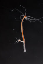
Hydra 04
2440
Hydra magnipapillata is an invertebrate animal used as a model organism to study developmental questions, for example the formation of the body axis. Hiroshi Shimizu, National Institute of Genetics in Mishima, Japan View Media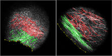
DDR2 Receptors Attach to Collagen in Breast Tumor
3478
On the left, the boundary of a breast tumor (yellow) attaches to collagen fibers that are closest to it (green) using DDR2. On the right, a tumor without DDR2 remains disconnected from the collagen. Callie Corsa and Suzanne Ponik, Washington University School of Medicine in St. Louis View Media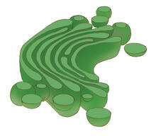
Golgi
1275
The Golgi complex, also called the Golgi apparatus or, simply, the Golgi. Judith Stoffer View Media
Centrioles anchor cilia in planaria
3292
Centrioles (green) anchor cilia (red), which project on the surface of pharynx cells of the freshwater planarian Schmidtea mediterranea. Juliette Azimzadeh, University of California, San Francisco View Media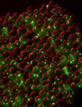
Protein clumping in zinc-deficient yeast cells
3550
The green spots in this image are clumps of protein inside yeast cells that are deficient in both zinc and a protein called Tsa1 that prevents clumping. Colin MacDiarmid and David Eide, University of Wisconsin--Madison View Media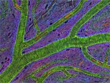
Small blood vessels in a mouse retina
3400
Blood vessels at the back of the eye (retina) are used to diagnose glaucoma and diabetic eye disease. They also display characteristic changes in people with high blood pressure. National Center for Microscopy and Imaging Research View Media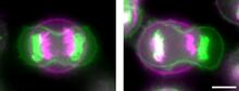
Symmetrically and asymmetrically elongating cells
3648
Merged fluorescent images of symmetrically (left) or asymmetrically (right) elongating HeLa cells at the end of early anaphase (magenta) and late anaphase (green). Tomomi Kiyomitsu and Iain M. Cheeseman, Whitehead Institute for Biomedical Research View Media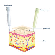
Drugs enter skin (with labels)
2532
Drugs enter different layers of skin via intramuscular, subcutaneous, or transdermal delivery methods. See image 2531 for an unlabeled version of this illustration. Crabtree + Company View Media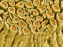
Myelinated axons 1
3396
Myelinated axons in a rat spinal root. Tom Deerinck, National Center for Microscopy and Imaging Research (NCMIR) View Media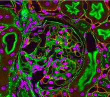
Fluorescent microscopy of kidney tissue--close-up
3725
This photograph of kidney tissue, taken using fluorescent light microscopy, shows a close-up view of part of image 3723. Tom Deerinck , National Center for Microscopy and Imaging Research View Media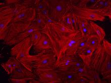
Mouse heart fibroblasts
3281
This image shows mouse fetal heart fibroblast cells. The muscle protein actin is stained red, and the cell nuclei are stained blue. Kara McCloskey lab, University of California, Merced, via CIRM View Media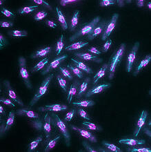
Yeast cells with nuclear envelopes and tubulin
6798
Yeast cells with nuclear envelopes shown in magenta and tubulin shown in light blue. The nuclear envelope defines the borders of the nucleus, which houses DNA. Alaina Willet, Kathy Gould’s lab, Vanderbilt University. View Media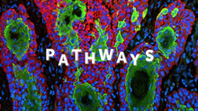
Pathways: What is It? | Why Scientists Study Cells
6540
Learn how curiosity about the world and our cells is key to scientific discoveries. National Institute of General Medical Sciences View Media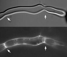
Z rings in bacterial division
2456
Lab-made liposomes contract where Z rings have gathered together and the constriction forces are greatest (arrows). Masaki Osawa, Duke University View Media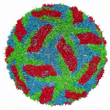
Cryo-electron microscopy of the dengue virus showing protective membrane and membrane proteins
3748
Dengue virus is a mosquito-borne illness that infects millions of people in the tropics and subtropics each year. Like many viruses, dengue is enclosed by a protective membrane. Hong Zhou, UCLA View Media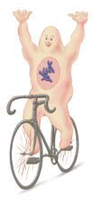
Bicycling cell
1337
A humorous treatment of the concept of a cycling cell. Judith Stoffer View Media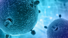
Pathways – Bacteria vs. Viruses: What's the Difference?
6597
Learn about how bacteria and viruses differ, how they each can make you sick, and how they can or cannot be treated. National Institute of General Medical Sciences View Media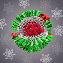
H1N1 Influenza Virus
6355
CellPack image of the H1N1 influenza virus, with hemagglutinin and neuraminidase glycoproteins in green and red, respectively, on the outer envelope (white); matrix protein in gray, and ribonucleoprot Dr. Rommie Amaro, University of California, San Diego View Media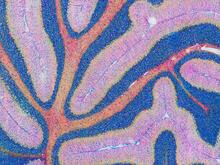
Mouse cerebellum in pink and blue
5800
The cerebellum is the brain's locomotion control center. Found at the base of your brain, the cerebellum is a single layer of tissue with deep folds like an accordion. National Center for Microscopy and Imaging Research (NCMIR) View Media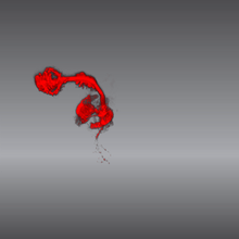
Color coding of the Drosophila brain - video
5843
This video results from a research project to visualize which regions of the adult fruit fly (Drosophila) brain derive from each neural stem cell. Yong Wan from Charles Hansen’s lab, University of Utah. Data preparation and visualization by Masayoshi Ito in the lab of Kei Ito, University of Tokyo. View Media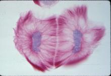
Lily mitosis 13
1019
A light microscope image of cells from the endosperm of an African globe lily (Scadoxus katherinae). This is one frame of a time-lapse sequence that shows cell division in action. Andrew S. Bajer, University of Oregon, Eugene View Media
Plasma membrane (with labels)
2524
The plasma membrane is a cell's protective barrier. See image 2523 for an unlabeled version of this illustration. Featured in The Chemistry of Health. Crabtree + Company View Media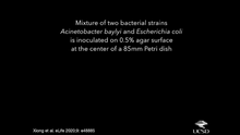
Time-lapse video of floral pattern in a mixture of two bacterial species, Acinetobacter baylyi and Escherichia coli, grown on a semi-solid agar for 24 hours
6550
This time-lapse video shows the emergence of a flower-like pattern in a mixture of two bacterial species, motile Acinetobacter baylyi and non-motile Escherichia coli (green), that are gr L. Xiong et al, eLife 2020;9: e48885 View Media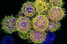
Pollen grains: male germ cells in plants and a cause of seasonal allergies
3609
Those of us who get sneezy and itchy-eyed every spring or fall may have pollen grains, like those shown here, to blame. Edna, Gil, and Amit Cukierman, Fox Chase Cancer Center, Philadelphia, Pa. View Media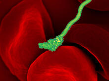
Human blood cells with Borrelia hermsii, a bacterium that causes relapsing fever
3586
Relapsing fever is caused by a bacterium and transmitted by certain soft-bodied ticks or body lice. The disease is seldom fatal in humans, but it can be very serious and prolonged. NIAID View Media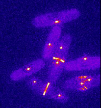
Dividing yeast cells with spindle pole bodies and contractile rings
6796
During cell division, spindle pole bodies (glowing dots) move toward the ends of yeast cells to separate copied genetic information. Alaina Willet, Kathy Gould’s lab, Vanderbilt University. View Media