Switch to Gallery View
Image and Video Gallery
This is a searchable collection of scientific photos, illustrations, and videos. The images and videos in this gallery are licensed under Creative Commons Attribution Non-Commercial ShareAlike 3.0. This license lets you remix, tweak, and build upon this work non-commercially, as long as you credit and license your new creations under identical terms.
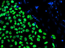
Human embryonic stem cells on feeder cells
3275
The nuclei stained green highlight human embryonic stem cells grown under controlled conditions in a laboratory. Blue represents the DNA of surrounding, supportive feeder cells. Julie Baker lab, Stanford University School of Medicine, via CIRM View Media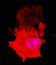
Polarized cells- 01
3332
Cells move forward with lamellipodia and filopodia supported by networks and bundles of actin filaments. Proper, controlled cell movement is a complex process. Rong Li and Praveen Suraneni, Stowers Institute for Medical Research View Media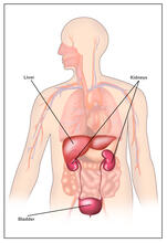
Body toxins (with labels)
2497
Body organs such as the liver and kidneys process chemicals and toxins. These "target" organs are susceptible to damage caused by these substances. Crabtree + Company View MediaTranslation
1281
Ribosomes manufacture proteins based on mRNA instructions. Each ribosome reads mRNA, recruits tRNA molecules to fetch amino acids, and assembles the amino acids in the proper order. Judith Stoffer View Media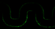
Biofilm blocking fluid flow
3446
This time-lapse movie shows that bacterial communities called biofilms can create blockages that prevent fluid flow in devices such as stents and catheters over a period of about 56 hours. Bonnie Bassler, Princeton University View Media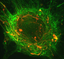
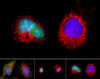
Human ES cells differentiating into neurons
3276
This image shows hundreds of human embryonic stem cells in various stages of differentiating into neurons. Guoping Fan lab, University of California, Los Angeles, via CIRM View Media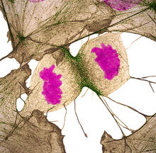
Human fibroblast undergoing cell division
6519
During cell division, cells physically divide after separating their genetic material to create two daughter cells that are genetically identical to the parent cell. Nilay Taneja, Vanderbilt University, and Dylan T. Burnette, Ph.D., Vanderbilt University School of Medicine. View Media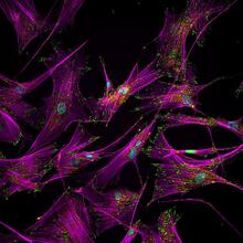
Sticky stem cells
3457
Like a group of barnacles hanging onto a rock, these human cells hang onto a matrix coated glass slide. Ankur Singh and Andrés García, Georgia Institute of Technology View Media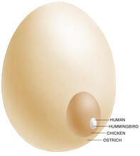
Egg comparison
1339
The largest human cell (by volume) is the egg. Human eggs are 150 micrometers in diameter and you can just barely see one with a naked eye. In comparison, consider the eggs of chickens...or ostriches! Judith Stoffer View Media
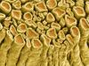
NCMIR Tongue 2
5811
Microscopy image of a tongue. One in a series of two, see image 5810 National Center for Microscopy and Imaging Research (NCMIR) View Media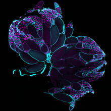
Fruit fly ovaries
6807
Fruit fly (Drosophila melanogaster) ovaries with DNA shown in magenta and actin filaments shown in light blue. This image was captured using a confocal laser scanning microscope.Vladimir I. Gelfand, Feinberg School of Medicine, Northwestern University. View Media
Lily mitosis 06
1016
A light microscope image of a cell from the endosperm of an African globe lily (Scadoxus katherinae). This is one frame of a time-lapse sequence that shows cell division in action. Andrew S. Bajer, University of Oregon, Eugene View Media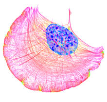
Crawling cell
6964
A crawling cell with DNA shown in blue and actin filaments, which are a major component of the cytoskeleton, visible in pink. Actin filaments help enable cells to crawl. Dylan T. Burnette, Vanderbilt University School of Medicine. View Media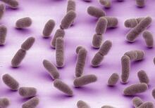

Telomerase illustration
1335
Reactivating telomerase in our cells does not appear to be a good way to extend the human lifespan. Cancer cells reactivate telomerase. Judith Stoffer View Media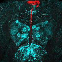
Fruit fly brain responds to adipokines
6985
Drosophila adult brain showing that an adipokine (fat hormone) generates a response from neurons (aqua) and regulates insulin-producing neurons (red).Akhila Rajan, Fred Hutchinson Cancer Center View Media

Circadian rhythm neurons in the fruit fly brain
3754
Some nerve cells (neurons) in the brain keep track of the daily cycle. This time-keeping mechanism, called the circadian clock, is found in all animals including us. Justin Blau, New York University View Media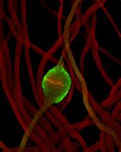
Fruit fly sperm cells
2433
Developing fruit fly spermatids require caspase activity (green) for the elimination of unwanted organelles and cytoplasm via apoptosis. Hermann Steller, Rockefeller University View Media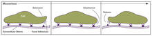
Focal adhesions (with labels)
2503
Cells walk along body surfaces via tiny "feet," called focal adhesions, that connect with the extracellular matrix. Crabtree + Company View Media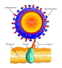
Influenza virus attaches to host membrane (with labels)
2505
Influenza A infects a host cell when hemagglutinin grips onto glycans on its surface. Crabtree + Company View Media
Yeast cells pack a punch
3788
Although they are tiny, microbes that are growing in confined spaces can generate a lot of pressure. In this video, yeast cells grow in a small chamber called a microfluidic bioreactor. Oskar Hallatschek, UC Berkeley View Media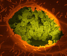
Q fever bacteria in an infected cell
3621
This image shows Q fever bacteria (yellow), which infect cows, sheep, and goats around the world and can infect humans, as well. When caught early, Q fever can be cured with antibiotics. Robert Heinzen, Elizabeth Fischer, and Anita Mora, National Institute of Allergy and Infectious Diseases, National Institutes of Health View Media
Fruit fly retina 02
2434
Section of a fruit fly retina showing the light-sensing molecules rhodopsin-5 (blue) and rhodopsin-6 (red). Hermann Steller, Rockefeller University View Media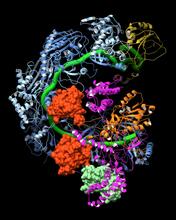
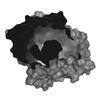
CRISPR surveillance complex
6352
This image shows how the CRISPR surveillance complex is disabled by two copies of anti-CRISPR protein AcrF1 (red) and one AcrF2 (light green). NRAMM National Resource for Automated Molecular Microscopy http://nramm.nysbc.org/nramm-images/ Source: Bridget Carragher View Media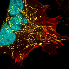
Multinucleated cancer cell
6967
A cancer cell with three nuclei, shown in turquoise. The abnormal number of nuclei indicates that the cell failed to go through cell division, probably more than once. Dylan T. Burnette, Vanderbilt University School of Medicine. View Media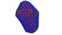
Atomic-level structure of the HIV capsid
6601
This animation shows atoms of the HIV capsid, the shell that encloses the virus's genetic material. Juan R. Perilla and the Theoretical and Computational Biophysics Group, University of Illinois at Urbana-Champaign View Media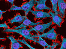
HeLa cells
3521
Multiphoton fluorescence image of HeLa cells stained with the actin binding toxin phalloidin (red), microtubules (cyan) and cell nuclei (blue). Nikon RTS2000MP custom laser scanning microscope. National Center for Microscopy and Imaging Research (NCMIR) View Media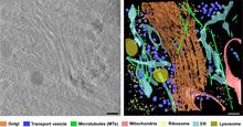
Cryo-ET cross-section of the Golgi apparatus
6606
On the left, a cross-section slice of a rat pancreas cell captured using cryo-electron tomography (cryo-ET). On the right, a 3D, color-coded version of the image highlighting cell structures. Xianjun Zhang, University of Southern California. View Media
A bundle of myelinated peripheral nerve cells (axons)
3737
The extracellular matrix (ECM) is most prevalent in connective tissues but also is present between the stems (axons) of nerve cells. Tom Deerinck, National Center for Microscopy and Imaging Research (NCMIR) View Media
Cell-like compartments emerging from scrambled frog eggs 4
6590
Cell-like compartments that spontaneously emerged from scrambled frog eggs, with nuclei (blue) from frog sperm. Endoplasmic reticulum (red) and microtubules (green) are also visible. Xianrui Cheng, Stanford University School of Medicine. View Media
Cell-like compartments emerging from scrambled frog eggs 3
6589
Cell-like compartments spontaneously emerge from scrambled frog eggs. Endoplasmic reticulum (red) and microtubules (green) are visible. Video created using epifluorescence microscopy. Xianrui Cheng, Stanford University School of Medicine. View Media
NCMIR mouse tail
3395
Stained cross section of a mouse tail. Tom Deerinck, National Center for Microscopy and Imaging Research (NCMIR) View Media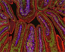
NCMIR Intestine-2
3390
The small intestine is where most of our nutrients from the food we eat are absorbed into the bloodstream. Tom Deerinck, National Center for Microscopy and Imaging Research (NCMIR) View Media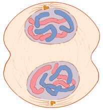
Mitosis - telophase
1332
Telophase during mitosis: Nuclear membranes form around each of the two sets of chromosomes, the chromosomes begin to spread out, and the spindle begins to break down. Judith Stoffer View Media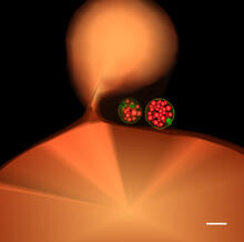
Multivesicular bodies containing intralumenal vesicles assemble at the vacuole 1
5769
Collecting and transporting cellular waste and sorting it into recylable and nonrecylable pieces is a complex business in the cell. Matthew West and Greg Odorizzi, University of Colorado View Media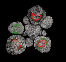
Developing Arabidopsis flower buds
3743
Flower development is a carefully orchestrated, genetically programmed process that ensures that the male (stamen) and female (pistil) organs form in the right place and at the right time in the flowe Nathanaël Prunet, Caltech View Media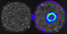
Cancer Cells Glowing from Luciferin
3480
The activator cancer cell culture, right, contains a chemical that causes the cells to emit light when in the presence of immune cells. Mark Sellmyer, Stanford University School of Medicine View Media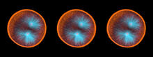
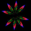
Sea urchin embryo 03
1049
Stereo triplet of a sea urchin embryo stained to reveal actin filaments (orange) and microtubules (blue). George von Dassow, University of Washington View Media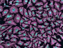
HeLa cells
3520
Multiphoton fluorescence image of HeLa cells with cytoskeletal microtubules (magenta) and DNA (cyan). Nikon RTS2000MP custom laser scanning microscope. National Center for Microscopy and Imaging Research (NCMIR) View Media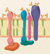
Animal cell membrane
1286
The membrane that surrounds a cell is made up of proteins and lipids. Judith Stoffer View Media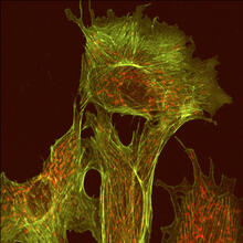
Endothelial cell
1102
This image shows two components of the cytoskeleton, microtubules (green) and actin filaments (red), in an endothelial cell derived from a cow lung. Tina Weatherby Carvalho, University of Hawaii at Manoa View Media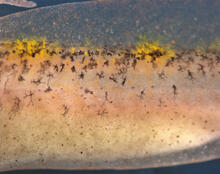
Migrating pigment cells
5758
Pigment cells are cells that give skin its color. David Parichy, University of Washington View Media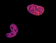
Cell division and cell death
6790
Two cells over a 2-hour period. The one on the bottom left goes through programmed cell death, also known as apoptosis. The one on the top right goes through cell division, also called mitosis. Dylan T. Burnette, Vanderbilt University School of Medicine. View Media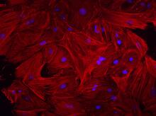
Mouse heart fibroblasts
3281
This image shows mouse fetal heart fibroblast cells. The muscle protein actin is stained red, and the cell nuclei are stained blue. Kara McCloskey lab, University of California, Merced, via CIRM View Media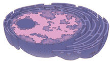
Nucleus and rough ER
1290
The nucleus contains the DNA of eukaryotic cells. Judith Stoffer View Media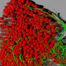
Fat cells (red) and blood vessels (green)
3600
A mouse's fat cells (red) are shown surrounded by a network of blood vessels (green). Daniela Malide, National Heart, Lung, and Blood Institute, National Institutes of Health View Media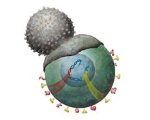
Immune cell attacks cell infected with a retrovirus
2489
T cells engulf and digest cells displaying markers (or antigens) for retroviruses, such as HIV. Kristy Whitehouse, science illustrator View Media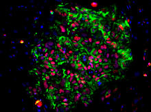
Induced pluripotent stem cells from skin
3278
These induced pluripotent stem cells (iPS cells) were derived from a woman's skin. Green and red indicate proteins found in reprogrammed cells but not in skin cells (TRA1-62 and NANOG). Kathrin Plath lab, University of California, Los Angeles, via CIRM View Media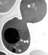
EM of yeast cell division
5770
Cell division is an incredibly coordinated process. Matthew West and Greg Odorizzi, University of Colorado View Media