Switch to Gallery View
Image and Video Gallery
This is a searchable collection of scientific photos, illustrations, and videos. The images and videos in this gallery are licensed under Creative Commons Attribution Non-Commercial ShareAlike 3.0. This license lets you remix, tweak, and build upon this work non-commercially, as long as you credit and license your new creations under identical terms.
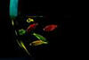
Staphylococcus aureus aggregating upon contact with synovial fluid
6805
Staphylococcus aureus bacteria (green) grouping together upon contact with synovial fluid—a viscous substance found in joints. Paul Stoodley, The Ohio State University. View Media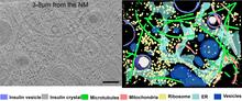
Cryo-ET cross-section of a rat pancreas cell
6608
On the left, a cross-section slice of a rat pancreas cell captured using cryo-electron tomography (cryo-ET). On the right, a 3D, color-coded version of the image highlighting cell structures. Xianjun Zhang, University of Southern California. View Media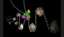
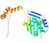
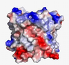
Intasome
6346
Salk researchers captured the structure of a protein complex called an intasome (center) that lets viruses similar to HIV establish permanent infection in their hosts. National Resource for Automated Molecular Microscopy http://nramm.nysbc.org/nramm-images/ Source: Bridget Carragher View Media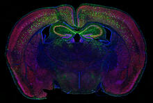
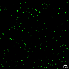
Calling Cards in a mouse brain
6780
The green spots in this mouse brain are cells labeled with Calling Cards, a technology that records molecular events in brain cells as they mature. Allen Yen, Lab of Joseph Dougherty, Washington University School of Medicine in St. Louis. View Media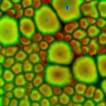
Cell-like compartments from frog eggs 2
6585
Cell-like compartments that spontaneously emerged from scrambled frog eggs, with nuclei (blue) from frog sperm. Endoplasmic reticulum (red) and microtubules (green) are also visible. Xianrui Cheng, Stanford University School of Medicine. View Media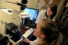
Research mentor and student
2767
A research mentor (Lori Eidson) and student (Nina Waldron, on the microscope) were 2009 members of the BRAIN (Behavioral Research Advancements In Neuroscience) program at Georgia State University in A Elizabeth Weaver, Georgia State University View Media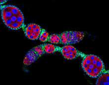
Confocal microscopy image of two Drosophila ovarioles
5772
Ovarioles in female insects are tubes in which egg cells (called oocytes) form at one end and complete their development as they reach the other end of the tube. 2004 Olympus BioScapes Competition View Media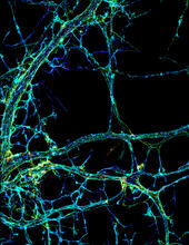
STORM image of axonal cytoskeleton
3678
This image shows the long, branched structures (axons) of nerve cells. Xiaowei Zhuang Laboratory, Howard Hughes Medical Institute, Harvard University View Media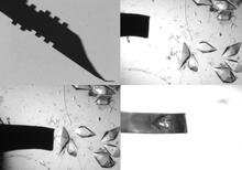
Mounting of protein crystals
2368
Automated methods using micromachined silicon are used at the Northeast Collaboratory for Structural Genomics to mount protein crystals for X-ray crystallography. The Northeast Collaboratory for Structural Genomics View Media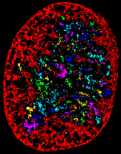
Chromatin in human fibroblast
6887
The nucleus of a human fibroblast cell with chromatin—a substance made up of DNA and proteins—shown in various colors. Melike Lakadamyali, Perelman School of Medicine at the University of Pennsylvania. View Media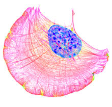
Crawling cell
6964
A crawling cell with DNA shown in blue and actin filaments, which are a major component of the cytoskeleton, visible in pink. Actin filaments help enable cells to crawl. Dylan T. Burnette, Vanderbilt University School of Medicine. View Media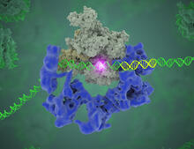
TFIID complex binds DNA to start gene transcription
3766
Gene transcription is a process by which the genetic information encoded in DNA is transcribed into RNA. Eva Nogales, Berkeley Lab View Media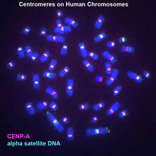
Centromeres on human chromosomes
3255
Human metaphase chromosomes are visible with fluorescence in vitro hybridization (FISH). Centromeric alpha satellite DNA (green) are found in the heterochromatin at each centromere. Peter Warburton, Mount Sinai School of Medicine View Media
Annotated TEM cross-section of C. elegans (roundworm)
5760
The worm Caenorhabditis elegans is a popular laboratory animal because its small size and fairly simple body make it easy to study. Piali Sengupta, Brandeis University View Media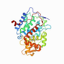
CCP enzyme
6762
The enzyme CCP is found in the mitochondria of baker’s yeast. Scientists study the chemical reactions that CCP triggers, which involve a water molecule, iron, and oxygen. Protein Data Bank. View Media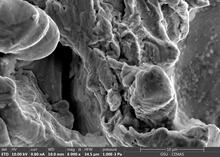
Staphylococcus aureus in the porous coating of a femoral hip stem
6804
Staphylococcus aureus bacteria (blue) on the porous coating of a femoral hip stem used in hip replacement surgery. Paul Stoodley, The Ohio State University. View Media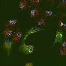
Endoplasmic reticulum abnormalities 2
6774
Human cells with the gene that codes for the protein FIT2 deleted. After an experimental intervention, they are expressing a nonfunctional version of FIT2, shown in green. Michel Becuwe, Harvard University. View Media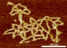
Microscopy image of bird-and-flower DNA origami
3690
An atomic force microscopy image shows DNA folded into an intricate, computer-designed structure. Hao Yan, Arizona State University View Media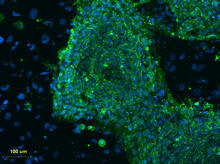
Human embryonic stem cells on feeder cells
3274
This fluorescent microscope image shows human embryonic stem cells whose nuclei are stained green. Blue staining shows the surrounding supportive feeder cells. Michael Longaker lab, Stanford University School of Medicine, via CIRM View Media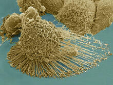
HeLa cells
3519
Scanning electron micrograph of an apoptotic HeLa cell. Zeiss Merlin HR-SEM. National Center for Microscopy and Imaging Research View Media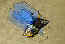
DNase
2410
Crystals of DNase protein created for X-ray crystallography, which can reveal detailed, three-dimensional protein structures. Alex McPherson, University of California, Irvine View Media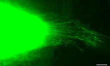
Cytonemes in developing fruit fly cells
3574
Scientists have long known that multicellular organisms use biological molecules produced by one cell and sensed by another to transmit messages that, for instance, guide proper development of organs Sougata Roy, University of California, San Francisco View Media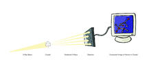
X-ray crystallography (with labels)
2512
X-ray crystallography allows researchers to see structures too small to be seen by even the most powerful microscopes. Crabtree + Company View Media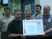
Scientists display X-ray diffraction pattern obtained with split X-ray beamline
2384
Scientists from Argonne National Laboratory's Advanced Photon Source (APS) display the first X-ray diffraction pattern obtained from a protein crystal using a split X-ray beam, the first of its kind a GM/CA Collaborative Access Team View Media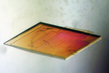
Pig trypsin crystal
2403
A crystal of pig trypsin protein created for X-ray crystallography, which can reveal detailed, three-dimensional protein structures. Alex McPherson, University of California, Irvine View Media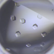
Crystals of CCD-1 in complex with cefotaxime
6764
CCD-1 is an enzyme produced by the bacterium Clostridioides difficile that helps it resist antibiotics. Keith Hodgson, Stanford University. View Media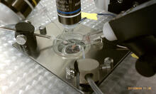
Electrode probe on mouse Huntington's muscle cell
3479
Using an electrode, researchers apply an electrical pulse onto a piece of muscle tissue affected by Huntington's disease. Grigor Varuzhanyan and Andrew A. Voss, California State Polytechnic University View Media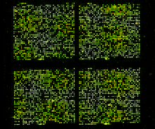
Microarray 01
1070
Microarrays, also called gene chips, are tools that let scientists track the activity of hundreds or thousands of genes simultaneously. Maggie Werner-Washburne, University of New Mexico, Albuquerque View Media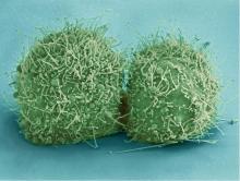
HeLa cells
3518
Scanning electron micrograph of just-divided HeLa cells. Zeiss Merlin HR-SEM. National Center for Microscopy and Imaging Research View Media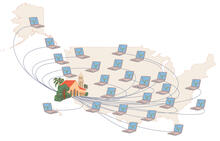
Folding@Home
1276
Stanford University scientist Vijay Pande decided to couple the power of computers with the help of the public. Judith Stoffer View Media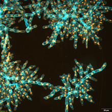
Snowflake yeast 3
6971
Multicellular yeast called snowflake yeast that researchers created through many generations of directed evolution from unicellular yeast. William Ratcliff, Georgia Institute of Technology. View Media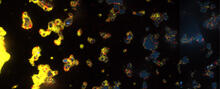
V. Cholerae Biofilm
3580
Industrious V. cholerae bacteria (yellow) tend to thrive in denser biofilms (left) while moochers (red) thrive in weaker biofilms (right). View Media
Adult Hawaiian bobtail squid burying in the sand
7012
Each morning, the nocturnal Hawaiian bobtail squid, Euprymna scolopes, hides from predators by digging into the sand. At dusk, it leaves the sand again to hunt. Margaret J. McFall-Ngai, Carnegie Institution for Science/California Institute of Technology, and Edward G. Ruby, California Institute of Technology. View Media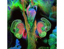
The nascent juvenile light organ of the Hawaiian bobtail squid
7017
A light organ (~0.5 mm across) of a Hawaiian bobtail squid, Euprymna scolopes, with different tissues are stained various colors. Margaret J. McFall-Ngai, Carnegie Institution for Science/California Institute of Technology, and Edward G. Ruby, California Institute of Technology. View Media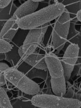
Flagellated bacterial cells
7014
Vibrio fischeri (2 mm in length) is the exclusive symbiotic partner of the Hawaiian bobtail squid, Euprymna scolopes. Margaret J. McFall-Ngai, Carnegie Institution for Science/California Institute of Technology, and Edward G. Ruby, California Institute of Technology. View Media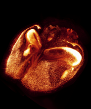
Mouse brain 2
6930
A mouse brain that was genetically modified so that subpopulations of its neurons glow. Prayag Murawala, MDI Biological Laboratory and Hannover Medical School. View Media
Mapping metabolic activity
2319
Like a map showing heavily traveled roads, this mathematical model of metabolic activity inside an E. coli cell shows the busiest pathway in white. Albert-László Barabási, University of Notre Dame View Media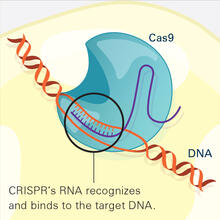
CRISPR Illustration Frame 2
6486
This illustration shows, in simplified terms, how the CRISPR-Cas9 system can be used as a gene-editing tool. National Institute of General Medical Sciences. View Media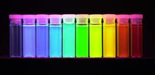
Nano-rainbow
2326
These vials may look like they're filled with colored water, but they really contain nanocrystals reflecting different colors under ultraviolet light. Shuming Nie, Emory University View Media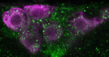
Insulin production and fat sensing in fruit flies
6982
Fourteen neurons (magenta) in the adult Drosophila brain produce insulin, and fat tissue sends packets of lipids to the brain via the lipoprotein carriers (green). Akhila Rajan, Fred Hutchinson Cancer Center View Media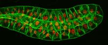
Salivary gland in the developing fruit fly
3603
For fruit flies, the salivary gland is used to secrete materials for making the pupal case, the protective enclosure in which a larva transforms into an adult fly. Richard Fehon, University of Chicago View Media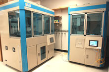
Cell-free protein synthesizers
2360
Both instruments shown were developed by CellFree Sciences of Yokohama, Japan. Center for Eukaryotic Structural Genomics View Media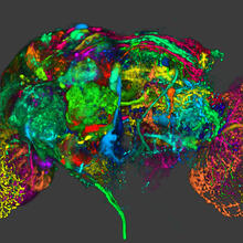
Color coding of the Drosophila brain - image
5838
This image results from a research project to visualize which regions of the adult fruit fly (Drosophila) brain derive from each neural stem cell. Yong Wan from Charles Hansen’s lab, University of Utah. Data preparation and visualization by Masayoshi Ito in the lab of Kei Ito, University of Tokyo. View Media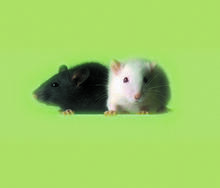
Lab mice
1069
Many researchers use the mouse (Mus musculus) as a model organism to study mammalian biology. Bill Branson, National Institutes of Health View Media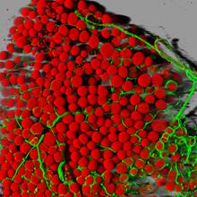
Fat cells (red) and blood vessels (green)
3600
A mouse's fat cells (red) are shown surrounded by a network of blood vessels (green). Daniela Malide, National Heart, Lung, and Blood Institute, National Institutes of Health View Media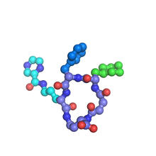
X-ray co-crystal structure of Src kinase bound to a DNA-templated macrocycle inhibitor 7
3419
X-ray co-crystal structure of Src kinase bound to a DNA-templated macrocycle inhibitor. Markus A. Seeliger, Stony Brook University Medical School and David R. Liu, Harvard University View Media
Cell-like compartments emerging from scrambled frog eggs 2
6588
Cell-like compartments spontaneously emerge from scrambled frog eggs, with nuclei (blue) from frog sperm. Endoplasmic reticulum (red) and microtubules (green) are also visible. Xianrui Cheng, Stanford University School of Medicine. View Media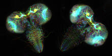
Fruit fly larvae brains showing tubulin
6808
Two fruit fly (Drosophila melanogaster) larvae brains with neurons expressing fluorescently tagged tubulin protein. Vladimir I. Gelfand, Feinberg School of Medicine, Northwestern University. View Media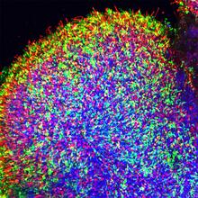
Human retinal organoid
6748
A replica of a human retina grown from stem cells. Kevin Eliceiri, University of Wisconsin-Madison. View Media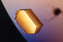
Pig trypsin (2)
2413
A crystal of porcine trypsin protein created for X-ray crystallography, which can reveal detailed, three-dimensional protein structures. Alex McPherson, University of California, Irvine View Media