Switch to Gallery View
Image and Video Gallery
This is a searchable collection of scientific photos, illustrations, and videos. The images and videos in this gallery are licensed under Creative Commons Attribution Non-Commercial ShareAlike 3.0. This license lets you remix, tweak, and build upon this work non-commercially, as long as you credit and license your new creations under identical terms.
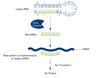
Staphylococcus aureus aggregating upon contact with synovial fluid
6805
Staphylococcus aureus bacteria (green) grouping together upon contact with synovial fluid—a viscous substance found in joints. Paul Stoodley, The Ohio State University. View Media
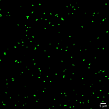
Fruit fly egg ooplasmic streaming
6809
Two fruit fly (Drosophila melanogaster) egg cells, one on each side of the central black line. Vladimir I. Gelfand, Feinberg School of Medicine, Northwestern University. View Media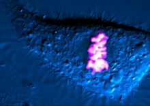
Dividing cell
6965
As this cell was undergoing cell division, it was imaged with two microscopy techniques: differential interference contrast (DIC) and confocal. The DIC view appears in blue and shows the entire cell. Dylan T. Burnette, Vanderbilt University School of Medicine. View Media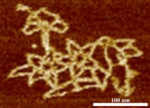
Microscopy image of bird-and-flower DNA origami
3690
An atomic force microscopy image shows DNA folded into an intricate, computer-designed structure. Hao Yan, Arizona State University View Media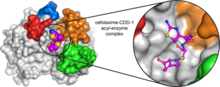
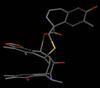
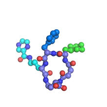
Space-filling model of a cefotaxime-CCD-1 complex
6767
CCD-1 is an enzyme produced by the bacterium Clostridioides difficile that helps it resist antibiotics. Keith Hodgson, Stanford University. View Media
Cytonemes in developing fruit fly cells
3574
Scientists have long known that multicellular organisms use biological molecules produced by one cell and sensed by another to transmit messages that, for instance, guide proper development of organs Sougata Roy, University of California, San Francisco View Media
Single-Molecule Imaging
3339
This is a super-resolution light microscope image taken by Hiro Hakozaki and Masa Hoshijima of NCMIR. Tom Deerinck, NCMIR View Media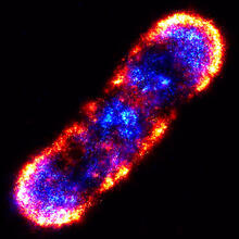
Self-organizing proteins
2771
Under the microscope, an E. coli cell lights up like a fireball. Each bright dot marks a surface protein that tells the bacteria to move toward or away from nearby food and toxins. View Media
Cell-like compartments from frog eggs 6
6593
Cell-like compartments that spontaneously emerged from scrambled frog eggs, with nuclei (blue) from frog sperm. Endoplasmic reticulum (red) and microtubules (green) are also visible. Xianrui Cheng, Stanford University School of Medicine. View Media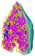
Soft X-ray tomography of a pancreatic beta cell
6605
A color-coded, 3D model of a rat pancreatic β cell. This type of cell produces insulin, a hormone that helps regulate blood sugar. Carolyn Larabell, University of California, San Francisco. View Media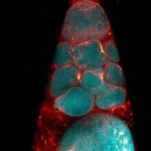
Fruit fly nurse cells during egg development
6753
In many animals, the egg cell develops alongside sister cells. Adam C. Martin, Massachusetts Institute of Technology. View Media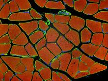
Human skeletal muscle
3677
Cross section of human skeletal muscle. Image taken with a confocal fluorescent light microscope. Tom Deerinck, National Center for Microscopy and Imaging Research (NCMIR) View Media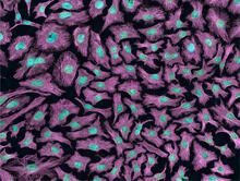
HeLa cells
3520
Multiphoton fluorescence image of HeLa cells with cytoskeletal microtubules (magenta) and DNA (cyan). Nikon RTS2000MP custom laser scanning microscope. National Center for Microscopy and Imaging Research (NCMIR) View Media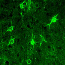
Confocal microscopy of perineuronal nets in the brain 1
3741
The photo shows a confocal microscopy image of perineuronal nets (PNNs), which are specialized extracellular matrix (ECM) structures in the brain. Tom Deerinck, National Center for Microscopy and Imaging Research (NCMIR) View Media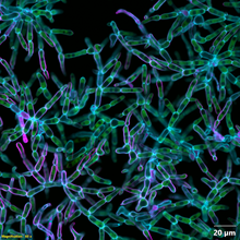
Snowflake yeast 2
6970
Multicellular yeast called snowflake yeast that researchers created through many generations of directed evolution from unicellular yeast. William Ratcliff, Georgia Institute of Technology. View Media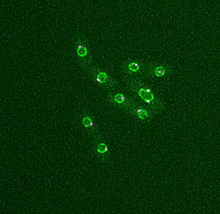
Dividing yeast cells with nuclear envelopes and spindle pole bodies
6795
Time-lapse video of yeast cells undergoing cell division. Nuclear envelopes are shown in green, and spindle pole bodies, which help pull apart copied genetic information, are shown in magenta. Alaina Willet, Kathy Gould’s lab, Vanderbilt University. View Media
Bence Jones protein MLE
2399
A crystal of Bence Jones protein created for X-ray crystallography, which can reveal detailed, three-dimensional protein structures. Alex McPherson, University of California, Irvine View Media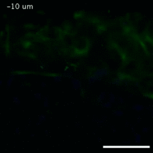
Vimentin in a quail embryo
2809
Video of high-resolution confocal images depicting vimentin immunofluorescence (green) and nuclei (blue) at the edge of a quail embryo yolk. Andrés Garcia, Georgia Tech View Media
Rabbit GPDA
2405
A crystal of rabbit GPDA protein created for X-ray crystallography, which can reveal detailed, three-dimensional protein structures. Alex McPherson, University of California, Irvine View Media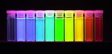
Nano-rainbow
2326
These vials may look like they're filled with colored water, but they really contain nanocrystals reflecting different colors under ultraviolet light. Shuming Nie, Emory University View Media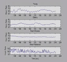
Heart rates time series image
3596
These time series show the heart rates of four different individuals. Madalena Costa and Ary Goldberger, Beth Israel Deaconess Medical Center View Media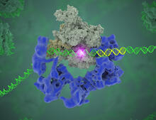
TFIID complex binds DNA to start gene transcription
3766
Gene transcription is a process by which the genetic information encoded in DNA is transcribed into RNA. Eva Nogales, Berkeley Lab View Media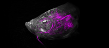
Zebrafish head vasculature video
6933
Various views of a zebrafish head with blood vessels shown in purple. Prayag Murawala, MDI Biological Laboratory and Hannover Medical School. View Media
X-ray co-crystal structure of Src kinase bound to a DNA-templated macrocycle inhibitor 7
3419
X-ray co-crystal structure of Src kinase bound to a DNA-templated macrocycle inhibitor. Markus A. Seeliger, Stony Brook University Medical School and David R. Liu, Harvard University View Media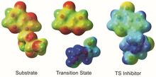
Enzyme transition states
3429
The molecule on the left is an electrostatic potential map of the van der Waals surface of the transition state for human purine nucleoside phosphorylase. Vern Schramm, Albert Einstein College of Medicine of Yeshiva University View Media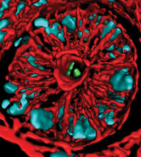
Mouse colon with gut bacteria
3566
A section of mouse colon with gut bacteria (center, in green) residing within a protective pocket. Sarkis K. Mazmanian, California Institute of Technology View Media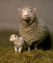
Dolly the sheep
2690
Scientists in Scotland were the first to clone an animal, this sheep named Dolly. She later gave birth to Bonnie, the lamb next to her. View MediaTracking embryonic zebrafish cells
6775
To better understand cell movements in developing embryos, researchers isolated cells from early zebrafish embryos and grew them as clusters. Liliana Solnica-Krezel, Washington University School of Medicine in St. Louis. View Media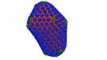
Atomic-level structure of the HIV capsid
6601
This animation shows atoms of the HIV capsid, the shell that encloses the virus's genetic material. Juan R. Perilla and the Theoretical and Computational Biophysics Group, University of Illinois at Urbana-Champaign View Media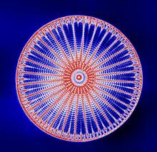
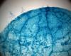
Arachnoidiscus diatom
6902
An Arachnoidiscus diatom with a diameter of 190µm. Michael Shribak, Marine Biological Laboratory/University of Chicago. View Media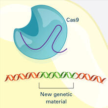
CRISPR Illustration Frame 4
6488
This illustration shows, in simplified terms, how the CRISPR-Cas9 system can be used as a gene-editing tool. National Institute of General Medical Sciences. View Media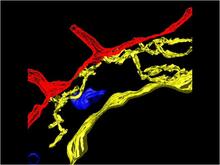
Computer model of cell membrane
2636
A computer model of the cell membrane, where the plasma membrane is red, endoplasmic reticulum is yellow, and mitochondria are blue. Bridget Wilson, University of New Mexico View Media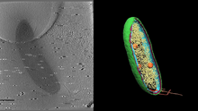
Cryo-electron tomography of a Caulobacter bacterium
6569
3D image of Caulobacter bacterium with various components highlighted: cell membranes (red and blue), protein shell (green), protein factories known as ribosomes (yellow), and storage granules Peter Dahlberg, Stanford University. View Media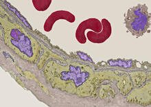
Transmission electron microscopy of coronary artery wall with elastin-rich ECM pseudocolored in light brown
3738
Elastin is a fibrous protein in the extracellular matrix (ECM). It is abundant in artery walls like the one shown here. As its name indicates, elastin confers elasticity. Tom Deerinck, National Center for Microscopy and Imaging Research (NCMIR) View Media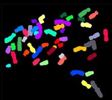
Color-coded chromosomes
2312
By mixing fluorescent dyes like an artist mixes paints, scientists are able to color code individual chromosomes. Anna Jauch, Institute of Human Genetics, Heidelberg, Germany View Media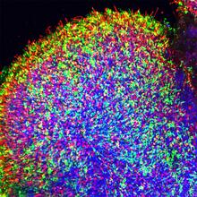
Human retinal organoid
6748
A replica of a human retina grown from stem cells. Kevin Eliceiri, University of Wisconsin-Madison. View Media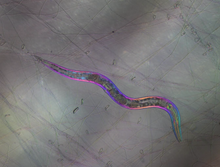
C. elegans trapped by carnivorous fungus
6963
Real-time footage of Caenorhabditis elegans, a tiny roundworm, trapped by a carnivorous fungus, Arthrobotrys dactyloides. Michael Shribak, Marine Biological Laboratory/University of Chicago. View Media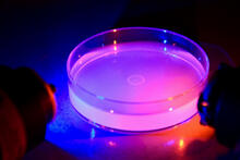
Petri dish containing C. elegans
6751
This Petri dish contains microscopic roundworms called Caenorhabditis elegans. Researchers used these particular worms to study how C. H. Robert Horvitz and Dipon Ghosh, Massachusetts Institute of Technology. View Media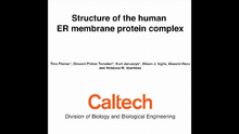
Human endoplasmic reticulum membrane protein complex
6777
A 3D model of the human endoplasmic reticulum membrane protein complex (EMC) that identifies its nine essential subunits. Rebecca Voorhees, California Institute of Technology. View Media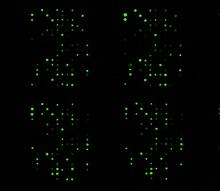
Glycan arrays
1265
The signal is obtained by allowing proteins in human serum to interact with glycan (polysaccharide) arrays. The arrays are shown in replicate so the pattern is clear. Ola Blixt, Scripps Research Institute View Media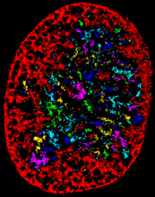
Chromatin in human fibroblast
6887
The nucleus of a human fibroblast cell with chromatin—a substance made up of DNA and proteins—shown in various colors. Melike Lakadamyali, Perelman School of Medicine at the University of Pennsylvania. View Media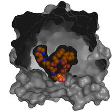
X-ray co-crystal structure of Src kinase bound to a DNA-templated macrocycle inhibitor 5
3417
X-ray co-crystal structure of Src kinase bound to a DNA-templated macrocycle inhibitor. Markus A. Seeliger, Stony Brook University Medical School and David R. Liu, Harvard University View Media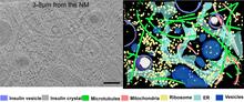
Cryo-ET cross-section of a rat pancreas cell
6608
On the left, a cross-section slice of a rat pancreas cell captured using cryo-electron tomography (cryo-ET). On the right, a 3D, color-coded version of the image highlighting cell structures. Xianjun Zhang, University of Southern California. View Media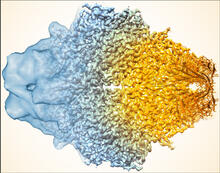
Beta-galactosidase montage showing cryo-EM improvement--gradient background
5883
Composite image of beta-galactosidase showing how cryo-EM’s resolution has improved dramatically in recent years. Older images to the left, more recent to the right. Veronica Falconieri, Sriram Subramaniam Lab, National Cancer Institute View Media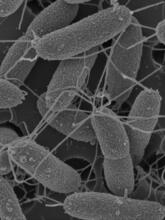
Flagellated bacterial cells
7014
Vibrio fischeri (2 mm in length) is the exclusive symbiotic partner of the Hawaiian bobtail squid, Euprymna scolopes. Margaret J. McFall-Ngai, Carnegie Institution for Science/California Institute of Technology, and Edward G. Ruby, California Institute of Technology. View Media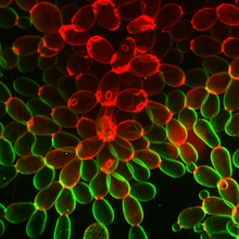
Snowflake yeast 1
6969
Multicellular yeast called snowflake yeast that researchers created through many generations of directed evolution from unicellular yeast. William Ratcliff, Georgia Institute of Technology. View Media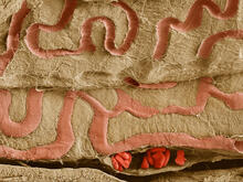
Scanning electron microscopy of the ECM on the surface of a calf muscle
3739
This image shows the extracellular matrix (ECM) on the surface of a soleus (lower calf) muscle in light brown and blood vessels in pink. Tom Deerinck, National Center for Microscopy and Imaging Research (NCMIR) View Media
Culex quinquefasciatus mosquito larvae
6771
Mosquito larvae with genes edited by CRISPR swimming in water. Valentino Gantz, University of California, San Diego. View Media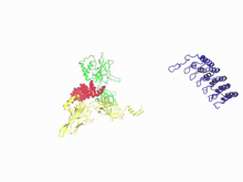
A molecular switch strips transcription factor from DNA
3729
In this video, Rice University scientists used molecular modeling with a mathematical algorithm called AWSEM (for associative memory, water-mediated, structure and energy model) and structural data to Davit Potoyan and Peter Wolynes View Media