Switch to Gallery View
Image and Video Gallery
This is a searchable collection of scientific photos, illustrations, and videos. The images and videos in this gallery are licensed under Creative Commons Attribution Non-Commercial ShareAlike 3.0. This license lets you remix, tweak, and build upon this work non-commercially, as long as you credit and license your new creations under identical terms.
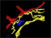
Endoplasmic reticulum abnormalities 2
6774
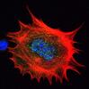
Human cells with the gene that codes for the protein FIT2 deleted. After an experimental intervention, they are expressing a nonfunctional version of FIT2, shown in green. Michel Becuwe, Harvard University. View Media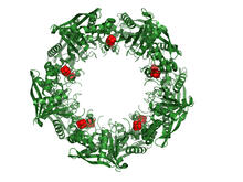
Cas4 nuclease protein structure
3720
This wreath represents the molecular structure of a protein, Cas4, which is part of a system, known as CRISPR, that bacteria use to protect themselves against viral invaders. Fred Dyda, NIDDK View Media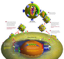
Life of an AIDS virus (with labels and stages)
2515
HIV is a retrovirus, a type of virus that carries its genetic material not as DNA but as RNA. Crabtree + Company View Media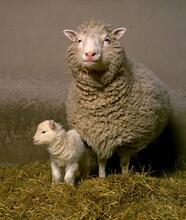
Dolly the sheep
2690
Scientists in Scotland were the first to clone an animal, this sheep named Dolly. She later gave birth to Bonnie, the lamb next to her. View Media
Cross section of a Drosophila melanogaster pupa lacking Draper
2759
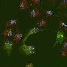
In the absence of the engulfment receptor Draper, salivary gland cells (light blue) persist in the thorax of a developing Drosophila melanogaster pupa. Christina McPhee and Eric Baehrecke, University of Massachusetts Medical School View Media
Introduction to Genome Editing Using CRISPR/Cas9
5815
Genome editing using CRISPR/Cas9 is a rapidly expanding field of scientific research with emerging applications in disease treatment, medical therapeutics and bioenergy, just to name a few. Janet Iwasa View Media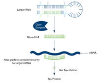
Dicer generates microRNAs (with labels)
2557
The enzyme Dicer generates microRNAs by chopping larger RNA molecules into tiny Velcro®-like pieces. MicroRNAs stick to mRNA molecules and prevent the mRNAs from being made into proteins. Crabtree + Company View Media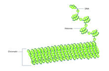
Histones in chromatin (with labels)
2561
Histone proteins loop together with double-stranded DNA to form a structure that resembles beads on a string. Crabtree + Company View Media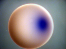
Xenopus laevis egg
2753
Xenopus laevis, the African clawed frog, has long been used as a model organism for studying embryonic development. Michael Klymkowsky, University of Colorado, Boulder View Media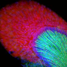
Developing zebrafish fin
3598
Originally from the waters of India, Nepal, and neighboring countries, zebrafish can now be found swimming in science labs (and home aquariums) throughout the world. Jessica Plavicki View Media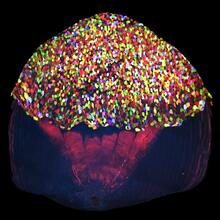
A multicolored fish scale 2
3783
Each of the tiny colored specs in this image is a cell on the surface of a fish scale. Chen-Hui Chen and Kenneth Poss, Duke University View Media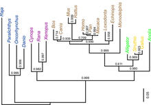
Dinosaur evolutionary tree
2474
Analysis of 68 million-year-old collagen molecule fragments preserved in a T. Chris Organ, Harvard University View Media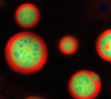
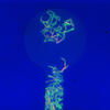
Nucleolus subcompartments spontaneously self-assemble 4
3793
What looks a little like distant planets with some mysterious surface features are actually assemblies of proteins normally found in the cell's nucleolus, a small but very important protein complex lo Nilesh Vaidya, Princeton University View Media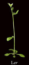
Mature, flowering Arabidopsis
2779
This is an adult flowering Arabidopsis thaliana plant with the inbred designation L-er. Arabidopsis is the most widely used model organism for researchers who study plant genetics. Jeff Dangl, University of North Carolina, Chapel Hill View Media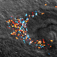
Planarian stem cell colony
3306
Planarians are freshwater flatworms that have powerful abilities to regenerate their bodies, which would seem to make them natural model organisms in which to study stem cells. Peter Reddien, Whitehead Institute View Media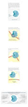
CRISPR illustration
3719
This illustration shows, in simplified terms, how the CRISPR-Cas9 system can be used as a gene-editing tool. National Institute of General Medical Sciences. View Media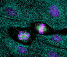
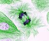
Highlighted cells
2429
The cytoskeleton (green) and DNA (purple) are highlighed in these cells by immunofluorescence. Torsten Wittmann, Scripps Research Institute View Media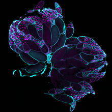
Fruit fly ovaries
6807
Fruit fly (Drosophila melanogaster) ovaries with DNA shown in magenta and actin filaments shown in light blue. This image was captured using a confocal laser scanning microscope.Vladimir I. Gelfand, Feinberg School of Medicine, Northwestern University. View Media
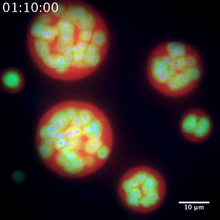
Nucleolus subcompartments spontaneously self-assemble 2
3791
The nucleolus is a small but very important protein complex located in the cell's nucleus. Nilesh Vaidya, Princeton University View Media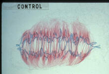
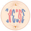
Lily mitosis 09
1022
A light microscope image of a cell from the endosperm of an African globe lily (Scadoxus katherinae). This is one frame of a time-lapse sequence that shows cell division in action. Andrew S. Bajer, University of Oregon, Eugene View Media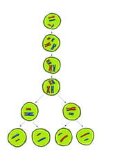
Meiosis illustration
2545
Meiosis is the process whereby a cell reduces its chromosomes from diploid to haploid in creating eggs or sperm. Crabtree + Company View MediaAssembly of the HIV capsid
5729
The HIV capsid is a pear-shaped structure that is made of proteins the virus needs to mature and become infective. John Grime and Gregory Voth, The University of Chicago View MediaCentral dogma, illustrated (with labels)
2548
DNA encodes RNA, which encodes protein. DNA is transcribed to make messenger RNA (mRNA). The mRNA sequence (dark red strand) is complementary to the DNA sequence (blue strand). Crabtree + Company View Media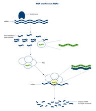
RNA interference (with labels)
2559
RNA interference or RNAi is a gene-silencing process in which double-stranded RNAs trigger the destruction of specific RNAs. Crabtree + Company View Media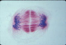
Lily mitosis 12
1018
A light microscope image of a cell from the endosperm of an African globe lily (Scadoxus katherinae). This is one frame of a time-lapse sequence that shows cell division in action. Andrew S. Bajer, University of Oregon, Eugene View MediaCentral dogma, illustrated (with labels and numbers for stages)
2549
DNA encodes RNA, which encodes protein. DNA is transcribed to make messenger RNA (mRNA). The mRNA sequence (dark red strand) is complementary to the DNA sequence (blue strand). Crabtree + Company View Media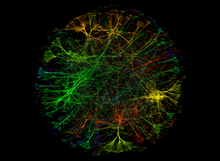
A molecular interaction network in yeast 3
3733
The image visualizes a part of the yeast molecular interaction network. Keiichiro Ono, UCSD View Media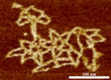
Microscopy image of bird-and-flower DNA origami
3690
An atomic force microscopy image shows DNA folded into an intricate, computer-designed structure. Hao Yan, Arizona State University View Media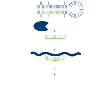
Dicer generates microRNAs
2556
The enzyme Dicer generates microRNAs by chopping larger RNA molecules into tiny Velcro®-like pieces. MicroRNAs stick to mRNA molecules and prevent the mRNAs from being made into proteins. Crabtree + Company View Media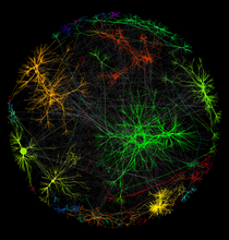
A molecular interaction network in yeast 1
3730
The image visualizes a part of the yeast molecular interaction network. Keiichiro Ono, UCSD View Media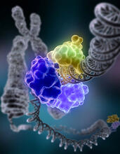
Repairing DNA
2330
Like a watch wrapped around a wrist, a special enzyme encircles the double helix to repair a broken strand of DNA. Tom Ellenberger, Washington University School of Medicine View Media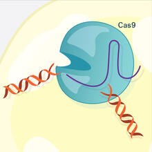
CRISPR Illustration Frame 3
6487
This illustration shows, in simplified terms, how the CRISPR-Cas9 system can be used as a gene-editing tool. National Institute of General Medical Sciences. View Media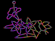
Computer sketch of bird-and-flower DNA origami
3689
A computer-generated sketch of a DNA origami folded into a flower-and-bird structure. See also related image 3690. Hao Yan, Arizona State University View Media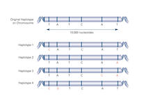
Haplotypes (with labels)
2567
Haplotypes are combinations of gene variants that are likely to be inherited together within the same chromosomal region. Crabtree + Company View Media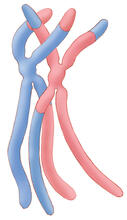
Chromosomes after crossing over
1314
Duplicated pair of chromosomes have exchanged material. Judith Stoffer View Media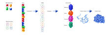
From DNA to Protein (labeled)
2510
The genetic code in DNA is transcribed into RNA, which is translated into proteins with specific sequences. Crabtree + Company View MediaCentral dogma, illustrated
2547
DNA encodes RNA, which encodes protein. DNA is transcribed to make messenger RNA (mRNA). The mRNA sequence (dark red strand) is complementary to the DNA sequence (blue strand). Crabtree + Company View Media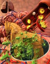
Host infection stimulates antibiotic resistance
5764
This illustration shows pathogenic bacteria behave like a Trojan horse: switching from antibiotic susceptibility to resistance during infection. View Media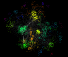
A molecular interaction network in yeast 2
3732
The image visualizes a part of the yeast molecular interaction network. Keiichiro Ono, UCSD View Media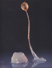
Dicty fruit
2684
Dictyostelium discoideum is a microscopic amoeba. A group of 100,000 form a mound as big as a grain of sand. Featured in The New Genetics. View Media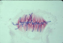
Lily mitosis 07
1017
A light microscope image of a cell from the endosperm of an African globe lily (Scadoxus katherinae). This is one frame of a time-lapse sequence that shows cell division in action. Andrew S. Bajer, University of Oregon, Eugene View Media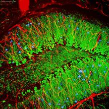
Brain showing hallmarks of Alzheimer's disease
3604
Along with blood vessels (red) and nerve cells (green), this mouse brain shows abnormal protein clumps known as plaques (blue). Alvin Gogineni, Genentech View Media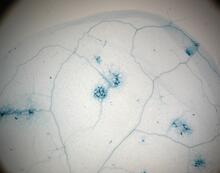
Disease-resistant Arabidopsis leaf
2781
This is a magnified view of an Arabidopsis thaliana leaf a few days after being exposed to the pathogen Hyaloperonospora arabidopsidis. Jeff Dangl, University of North Carolina, Chapel Hill View Media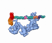
Dynamic cryo-EM model of the human transcription preinitiation complex
5730
Gene transcription is a process by which information encoded in DNA is transcribed into RNA. Eva Nogales, Berkeley Lab View Media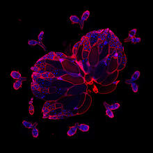
Wild-type and mutant fruit fly ovaries
6806
The two large, central, round shapes are ovaries from a typical fruit fly (Drosophila melanogaster). Vladimir I. Gelfand, Feinberg School of Medicine, Northwestern University. View Media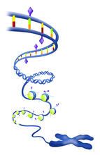
Epigenetic code
2562
The "epigenetic code" controls gene activity with chemical tags that mark DNA (purple diamonds) and the "tails" of histone proteins (purple triangles). Crabtree + Company View Media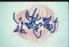
Lily mitosis 06
1016
A light microscope image of a cell from the endosperm of an African globe lily (Scadoxus katherinae). This is one frame of a time-lapse sequence that shows cell division in action. Andrew S. Bajer, University of Oregon, Eugene View Media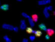
Painted chromosomes
2764
Like a paint-by-numbers picture, painted probes tint individual human chromosomes by targeting specific DNA sequences. Beth A. Sullivan, Duke University View Media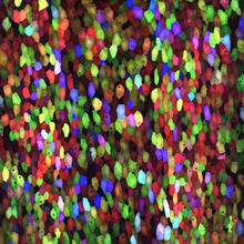
A multicolored fish scale 1
3782
Each of the colored specs in this image is a cell on the surface of a fish scale. Chen-Hui Chen and Kenneth Poss, Duke University View Media
Fluorescence in situ hybridization (FISH) in mouse ES cells shows DNA interactions
3296
Researchers used fluorescence in situ hybridization (FISH) to confirm the presence of long range DNA-DNA interactions in mouse embryonic stem cells. Kathrin Plath, University of California, Los Angeles View Media