Switch to Gallery View
Image and Video Gallery
This is a searchable collection of scientific photos, illustrations, and videos. The images and videos in this gallery are licensed under Creative Commons Attribution Non-Commercial ShareAlike 3.0. This license lets you remix, tweak, and build upon this work non-commercially, as long as you credit and license your new creations under identical terms.
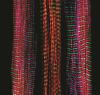
Tracking embryonic zebrafish cells
6775
To better understand cell movements in developing embryos, researchers isolated cells from early zebrafish embryos and grew them as clusters. Liliana Solnica-Krezel, Washington University School of Medicine in St. Louis. View Media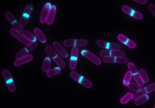
Yeast cells with accumulated cell wall material
6797
Yeast cells that abnormally accumulate cell wall material (blue) at their ends and, when preparing to divide, in their middles. This image was captured using wide-field microscopy with deconvolution. Alaina Willet, Kathy Gould’s lab, Vanderbilt University. View Media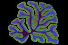
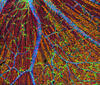
Cerebellum: the brain's locomotion control center
3639
The cerebellum of a mouse is shown here in cross-section. The cerebellum is the brain's locomotion control center. Thomas Deerinck, National Center for Microscopy and Imaging Research, University of California, San Diego View Media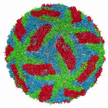
Cryo-electron microscopy of the dengue virus showing protective membrane and membrane proteins
3748
Dengue virus is a mosquito-borne illness that infects millions of people in the tropics and subtropics each year. Like many viruses, dengue is enclosed by a protective membrane. Hong Zhou, UCLA View Media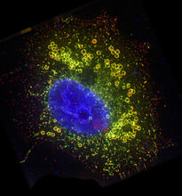
Cell Nucleus and Lipid Droplets
6547
A cell nucleus (blue) surrounded by lipid droplets (yellow). James Olzmann, University of California, Berkeley View Media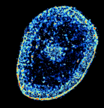
Chromatin in human tenocyte
6893
The nucleus of a degenerating human tendon cell, also known as a tenocyte. It has been color-coded based on the density of chromatin—a substance made up of DNA and proteins. Melike Lakadamyali, Perelman School of Medicine at the University of Pennsylvania. View Media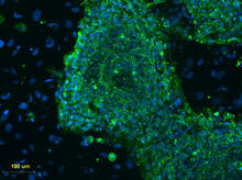
Human embryonic stem cells on feeder cells
3274
This fluorescent microscope image shows human embryonic stem cells whose nuclei are stained green. Blue staining shows the surrounding supportive feeder cells. Michael Longaker lab, Stanford University School of Medicine, via CIRM View Media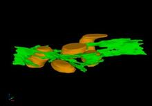
Mitochondria and endoplasmic reticulum
2635
A computer model shows how the endoplasmic reticulum is close to and almost wraps around mitochondria in the cell. The endoplasmic reticulum is lime green and the mitochondria are yellow. Bridget Wilson, University of New Mexico View Media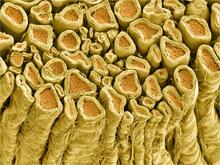
Myelinated axons 1
3396
Myelinated axons in a rat spinal root. Tom Deerinck, National Center for Microscopy and Imaging Research (NCMIR) View Media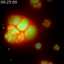
Nucleolus subcompartments spontaneously self-assemble 1
3789
The nucleolus is a small but very important protein complex located in the cell's nucleus. Nilesh Vaidya, Princeton University View Media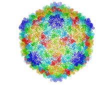
Bacteriophage P22 capsid
5874
Cryo-electron microscopy (cryo-EM) has the power to capture details of proteins and other small biological structures at the molecular level. This image shows proteins in the capsid, or outer co Dr. Wah Chiu, Baylor College of Medicine View Media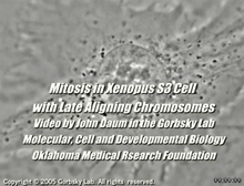
Cell division with late aligning chromosomes
2747
This video shows an instance of abnormal mitosis where chromosomes are late to align. Gary Gorbsky, Oklahoma Medical Research Foundation View Media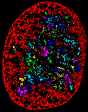
Chromatin in human fibroblast
6887
The nucleus of a human fibroblast cell with chromatin—a substance made up of DNA and proteins—shown in various colors. Melike Lakadamyali, Perelman School of Medicine at the University of Pennsylvania. View Media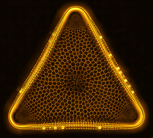
Trigonium diatom
6962
A Trigonium diatom imaged by a quantitative orientation-independent differential interference contrast (OI-DIC) microscope. Michael Shribak, Marine Biological Laboratory/University of Chicago. View Media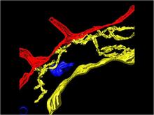
Computer model of cell membrane
2636
A computer model of the cell membrane, where the plasma membrane is red, endoplasmic reticulum is yellow, and mitochondria are blue. Bridget Wilson, University of New Mexico View Media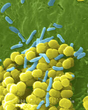
Bacteria shapes
1158
A colorized scanning electron micrograph of bacteria. Scanning electron microscopes allow scientists to see the three-dimensional surface of their samples. Tina Weatherby Carvalho, University of Hawaii at Manoa View Media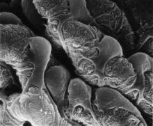
Podocytes from a chronically diseased kidney
3565
This scanning electron microscope (SEM) image shows podocytes--cells in the kidney that play a vital role in filtering waste from the bloodstream--from a patient with chronic kidney disease. Olga Troyanskaya, Princeton University and Matthias Kretzler, University of Michigan View Media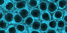
Movements of myosin
2324
Inside the fertilized egg cell of a fruit fly, we see a type of myosin (related to the protein that helps muscles contract) made to glow by attaching a fluorescent protein. Victoria Foe, University of Washington View Media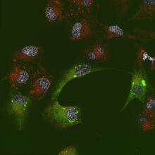
Endoplasmic reticulum abnormalities 2
6774
Human cells with the gene that codes for the protein FIT2 deleted. After an experimental intervention, they are expressing a nonfunctional version of FIT2, shown in green. Michel Becuwe, Harvard University. View Media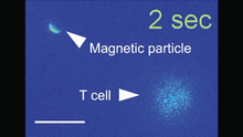
Magnetic Janus particle activating a T cell
6800
A Janus particle being used to activate a T cell, a type of immune cell. Yan Yu, Indiana University, Bloomington. View Media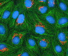
HeLa cells
3522
Multiphoton fluorescence image of cultured HeLa cells with a fluorescent protein targeted to the Golgi apparatus (orange), microtubules (green) and counterstained for DNA (cyan). National Center for Microscopy and Imaging Research (NCMIR) View Media
Dolly the sheep
2690
Scientists in Scotland were the first to clone an animal, this sheep named Dolly. She later gave birth to Bonnie, the lamb next to her. View Media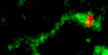
Shiga toxin being sorted inside a cell
3488
Shiga toxin (green) is sorted from the endosome into membrane tubules (red), which then pinch off and move to the Golgi apparatus. Somshuvra Mukhopadhyay, The University of Texas at Austin, and Adam D. Linstedt, Carnegie Mellon University View Media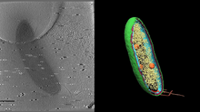
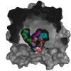
Cryo-electron tomography of a Caulobacter bacterium
6569
3D image of Caulobacter bacterium with various components highlighted: cell membranes (red and blue), protein shell (green), protein factories known as ribosomes (yellow), and storage granules Peter Dahlberg, Stanford University. View Media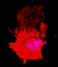
Polarized cells- 01
3332
Cells move forward with lamellipodia and filopodia supported by networks and bundles of actin filaments. Proper, controlled cell movement is a complex process. Rong Li and Praveen Suraneni, Stowers Institute for Medical Research View Media
Brains of sleep-deprived and well-rested fruit flies
3490
On top, the brain of a sleep-deprived fly glows orange because of Bruchpilot, a communication protein between brain cells. These bright orange brain areas are associated with learning. Chiara Cirelli, University of Wisconsin-Madison View Media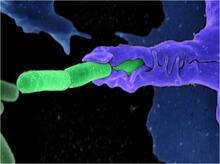
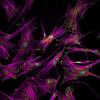
Anthrax bacteria (green) being swallowed by an immune system cell
3612
Multiple anthrax bacteria (green) being enveloped by an immune system cell (purple). Anthrax bacteria live in soil and form dormant spores that can survive for decades. Camenzind G. Robinson, Sarah Guilman, and Arthur Friedlander, United States Army Medical Research Institute of Infectious Diseases View Media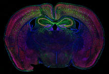
Calling Cards in a mouse brain
6780
The green spots in this mouse brain are cells labeled with Calling Cards, a technology that records molecular events in brain cells as they mature. Allen Yen, Lab of Joseph Dougherty, Washington University School of Medicine in St. Louis. View Media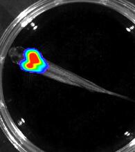
Bioluminescent imaging in adult zebrafish - overhead view
3557
Luciferase-based imaging enables visualization and quantification of internal organs and transplanted cells in live adult zebrafish. Kenneth Poss, Duke University View Media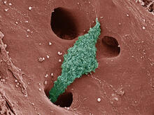
Kupffer cell residing in the liver
6535
Kupffer cells appear in the liver during the early stages of mammalian development and stay put throughout life to protect liver cells, clean up old red blood cells, and regulate iron levels. Thomas Deerinck, National Center for Microscopy and Imaging Research, University of California, San Diego. View Media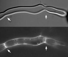
Z rings in bacterial division
2456
Lab-made liposomes contract where Z rings have gathered together and the constriction forces are greatest (arrows). Masaki Osawa, Duke University View Media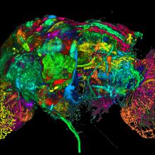
Color coding of the Drosophila brain - black background
5868
This image results from a research project to visualize which regions of the adult fruit fly (Drosophila) brain derive from each neural stem cell. Yong Wan from Charles Hansen’s lab, University of Utah. Data preparation and visualization by Masayoshi Ito in the lab of Kei Ito, University of Tokyo. View Media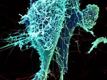
String-like Ebola virus peeling off an infected cell
3619
After multiplying inside a host cell, the stringlike Ebola virus is emerging to infect more cells. Heinz Feldmann, Peter Jahrling, Elizabeth Fischer and Anita Mora, National Institute of Allergy and Infectious Diseases, National Institutes of Health View Media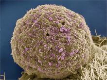
HIV Infected Cell
3386
The human immunodeficiency virus (HIV), shown here as tiny purple spheres, causes the disease known as AIDS (for acquired immunodeficiency syndrome). Tom Deerinck, National Center for Microscopy and Imaging Research (NCMIR) View Media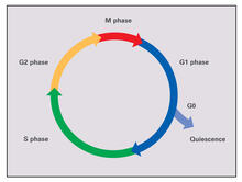
Cell cycle (with labels)
2499
Cells progress through a cycle that consists of phases for growth (G1, S, and G2) and division (M). Cells become quiescent when they exit this cycle (G0). Crabtree + Company View Media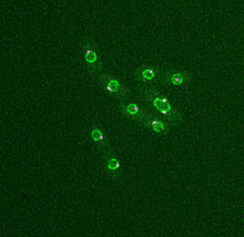
Dividing yeast cells with nuclear envelopes and spindle pole bodies
6795
Time-lapse video of yeast cells undergoing cell division. Nuclear envelopes are shown in green, and spindle pole bodies, which help pull apart copied genetic information, are shown in magenta. Alaina Willet, Kathy Gould’s lab, Vanderbilt University. View Media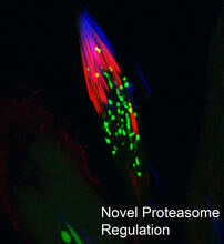
Proteasome
3451
This fruit fly spermatid recycles various molecules, including malformed or damaged proteins. Sigi Benjamin-Hong, Rockefeller University View Media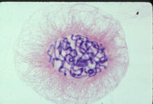
Lily mitosis 04
1014
A light microscope image of a cell from the endosperm of an African globe lily (Scadoxus katherinae). This is one frame of a time-lapse sequence that shows cell division in action. Andrew S. Bajer, University of Oregon, Eugene View MediaTranslation
1281
Ribosomes manufacture proteins based on mRNA instructions. Each ribosome reads mRNA, recruits tRNA molecules to fetch amino acids, and assembles the amino acids in the proper order. Judith Stoffer View Media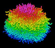
A Growing Bacterial Biofilm
5825
A growing Vibrio cholerae (cholera) biofilm. Cholera bacteria form colonies called biofilms that enable them to resist antibiotic therapy within the body and other challenges to their growth. Jing Yan, Ph.D., and Bonnie Bassler, Ph.D., Department of Molecular Biology, Princeton University, Princeton, NJ. View Media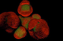
Arabidopsis Thaliana: Flowers Spring to Life
6503
This image capture shows how a single gene, STM, plays a starring role in plant development. Nathanaёl Prunet NIH Support: National Institute of General Medical Sciences View Media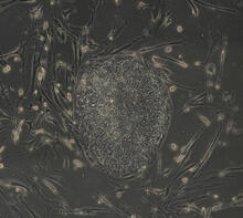
Induced stem cells from adult skin 03
2605
The human skin cells pictured contain genetic modifications that make them pluripotent, essentially equivalent to embryonic stem cells. James Thomson, University of Wisconsin-Madison View Media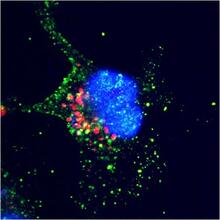
ARTS triggers apoptosis
2432
Cell showing overproduction of the ARTS protein (red). ARTS triggers apoptosis, as shown by the activation of caspase-3 (green) a key tool in the cell's destruction. The nucleus is shown in blue. Hermann Steller, Rockefeller University View Media
Aging book of life
1334
Damage to each person's genome, often called the "Book of Life," accumulates with time. Judith Stoffer View Media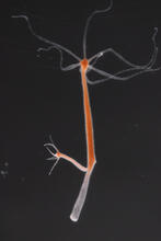
Hydra 01
2437
Hydra magnipapillata is an invertebrate animal used as a model organism to study developmental questions, for example the formation of the body axis. Hiroshi Shimizu, National Institute of Genetics in Mishima, Japan View Media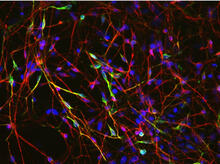
Neurons from human ES cells 02
3285
These neurons were derived from human embryonic stem cells. The neural cell bodies with axonal projections are visible in red, and the nuclei in blue. Xianmin Zeng lab, Buck Institute for Age Research, via CIRM View Media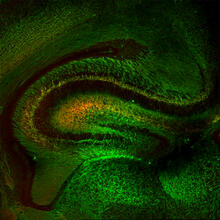
Mouse Brain Cross Section
5886
The brain sections are treated with fluorescent antibodies specific to a particular protein and visualized using serial electron microscopy (SEM). Anton Maximov, The Scripps Research Institute, La Jolla, CA View Media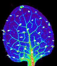
Zinc levels in a plant leaf
3727
Zinc is required for the function of more than 300 enzymes, including those that help regulate gene expression, in various organisms including humans. Suzana Car, Dartmouth College View MediaGenetic mosaicism in fruit flies
6983
Fat tissue from the abdomen of a genetically mosaic adult fruit fly. Genetic mosaicism means that the fly has cells with different genotypes even though it formed from a single zygote. Akhila Rajan, Fred Hutchinson Cancer Center View Media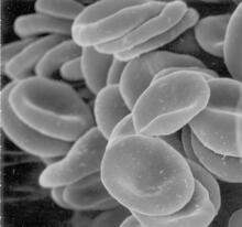
Red blood cells
1101
This image of human red blood cells was obtained with the help of a scanning electron microscope, an instrument that uses a finely focused electron beam to yield detailed images of the surface of a sa Tina Weatherby Carvalho, University of Hawaii at Manoa View Media