Switch to Gallery View
Image and Video Gallery
This is a searchable collection of scientific photos, illustrations, and videos. The images and videos in this gallery are licensed under Creative Commons Attribution Non-Commercial ShareAlike 3.0. This license lets you remix, tweak, and build upon this work non-commercially, as long as you credit and license your new creations under identical terms.
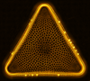
Cell-like compartments emerging from scrambled frog eggs 4
6590
Cell-like compartments that spontaneously emerged from scrambled frog eggs, with nuclei (blue) from frog sperm. Endoplasmic reticulum (red) and microtubules (green) are also visible. Xianrui Cheng, Stanford University School of Medicine. View Media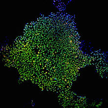
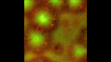
Induced stem cells from adult skin 02
2604
These cells are induced stem cells made from human adult skin cells that were genetically reprogrammed to mimic embryonic stem cells. James Thomson, University of Wisconsin-Madison View Media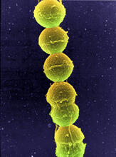
Streptococcus bacteria
1157
Image of Streptococcus, a type (genus) of spherical bacteria that can colonize the throat and back of the mouth. Stroptococci often occur in pairs or in chains, as shown here. Tina Weatherby Carvalho, University of Hawaii at Manoa View Media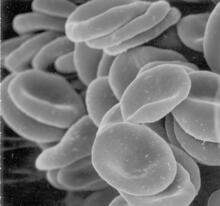
Red blood cells
1101
This image of human red blood cells was obtained with the help of a scanning electron microscope, an instrument that uses a finely focused electron beam to yield detailed images of the surface of a sa Tina Weatherby Carvalho, University of Hawaii at Manoa View Media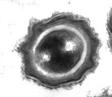
Bacterial spore
2752
A spore from the bacterium Bacillus subtilis shows four outer layers that protect the cell from harsh environmental conditions. Patrick Eichenberger, New York University View Media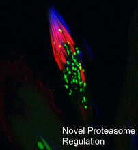
Proteasome
3451
This fruit fly spermatid recycles various molecules, including malformed or damaged proteins. Sigi Benjamin-Hong, Rockefeller University View Media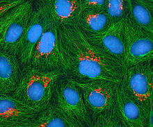
HeLa cells
3522
Multiphoton fluorescence image of cultured HeLa cells with a fluorescent protein targeted to the Golgi apparatus (orange), microtubules (green) and counterstained for DNA (cyan). National Center for Microscopy and Imaging Research (NCMIR) View Media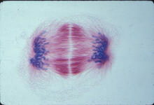
Lily mitosis 12
1018
A light microscope image of a cell from the endosperm of an African globe lily (Scadoxus katherinae). This is one frame of a time-lapse sequence that shows cell division in action. Andrew S. Bajer, University of Oregon, Eugene View Media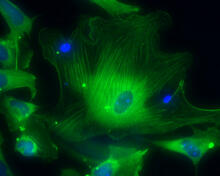
Smooth muscle from human ES cells
3288
These smooth muscle cells were derived from human embryonic stem cells. The nuclei are stained blue, and the proteins of the cytoskeleton are stained green. Alexey Terskikh lab, Burnham Institute for Medical Research, via CIRM View Media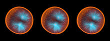
Sea urchin embryo 03
1049
Stereo triplet of a sea urchin embryo stained to reveal actin filaments (orange) and microtubules (blue). George von Dassow, University of Washington View Media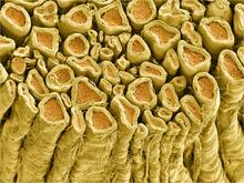
Myelinated axons 1
3396
Myelinated axons in a rat spinal root. Tom Deerinck, National Center for Microscopy and Imaging Research (NCMIR) View Media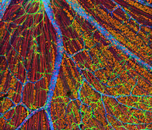
Mouse Retina
3309
A genetic disorder of the nervous system, neurofibromatosis causes tumors to form on nerves throughout the body, including a type of tumor called an optic nerve glioma that can result in childhood bli Tom Deerinck, NCMIR View Media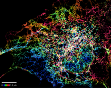
Dense tubular matrices in the peripheral endoplasmic reticulum (ER) 1
5855
Superresolution microscopy work on endoplasmic reticulum (ER) in the peripheral areas of the cell showing details of the structure and arrangement in a complex web of tubes. Jennifer Lippincott-Schwartz, Howard Hughes Medical Institute Janelia Research Campus, Virginia View Media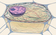
Cytoskeleton
1272
The three fibers of the cytoskeleton--microtubules in blue, intermediate filaments in red, and actin in green--play countless roles in the cell. Judith Stoffer View Media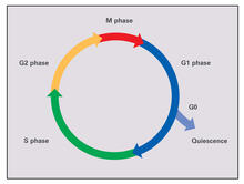
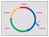
Cell cycle (with labels)
2499
Cells progress through a cycle that consists of phases for growth (G1, S, and G2) and division (M). Cells become quiescent when they exit this cycle (G0). Crabtree + Company View Media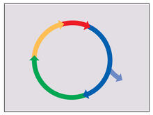
Cell cycle
2498
Cells progress through a cycle that consists of phases for growth (blue, green, yellow) and division (red). Cells become quiescent when they exit this cycle (purple). Crabtree + Company View Media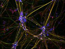
Peripheral nerve cells derived from ES cells
3263
Peripheral nerve cells made from human embryonic stem cell-derived neural crest stem cells. Stephen Dalton, University of Georgia View Media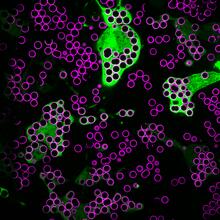
Hungry, hungry macrophages
7009
Macrophages (green) are the professional eaters of our immune system. Meghan Morrissey, University of California, Santa Barbara. View Media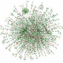
Protein map
2423
Network diagram showing a map of protein-protein interactions in a yeast (Saccharomyces cerevisiae) cell. This cluster includes 78 percent of the proteins in the yeast proteome. Hawoong Jeong, KAIST, Korea View Media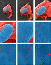
Crab larva eye
1251
Colorized scanning electron micrographs progressively zoom in on the eye of a crab larva. In the higher-resolution frames, bacteria are visible on the eye. Tina Weatherby Carvalho, University of Hawaii at Manoa View Media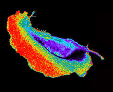
Seeing signaling protein activation in cells 02
2452
Cdc42, a member of the Rho family of small guanosine triphosphatase (GTPase) proteins, regulates multiple cell functions, including motility, proliferation, apoptosis, and cell morphology. Klaus Hahn, University of North Carolina, Chapel Hill Medical School View Media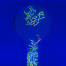
Genetically identical mycobacteria respond differently to antibiotic 1
5751
Antibiotic resistance in microbes is a serious health concern. So researchers have turned their attention to how bacteria undo the action of some antibiotics. Bree Aldridge, Tufts University View Media
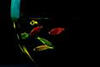
Circadian rhythm neurons in the fruit fly brain
3754
Some nerve cells (neurons) in the brain keep track of the daily cycle. This time-keeping mechanism, called the circadian clock, is found in all animals including us. Justin Blau, New York University View Media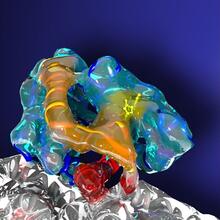
Kinesin moves cellular cargo
3491
A protein called kinesin (blue) is in charge of moving cargo around inside cells and helping them divide. Charles Sindelar, Yale University View Media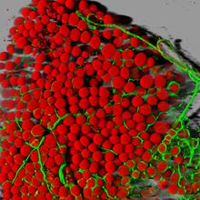
Fat cells (red) and blood vessels (green)
3600
A mouse's fat cells (red) are shown surrounded by a network of blood vessels (green). Daniela Malide, National Heart, Lung, and Blood Institute, National Institutes of Health View Media
Electrode probe on mouse Huntington's muscle cell
3479
Using an electrode, researchers apply an electrical pulse onto a piece of muscle tissue affected by Huntington's disease. Grigor Varuzhanyan and Andrew A. Voss, California State Polytechnic University View Media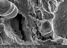
Staphylococcus aureus in the porous coating of a femoral hip stem
6804
Staphylococcus aureus bacteria (blue) on the porous coating of a femoral hip stem used in hip replacement surgery. Paul Stoodley, The Ohio State University. View Media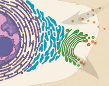
Vesicle traffic
1283
This illustration shows vesicle traffic inside a cell. Judith Stoffer View Media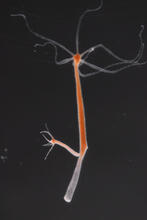
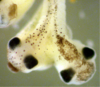
Hydra 01
2437
Hydra magnipapillata is an invertebrate animal used as a model organism to study developmental questions, for example the formation of the body axis. Hiroshi Shimizu, National Institute of Genetics in Mishima, Japan View Media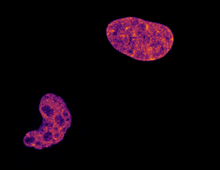
Cell division and cell death
6790
Two cells over a 2-hour period. The one on the bottom left goes through programmed cell death, also known as apoptosis. The one on the top right goes through cell division, also called mitosis. Dylan T. Burnette, Vanderbilt University School of Medicine. View Media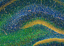
Rat Hippocampus
3308
This image of the hippocampus was taken with an ultra-widefield high-speed multiphoton laser microscope. Tom Deerinck, NCMIR View Media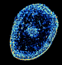
Chromatin in human tenocyte
6893
The nucleus of a degenerating human tendon cell, also known as a tenocyte. It has been color-coded based on the density of chromatin—a substance made up of DNA and proteins. Melike Lakadamyali, Perelman School of Medicine at the University of Pennsylvania. View Media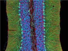
Mouse cerebellum close-up
3371
The cerebellum is the brain's locomotion control center. Every time you shoot a basketball, tie your shoe or chop an onion, your cerebellum fires into action. National Center for Microscopy and Imaging Research (NCMIR) View Media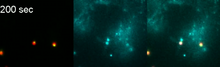
Phagosome in macrophage cell
6799
A sensor particle being engulfed by a macrophage—an immune cell—and encapsuled in a compartment called a phagosome. The phagosome then fuses with lysosomes—another type of compartment. Yan Yu, Indiana University, Bloomington. View Media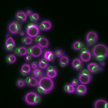
Yeast cells responding to a glucose shortage
6772
These yeast cells were exposed to a glucose (sugar) shortage. Mike Henne, University of Texas Southwestern Medical Center. View Media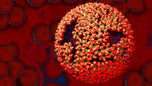
Molecular model of freshly made Rous sarcoma virus (RSV)
3771
Viruses have been the foes of animals and other organisms for time immemorial. Boon Chong Goh, University of Illinois at Urbana-Champaign View Media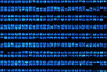
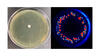
Biopixels
3266
Bioengineers were able to coax bacteria to blink in unison on microfluidic chips. This image shows a small chip with about 500 blinking bacterial colonies or biopixels. Jeff Hasty Lab, UC San Diego View Media