Switch to Gallery View
Image and Video Gallery
This is a searchable collection of scientific photos, illustrations, and videos. The images and videos in this gallery are licensed under Creative Commons Attribution Non-Commercial ShareAlike 3.0. This license lets you remix, tweak, and build upon this work non-commercially, as long as you credit and license your new creations under identical terms.
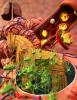
Growing hair follicle stem cells
3499
Wound healing requires the action of stem cells. Hermann Steller, Rockefeller University View Media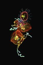
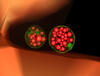
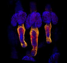
Jellyfish, viewed with ZEISS Lightsheet Z.1 microscope
3636
Jellyfish are especially good models for studying the evolution of embryonic tissue layers. Despite being primitive, jellyfish have a nervous system (stained green here) and musculature (red). Helena Parra, Pompeu Fabra University, Spain View Media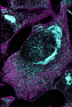
Microtubules and tau aggregates
6892
Microtubules (magenta) and tau protein (light blue) in a cell model of tauopathy. Melike Lakadamyali, Perelman School of Medicine at the University of Pennsylvania. View Media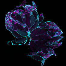
Fruit fly ovaries
6807
Fruit fly (Drosophila melanogaster) ovaries with DNA shown in magenta and actin filaments shown in light blue. This image was captured using a confocal laser scanning microscope.Vladimir I. Gelfand, Feinberg School of Medicine, Northwestern University. View Media
Focal adhesions
2502
Cells walk along body surfaces via tiny "feet," called focal adhesions, that connect with the extracellular matrix. Crabtree + Company View Media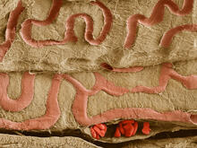
Scanning electron microscopy of the ECM on the surface of a calf muscle
3739
This image shows the extracellular matrix (ECM) on the surface of a soleus (lower calf) muscle in light brown and blood vessels in pink. Tom Deerinck, National Center for Microscopy and Imaging Research (NCMIR) View Media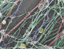
Cells use bubble-like structures called vesicles to transport cargo
3634
Cells use bubble-like structures called vesicles (yellow) to import, transport, and export cargo and in cellular communication. A single cell may be filled with thousands of moving vesicles.Tatyana Svitkina, University of Pennsylvania View Media
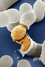
Birth of a yeast cell
3614
Yeast make bread, beer, and wine. And like us, yeast can reproduce sexually. A mother and father cell fuse and create one large cell that contains four offspring. Juergen Berger, Max Planck Institute for Developmental Biology, and Maria Langegger, Friedrich Miescher Laboratory of the Max Planck Society, Germany View Media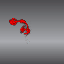
Color coding of the Drosophila brain - video
5843
This video results from a research project to visualize which regions of the adult fruit fly (Drosophila) brain derive from each neural stem cell. Yong Wan from Charles Hansen’s lab, University of Utah. Data preparation and visualization by Masayoshi Ito in the lab of Kei Ito, University of Tokyo. View Media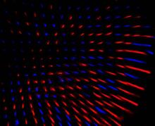
Fruit fly retina 02
2434
Section of a fruit fly retina showing the light-sensing molecules rhodopsin-5 (blue) and rhodopsin-6 (red). Hermann Steller, Rockefeller University View Media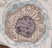
Transmission electron microscopy showing cross-section of the node of Ranvier
3740
Nodes of Ranvier are short gaps in the myelin sheath surrounding myelinated nerve cells (axons). Tom Deerinck, National Center for Microscopy and Imaging Research (NCMIR) View Media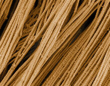
Scanning electron microscopy of collagen fibers
3735
This image shows collagen, a fibrous protein that's the main component of the extracellular matrix (ECM). Collagen is a strong, ropelike molecule that forms stretch-resistant fibers. Tom Deerinck, National Center for Microscopy and Imaging Research (NCMIR) View Media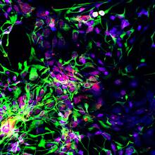
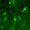
Motor neuron progenitors derived from human ES cells
3280
Motor neuron progenitors (green) were derived from human embryonic stem cells. Image and caption information courtesy of the California Institute for Regenerative Medicine. Hans Keirstead lab, University of California, Irvine, via CIRM View Media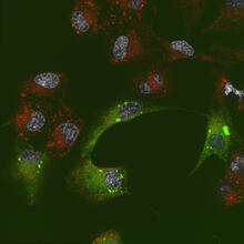
Endoplasmic reticulum abnormalities 2
6774
Human cells with the gene that codes for the protein FIT2 deleted. After an experimental intervention, they are expressing a nonfunctional version of FIT2, shown in green. Michel Becuwe, Harvard University. View Media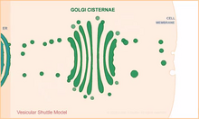
Vesicular shuttle model
1306
Animation for the vesicular shuttle model of Golgi transport. Judith Stoffer View Media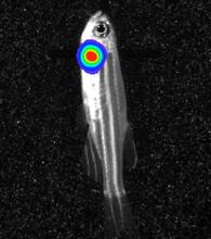
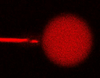
Bioluminescent imaging in adult zebrafish - lateral view
3558
Luciferase-based imaging enables visualization and quantification of internal organs and transplanted cells in live adult zebrafish. Kenneth Poss, Duke University View Media
The Proteasome: The Cell's Trash Processor in Action
3772
Our cells are constantly removing and recycling molecular waste. This video shows one way cells process their trash. View Media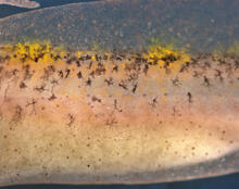
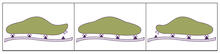
Migrating pigment cells
5758
Pigment cells are cells that give skin its color. David Parichy, University of Washington View Media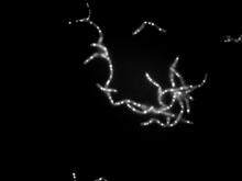
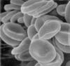
Bacillus anthracis being killed
3481
Bacillus anthracis (anthrax) cells being killed by a fluorescent trans-translation inhibitor, which disrupts bacterial protein synthesis. John Alumasa, Keiler Laboratory, Pennsylvania State University View Media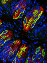
Taste buds signal different tastes through ATP release
3444
Taste buds in a mouse tongue epithelium with types I, II, and III taste cells visualized by cell-type-specific fluorescent antibodies. Aki Taruno, Perelman School of Medicine, University of Pennsylvania View Media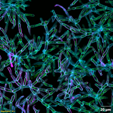
Snowflake yeast 2
6970
Multicellular yeast called snowflake yeast that researchers created through many generations of directed evolution from unicellular yeast. William Ratcliff, Georgia Institute of Technology. View Media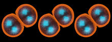
Sea urchin embryo 01
1047
Stereo triplet of a sea urchin embryo stained to reveal actin filaments (orange) and microtubules (blue). George von Dassow, University of Washington View Media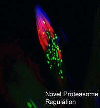
Proteasome
3451
This fruit fly spermatid recycles various molecules, including malformed or damaged proteins. Sigi Benjamin-Hong, Rockefeller University View Media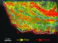
Intracellular forces
2799
Force vectors computed from actin cytoskeleton flow. This is an example of NIH-supported research on single-cell analysis. Gaudenz Danuser, Harvard Medical School View Media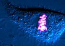
Dividing cell
6965
As this cell was undergoing cell division, it was imaged with two microscopy techniques: differential interference contrast (DIC) and confocal. The DIC view appears in blue and shows the entire cell. Dylan T. Burnette, Vanderbilt University School of Medicine. View Media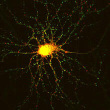
Neuron with labeled synapses
3509
In this image, recombinant probes known as FingRs (Fibronectin Intrabodies Generated by mRNA display) were expressed in a cortical neuron, where they attached fluorescent proteins to either PSD95 (gre Don Arnold and Richard Roberts, University of Southern California. View Media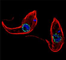
Trypanosoma brucei, the cause of sleeping sickness
3765
Trypanosoma brucei is a single-cell parasite that causes sleeping sickness in humans. Michael Rout, Rockefeller University View Media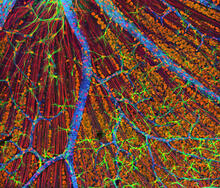
Mouse Retina
3309
A genetic disorder of the nervous system, neurofibromatosis causes tumors to form on nerves throughout the body, including a type of tumor called an optic nerve glioma that can result in childhood bli Tom Deerinck, NCMIR View Media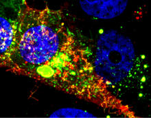
RSV-Infected Cell
3567
Viral RNA (red) in an RSV-infected cell. Eric Alonas and Philip Santangelo, Georgia Institute of Technology and Emory University View Media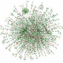
Protein map
2423
Network diagram showing a map of protein-protein interactions in a yeast (Saccharomyces cerevisiae) cell. This cluster includes 78 percent of the proteins in the yeast proteome. Hawoong Jeong, KAIST, Korea View Media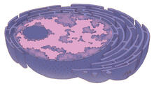
Nucleus and rough ER
1290
The nucleus contains the DNA of eukaryotic cells. Judith Stoffer View Media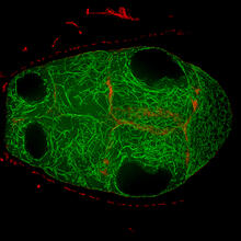
Fruit fly egg chamber
6811
A fruit fly (Drosophila melanogaster) egg chamber with microtubules shown in green and actin filaments shown in red. Vladimir I. Gelfand, Feinberg School of Medicine, Northwestern University. View Media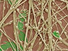
Synapses in culture
3399
Cultured hippocampal neurons grown on a substrate of glial cells (astrocytes). The glial cells form the pink/brown underlayment in this image. The tan threads are the neurons. National Center for Microscopy and Imaging Research View MediaGenetic mosaicism in fruit flies
6983
Fat tissue from the abdomen of a genetically mosaic adult fruit fly. Genetic mosaicism means that the fly has cells with different genotypes even though it formed from a single zygote. Akhila Rajan, Fred Hutchinson Cancer Center View Media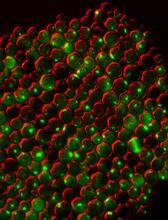
Protein clumping in zinc-deficient yeast cells
3550
The green spots in this image are clumps of protein inside yeast cells that are deficient in both zinc and a protein called Tsa1 that prevents clumping. Colin MacDiarmid and David Eide, University of Wisconsin--Madison View Media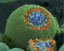
Nerve ending
1244
A scanning electron microscope picture of a nerve ending. It has been broken open to reveal vesicles (orange and blue) containing chemicals used to pass messages in the nervous system. Tina Weatherby Carvalho, University of Hawaii at Manoa View Media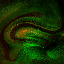
Mouse Brain Cross Section
5886
The brain sections are treated with fluorescent antibodies specific to a particular protein and visualized using serial electron microscopy (SEM). Anton Maximov, The Scripps Research Institute, La Jolla, CA View Media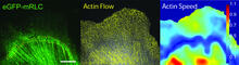
Actin flow
2798
Speckle microscopy analysis of actin cytoskeleton force. This is an example of NIH-supported research on single-cell analysis. Gaudenz Danuser, Harvard Medical School View Media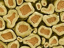
Myelinated axons 2
3397
Top view of myelinated axons in a rat spinal root. Tom Deerinck, National Center for Microscopy and Imaging Research (NCMIR) View Media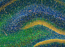
Rat Hippocampus
3308
This image of the hippocampus was taken with an ultra-widefield high-speed multiphoton laser microscope. Tom Deerinck, NCMIR View Media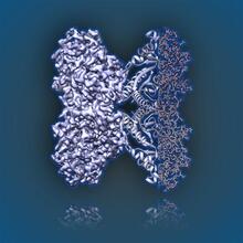
Aldolase
6350
2.5Å resolution reconstruction of rabbit muscle aldolase collected on a FEI/Thermo Fisher Titan Krios with energy filter and image corrector. National Resource for Automated Molecular Microscopy http://nramm.nysbc.org/nramm-images/ Source: Bridget Carragher View Media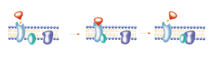
G switch
2536
The G switch allows our bodies to respond rapidly to hormones. See images 2537 and 2538 for labeled versions of this image. Crabtree + Company View Media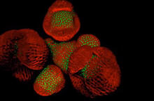
Arabidopsis Thaliana: Flowers Spring to Life
6503
This image capture shows how a single gene, STM, plays a starring role in plant development. Nathanaёl Prunet NIH Support: National Institute of General Medical Sciences View Media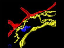
Computer model of cell membrane
2636
A computer model of the cell membrane, where the plasma membrane is red, endoplasmic reticulum is yellow, and mitochondria are blue. Bridget Wilson, University of New Mexico View Media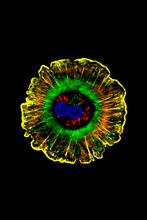
Human liver cell (hepatocyte)
3610
Hepatocytes, like the one shown here, are the most abundant type of cell in the human liver. Donna Beer Stolz, University of Pittsburgh View Media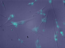
Molecules blocking Huntington's protein production
2600
The molecules that glow blue in these cultured cells prevent the expression of the mutant proteins that cause Huntington's disease. Jiaxin Hu, David W. Dodd and Robert H. E. Hudson, UT Southwestern Medical Center View Media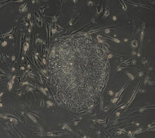
Induced stem cells from adult skin 04
2606
The human skin cells pictured contain genetic modifications that make them pluripotent, essentially equivalent to embryonic stem cells. James Thomson, University of Wisconsin-Madison View Media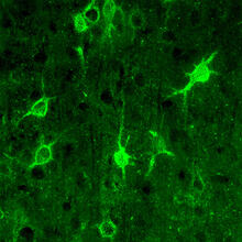
Confocal microscopy of perineuronal nets in the brain 1
3741
The photo shows a confocal microscopy image of perineuronal nets (PNNs), which are specialized extracellular matrix (ECM) structures in the brain. Tom Deerinck, National Center for Microscopy and Imaging Research (NCMIR) View Media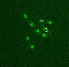
Dividing yeast cells with nuclear envelopes and spindle pole bodies
6795
Time-lapse video of yeast cells undergoing cell division. Nuclear envelopes are shown in green, and spindle pole bodies, which help pull apart copied genetic information, are shown in magenta. Alaina Willet, Kathy Gould’s lab, Vanderbilt University. View Media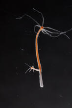
Hydra 04
2440
Hydra magnipapillata is an invertebrate animal used as a model organism to study developmental questions, for example the formation of the body axis. Hiroshi Shimizu, National Institute of Genetics in Mishima, Japan View Media