Switch to Gallery View
Image and Video Gallery
This is a searchable collection of scientific photos, illustrations, and videos. The images and videos in this gallery are licensed under Creative Commons Attribution Non-Commercial ShareAlike 3.0. This license lets you remix, tweak, and build upon this work non-commercially, as long as you credit and license your new creations under identical terms.

HeLa cell undergoing division into two daughter cells
6520
Here, a human HeLa cell (a type of immortal cell line used in laboratory experiments) is undergoing cell division. Dylan T. Burnette, Ph.D., Vanderbilt University School of Medicine. View Media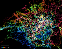
Dense tubular matrices in the peripheral endoplasmic reticulum (ER) 1
5855
Superresolution microscopy work on endoplasmic reticulum (ER) in the peripheral areas of the cell showing details of the structure and arrangement in a complex web of tubes. Jennifer Lippincott-Schwartz, Howard Hughes Medical Institute Janelia Research Campus, Virginia View Media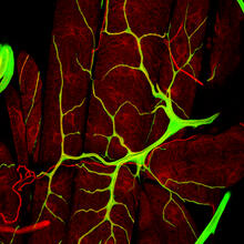
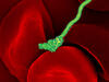
An insect tracheal cell delivers air to muscles
3615
Insects like the fruit fly use an elaborate network of branching tubes called trachea (green) to transport oxygen throughout their bodies. Jayan Nair and Maria Leptin, European Molecular Biology Laboratory, Heidelberg, Germany View Media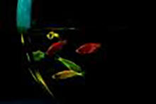
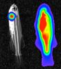
Glowing fish
2667
Professor Marc Zimmer's family pets, including these fish, glow in the dark in response to blue light. Featured in the September 2009 issue of Findings. View Media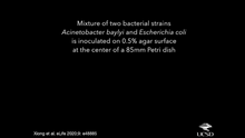
Time-lapse video of floral pattern in a mixture of two bacterial species, Acinetobacter baylyi and Escherichia coli, grown on a semi-solid agar for 24 hours
6550
This time-lapse video shows the emergence of a flower-like pattern in a mixture of two bacterial species, motile Acinetobacter baylyi and non-motile Escherichia coli (green), that are gr L. Xiong et al, eLife 2020;9: e48885 View Media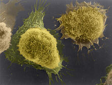
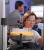
Cultured cells
1178
This image of laboratory-grown cells was taken with the help of a scanning electron microscope, which yields detailed images of cell surfaces. Tina Weatherby Carvalho, University of Hawaii at Manoa View Media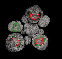
Developing Arabidopsis flower buds
3743
Flower development is a carefully orchestrated, genetically programmed process that ensures that the male (stamen) and female (pistil) organs form in the right place and at the right time in the flowe Nathanaël Prunet, Caltech View MediaTranslation
1281
Ribosomes manufacture proteins based on mRNA instructions. Each ribosome reads mRNA, recruits tRNA molecules to fetch amino acids, and assembles the amino acids in the proper order. Judith Stoffer View Media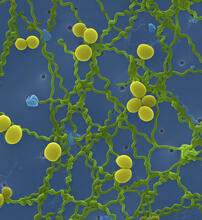
Leptospira bacteria
1166
Leptospira, shown here in green, is a type (genus) of elongated, spiral-shaped bacteria. Infection can cause Weil's disease, a kind of jaundice, in humans. Tina Weatherby Carvalho, University of Hawaii at Manoa View Media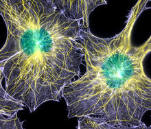
Colorful cells
2428
Actin (purple), microtubules (yellow), and nuclei (green) are labeled in these cells by immunofluorescence. This image won first place in the Nikon 2003 Small World photo competition. Torsten Wittmann, Scripps Research Institute View Media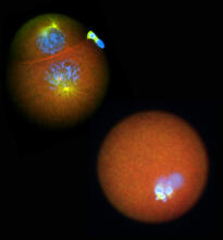
Nucleolinus
2762
The nucleolinus is a cellular compartment that has been a lonely bystander in scientific endeavors. Mary Anne Alliegro, Marine Biological Laboratory View Media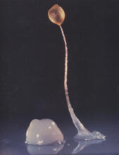
Dicty fruit
2684
Dictyostelium discoideum is a microscopic amoeba. A group of 100,000 form a mound as big as a grain of sand. Featured in The New Genetics. View Media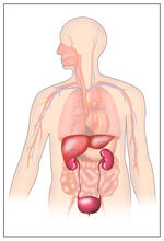
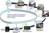
Body toxins
2496
Body organs such as the liver and kidneys process chemicals and toxins. These "target" organs are susceptible to damage caused by these substances. Crabtree + Company View Media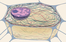
Cytoskeleton
1272
The three fibers of the cytoskeleton--microtubules in blue, intermediate filaments in red, and actin in green--play countless roles in the cell. Judith Stoffer View Media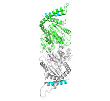
Dynamin structure
2744
When a molecule arrives at a cell's outer membrane, the membrane creates a pouch around the molecule that protrudes inward. Josh Chappie, National Institute of Diabetes and Digestive and Kidney Diseases, NIH View Media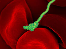
Human blood cells with Borrelia hermsii, a bacterium that causes relapsing fever
3586
Relapsing fever is caused by a bacterium and transmitted by certain soft-bodied ticks or body lice. The disease is seldom fatal in humans, but it can be very serious and prolonged. NIAID View Media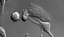
Dying melanoma cells
6966
Melanoma (skin cancer) cells undergoing programmed cell death, also called apoptosis. This process was triggered by raising the pH of the medium that the cells were growing in. Dylan T. Burnette, Vanderbilt University School of Medicine. View Media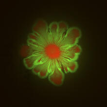
Floral pattern in a mixture of two bacterial species, Acinetobacter baylyi and Escherichia coli, grown on a semi-solid agar for 48 hours (photo 1)
6553
Floral pattern emerging as two bacterial species, motile Acinetobacter baylyi (red) and non-motile Escherichia coli (green), are grown together for 48 hours on 1% agar surface from a sma L. Xiong et al, eLife 2020;9: e48885 View Media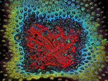
Tongue 1
5810
Microscopy image of tongue. One in a series of two, see image 5811 National Center for Microscopy and Imaging Research (NCMIR) View Media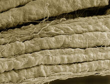
A bundle of myelinated peripheral nerve cells (axons)
3737
The extracellular matrix (ECM) is most prevalent in connective tissues but also is present between the stems (axons) of nerve cells. Tom Deerinck, National Center for Microscopy and Imaging Research (NCMIR) View Media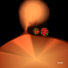
Multivesicular bodies containing intralumenal vesicles assemble at the vacuole 1
5769
Collecting and transporting cellular waste and sorting it into recylable and nonrecylable pieces is a complex business in the cell. Matthew West and Greg Odorizzi, University of Colorado View Media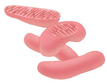
Mitochondria
1287
Bean-shaped mitochondria are cells' power plants. These organelles have their own DNA and replicate independently. The highly folded inner membranes are the site of energy generation. Judith Stoffer View Media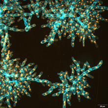
Snowflake yeast 3
6971
Multicellular yeast called snowflake yeast that researchers created through many generations of directed evolution from unicellular yeast. William Ratcliff, Georgia Institute of Technology. View Media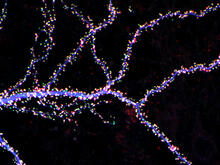
Hippocampal neuron from rodent brain
3686
Hippocampal neuron from rodent brain with dendrites shown in blue. The hundreds of tiny magenta, green and white dots are the dendritic spines of excitatory synapses. Shelley Halpain, UC San Diego View Media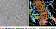
Cryo-ET cross-section of the Golgi apparatus
6606
On the left, a cross-section slice of a rat pancreas cell captured using cryo-electron tomography (cryo-ET). On the right, a 3D, color-coded version of the image highlighting cell structures. Xianjun Zhang, University of Southern California. View Media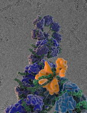
Flu virus proteins during self-replication
3434
Influenza (flu) virus proteins in the act of self-replication. Viral nucleoprotein (blue) encapsidates [encapsulates] the RNA genome (green). Scripps Research Institute in La Jolla, CA View Media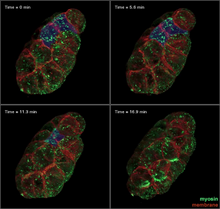
Four timepoints in gastrulation
3334
It has been said that gastrulation is the most important event in a person's life. Bob Goldstein, University of North Carolina, Chapel Hill View Media
Centrioles anchor cilia in planaria
3292
Centrioles (green) anchor cilia (red), which project on the surface of pharynx cells of the freshwater planarian Schmidtea mediterranea. Juliette Azimzadeh, University of California, San Francisco View Media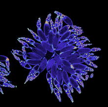
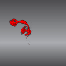
Fruit fly ovary
3607
A fruit fly ovary, shown here, contains as many as 20 eggs. Fruit flies are not merely tiny insects that buzz around overripe fruit—they are a venerable scientific tool. Denise Montell, Johns Hopkins University and University of California, Santa Barbara View Media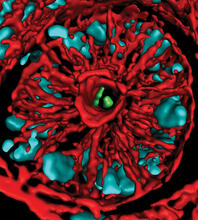
Mouse colon with gut bacteria
3566
A section of mouse colon with gut bacteria (center, in green) residing within a protective pocket. Sarkis K. Mazmanian, California Institute of Technology View Media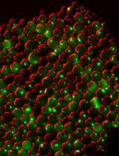
Protein clumping in zinc-deficient yeast cells
3550
The green spots in this image are clumps of protein inside yeast cells that are deficient in both zinc and a protein called Tsa1 that prevents clumping. Colin MacDiarmid and David Eide, University of Wisconsin--Madison View Media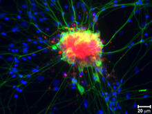
Spinal nerve cells
3251
Neurons (green) and glial cells from isolated dorsal root ganglia express COX-2 (red) after exposure to an inflammatory stimulus (cell nuclei are blue). Lawrence Marnett, Vanderbilt University View Media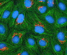
HeLa cells
3522
Multiphoton fluorescence image of cultured HeLa cells with a fluorescent protein targeted to the Golgi apparatus (orange), microtubules (green) and counterstained for DNA (cyan). National Center for Microscopy and Imaging Research (NCMIR) View Media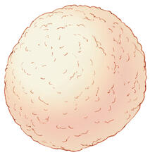
Color coding of the Drosophila brain - video
5843
This video results from a research project to visualize which regions of the adult fruit fly (Drosophila) brain derive from each neural stem cell. Yong Wan from Charles Hansen’s lab, University of Utah. Data preparation and visualization by Masayoshi Ito in the lab of Kei Ito, University of Tokyo. View Media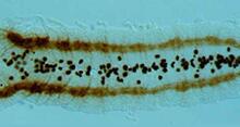
Developing fruit fly nerve cord
2435
The glial cells (black dots) and nerve cells (brown bands) in this developing fruit fly nerve cord formed normally despite the absence of the SPITZ protein, which blocks their impending suicide. Hermann Steller, Rockefeller University View Media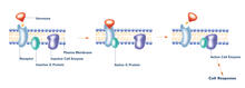
G switch (with labels)
2537
The G switch allows our bodies to respond rapidly to hormones. G proteins act like relay batons to pass messages from circulating hormones into cells. Crabtree + Company View Media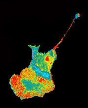
Seeing signaling protein activation in cells 04
2454
Cdc42, a member of the Rho family of small guanosine triphosphatase (GTPase) proteins, regulates multiple cell functions, including motility, proliferation, apoptosis, and cell morphology. Klaus Hahn, University of North Carolina, Chapel Hill Medical School View Media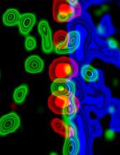
Molecular interactions at the astrocyte nuclear membrane
3734
These ripples of color represent the outer membrane of the nucleus inside an astrocyte, a star-shaped cell inside the brain. Katerina Akassoglou, Gladstone Institute for Neurological Disease & UCSF View Media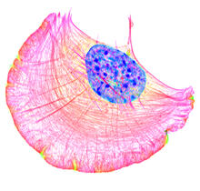
Crawling cell
6964
A crawling cell with DNA shown in blue and actin filaments, which are a major component of the cytoskeleton, visible in pink. Actin filaments help enable cells to crawl. Dylan T. Burnette, Vanderbilt University School of Medicine. View Media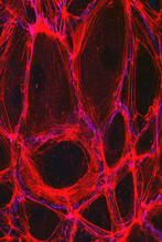
Cells lining the blood vessel walls
3633
The structure of the endothelium, the thin layer of cells that line our arteries and veins, is visible here. Christopher V. Carman and Roberta Martinelli, Harvard Medical School. View Media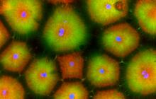
Cell-like compartments from frog eggs 5
6592
Cell-like compartments that spontaneously emerged from scrambled frog eggs, with nuclei (blue) from frog sperm. Endoplasmic reticulum (red) and microtubules (green) are also visible. Xianrui Cheng, Stanford University School of Medicine. View Media
NCMIR mouse tail
3395
Stained cross section of a mouse tail. Tom Deerinck, National Center for Microscopy and Imaging Research (NCMIR) View Media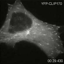
Microtubule dynamics in real time
2784
Cytoplasmic linker protein (CLIP)-170 is a microtubule plus-end-tracking protein that regulates microtubule dynamics and links microtubule ends to different intracellular structures. Gary Borisy, Marine Biology Laboratory View Media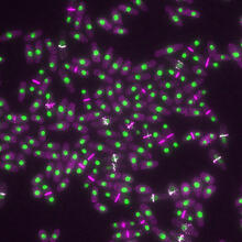
Yeast cells with nuclei and contractile rings
6792
Yeast cells with nuclei shown in green and contractile rings shown in magenta. Nuclei store DNA, and contractile rings help cells divide. Alaina Willet, Kathy Gould’s lab, Vanderbilt University. View Media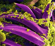
Bubonic plague bacteria on part of the digestive system in a rat flea
3576
Here, bubonic plague bacteria (yellow) are shown in the digestive system of a rat flea (purple). The bubonic plague killed a third of Europeans in the mid-14th century. NIAID View Media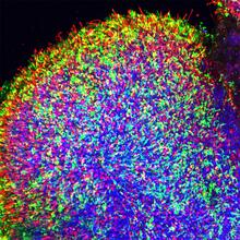
Human retinal organoid
6748
A replica of a human retina grown from stem cells. Kevin Eliceiri, University of Wisconsin-Madison. View Media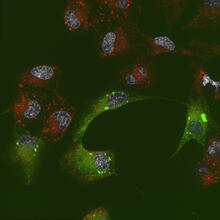
Endoplasmic reticulum abnormalities 2
6774
Human cells with the gene that codes for the protein FIT2 deleted. After an experimental intervention, they are expressing a nonfunctional version of FIT2, shown in green. Michel Becuwe, Harvard University. View Media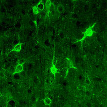
Confocal microscopy of perineuronal nets in the brain 1
3741
The photo shows a confocal microscopy image of perineuronal nets (PNNs), which are specialized extracellular matrix (ECM) structures in the brain. Tom Deerinck, National Center for Microscopy and Imaging Research (NCMIR) View Media
Cell-like compartments from frog eggs 6
6593
Cell-like compartments that spontaneously emerged from scrambled frog eggs, with nuclei (blue) from frog sperm. Endoplasmic reticulum (red) and microtubules (green) are also visible. Xianrui Cheng, Stanford University School of Medicine. View Media