Switch to Gallery View
Image and Video Gallery
This is a searchable collection of scientific photos, illustrations, and videos. The images and videos in this gallery are licensed under Creative Commons Attribution Non-Commercial ShareAlike 3.0. This license lets you remix, tweak, and build upon this work non-commercially, as long as you credit and license your new creations under identical terms.
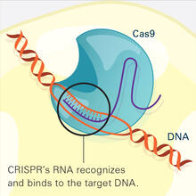
CRISPR Illustration Frame 2
6486
This illustration shows, in simplified terms, how the CRISPR-Cas9 system can be used as a gene-editing tool. National Institute of General Medical Sciences. View Media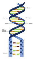
Nucleotides make up DNA (with labels)
2542
DNA consists of two long, twisted chains made up of nucleotides. Each nucleotide contains one base, one phosphate molecule, and the sugar molecule deoxyribose. Crabtree + Company View Media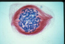
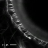
Lily mitosis 05
1015
A light microscope image of a cell from the endosperm of an African globe lily (Scadoxus katherinae). This is one frame of a time-lapse sequence that shows cell division in action. Andrew S. Bajer, University of Oregon, Eugene View Media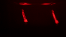
Culex quinquefasciatus mosquito larvae
6771
Mosquito larvae with genes edited by CRISPR swimming in water. Valentino Gantz, University of California, San Diego. View Media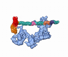
Dynamic cryo-EM model of the human transcription preinitiation complex
5730
Gene transcription is a process by which information encoded in DNA is transcribed into RNA. Eva Nogales, Berkeley Lab View Media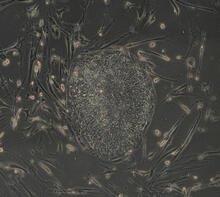
Induced stem cells from adult skin 03
2605
The human skin cells pictured contain genetic modifications that make them pluripotent, essentially equivalent to embryonic stem cells. James Thomson, University of Wisconsin-Madison View Media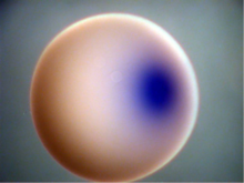
Xenopus laevis egg
2753
Xenopus laevis, the African clawed frog, has long been used as a model organism for studying embryonic development. Michael Klymkowsky, University of Colorado, Boulder View Media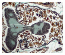
Cross section of a Drosophila melanogaster pupa
2758
This photograph shows a magnified view of a Drosophila melanogaster pupa in cross section. Compare this normal pupa to one that lacks an important receptor, shown in image 2759. Christina McPhee and Eric Baehrecke, University of Massachusetts Medical School View Media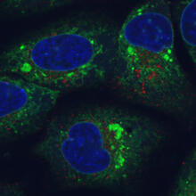
Endoplasmic reticulum abnormalities
6773
Human cells with the gene that codes for the protein FIT2 deleted. Green indicates an endoplasmic reticulum (ER) resident protein. Michel Becuwe, Harvard University. View Media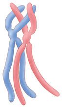
Chromosomes before crossing over
1315
Duplicated pair of chromosomes lined up and ready to cross over. Judith Stoffer View Media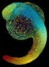
Zebrafish embryo
3644
Just 22 hours after fertilization, this zebrafish embryo is already taking shape. By 36 hours, all of the major organs will have started to form. Philipp Keller, Bill Lemon, Yinan Wan, and Kristin Branson, Janelia Farm Research Campus, Howard Hughes Medical Institute, Ashburn, Va. View Media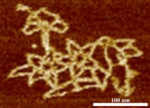
Microscopy image of bird-and-flower DNA origami
3690
An atomic force microscopy image shows DNA folded into an intricate, computer-designed structure. Hao Yan, Arizona State University View Media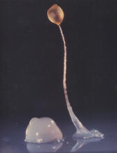
Dicty fruit
2684
Dictyostelium discoideum is a microscopic amoeba. A group of 100,000 form a mound as big as a grain of sand. Featured in The New Genetics. View Media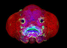
Zebrafish larva
5881
You are face to face with a 6-day-old zebrafish larva. What look like eyes will become nostrils, and the bulges on either side will become eyes. Oscar Ruiz and George Eisenhoffer, University of Texas MD Anderson Cancer Center, Houston View Media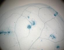
Disease-resistant Arabidopsis leaf
2781
This is a magnified view of an Arabidopsis thaliana leaf a few days after being exposed to the pathogen Hyaloperonospora arabidopsidis. Jeff Dangl, University of North Carolina, Chapel Hill View Media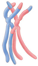
Chromosomes after crossing over
1314
Duplicated pair of chromosomes have exchanged material. Judith Stoffer View Media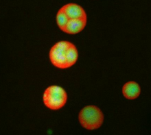
Nucleolus subcompartments spontaneously self-assemble 3
3792
What looks a little like distant planets with some mysterious surface features are actually assemblies of proteins normally found in the cell's nucleolus, a small but very important protein complex lo Nilesh Vaidya, Princeton University View Media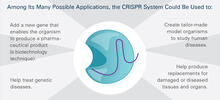
CRISPR Illustration Frame 5
6489
This illustration shows, in simplified terms, how the CRISPR-Cas9 system can be used as a gene-editing tool. This is the fifthframe in a series of five. View Media
Fluorescence in situ hybridization (FISH) in mouse ES cells shows DNA interactions
3296
Researchers used fluorescence in situ hybridization (FISH) to confirm the presence of long range DNA-DNA interactions in mouse embryonic stem cells. Kathrin Plath, University of California, Los Angeles View Media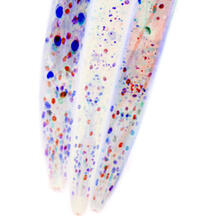
Mosaicism in C. elegans (White Background)
6534
In the worm C. elegans, double-stranded RNA made in neurons can silence matching genes in a variety of cell types through the transport of RNA between cells. Snusha Ravikumar, Ph.D., University of Maryland, College Park, and Antony M. Jose, Ph.D., University of Maryland, College Park View Media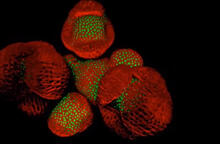
Arabidopsis Thaliana: Flowers Spring to Life
6503
This image capture shows how a single gene, STM, plays a starring role in plant development. Nathanaёl Prunet NIH Support: National Institute of General Medical Sciences View Media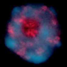
Gene silencing
2318
Pretty in pink, the enzyme histone deacetylase (HDA6) stands out against a background of blue-tinted DNA in the nucleus of an Arabidopsis plant cell. Olga Pontes and Craig Pikaard, Washington University View Media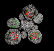
Developing Arabidopsis flower buds
3743
Flower development is a carefully orchestrated, genetically programmed process that ensures that the male (stamen) and female (pistil) organs form in the right place and at the right time in the flowe Nathanaël Prunet, Caltech View Media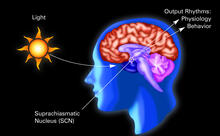
Circadian rhythm (with labels)
2569
The human body keeps time with a master clock called the suprachiasmatic nucleus or SCN. Crabtree + Company View Media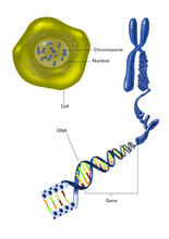
Chromosome inside nucleus (with labels)
2540
The long, stringy DNA that makes up genes is spooled within chromosomes inside the nucleus of a cell. Crabtree + Company View Media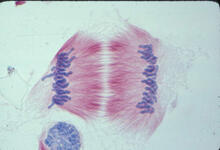
Lily mitosis 11
1011
A light microscope image of cells from the endosperm of an African globe lily (Scadoxus katherinae). This is one frame of a time-lapse sequence that shows cell division in action. Andrew S. Bajer, University of Oregon, Eugene View Media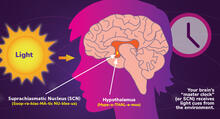
Circadian rhythms and the SCN
6613
Circadian rhythms are physical, mental, and behavioral changes that follow a 24-hour cycle. NIGMS View Media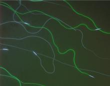
GFP sperm
2683
Fruit fly sperm cells glow bright green when they express the gene for green fluorescent protein (GFP). View Media
Repairing DNA
3493
Like a watch wrapped around a wrist, a special enzyme encircles the double helix to repair a broken strand of DNA. Tom Ellenberger, Washington University School of Medicine View Media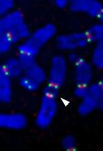
Fused, dicentric chromosomes
2763
This fused chromosome has two functional centromeres, shown as two sets of red and green dots. Beth A. Sullivan, Duke University View Media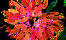
Fruit fly ovary_2
3656
A fruit fly ovary, shown here, contains as many as 20 eggs. Fruit flies are not merely tiny insects that buzz around overripe fruit--they are a venerable scientific tool. Denise Montell, University of California, Santa Barbara View Media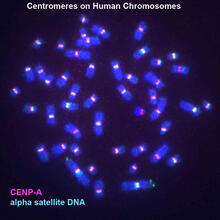
Centromeres on human chromosomes
3255
Human metaphase chromosomes are visible with fluorescence in vitro hybridization (FISH). Centromeric alpha satellite DNA (green) are found in the heterochromatin at each centromere. Peter Warburton, Mount Sinai School of Medicine View Media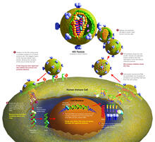
Life of an AIDS virus (with labels and stages)
2515
HIV is a retrovirus, a type of virus that carries its genetic material not as DNA but as RNA. Crabtree + Company View Media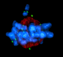
Mitotic cell awaits chromosome alignment
5765
During mitosis, spindle microtubules (red) attach to chromosome pairs (blue), directing them to the spindle equator. View Media
Katanin protein regulates anaphase
2594
The microtubule severing protein, katanin, localizes to chromosomes and regulates anaphase A in mitosis. David Sharp, Albert Einstein College of Medicine View Media
Genetically identical mycobacteria respond differently to antibiotic 2
5752
Antibiotic resistance in microbes is a serious health concern. So researchers have turned their attention to how bacteria undo the action of some antibiotics. Bree Aldridge, Tufts University View Media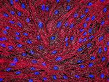
DNA and actin in cultured fibroblast cells
3670
DNA (blue) and actin (red) in cultured fibroblast cells. Tom Deerinck, National Center for Microscopy and Imaging Research (NCMIR) View Media
Introduction to Genome Editing Using CRISPR/Cas9
5815
Genome editing using CRISPR/Cas9 is a rapidly expanding field of scientific research with emerging applications in disease treatment, medical therapeutics and bioenergy, just to name a few. Janet Iwasa View Media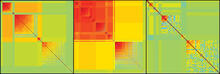
Genetic patchworks
2588
Each point in these colorful patchworks represents the correlation between two sleep-associated genes in fruit flies. Susan Harbison and Trudy Mackay, North Carolina State University View Media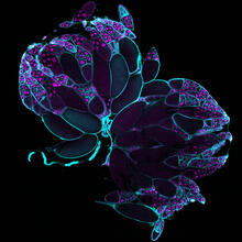
Fruit fly ovaries
6807
Fruit fly (Drosophila melanogaster) ovaries with DNA shown in magenta and actin filaments shown in light blue. This image was captured using a confocal laser scanning microscope.Vladimir I. Gelfand, Feinberg School of Medicine, Northwestern University. View Media
Assembly of the HIV capsid
5729
The HIV capsid is a pear-shaped structure that is made of proteins the virus needs to mature and become infective. John Grime and Gregory Voth, The University of Chicago View Media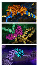
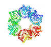
HIV enzyme
6999
These images model the molecular structures of three enzymes with critical roles in the life cycle of the human immunodeficiency virus (HIV). Amy Wu and Christine Zardecki, RCSB Protein Data Bank. View Media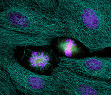
Highlighted cells
2429
The cytoskeleton (green) and DNA (purple) are highlighed in these cells by immunofluorescence. Torsten Wittmann, Scripps Research Institute View Media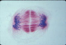
Lily mitosis 12
1018
A light microscope image of a cell from the endosperm of an African globe lily (Scadoxus katherinae). This is one frame of a time-lapse sequence that shows cell division in action. Andrew S. Bajer, University of Oregon, Eugene View Media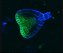
Early development in Arabidopsis
2733
Early on, this Arabidopsis plant embryo picks sides: While one end will form the shoot, the other will take root underground. Zachery R. Smith, Jeff Long lab at the Salk Institute for Biological Studies View Media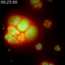
Nucleolus subcompartments spontaneously self-assemble 1
3789
The nucleolus is a small but very important protein complex located in the cell's nucleus. Nilesh Vaidya, Princeton University View Media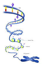
Epigenetic code (with labels)
2563
The "epigenetic code" controls gene activity with chemical tags that mark DNA (purple diamonds) and the "tails" of histone proteins (purple triangles). Crabtree + Company View Media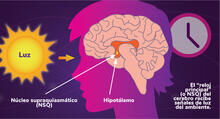
Los ritmos circadianos y el núcleo supraquiasmático
6614
Los ritmos circadianos son cambios físicos, mentales y de comportamiento que siguen un ciclo de 24 horas. NIGMS View Media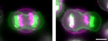
Symmetrically and asymmetrically elongating cells
3648
Merged fluorescent images of symmetrically (left) or asymmetrically (right) elongating HeLa cells at the end of early anaphase (magenta) and late anaphase (green). Tomomi Kiyomitsu and Iain M. Cheeseman, Whitehead Institute for Biomedical Research View Media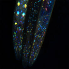
Mosaicism in C. elegans (Black Background)
6532
In the worm C. elegans, double-stranded RNA made in neurons can silence matching genes in a variety of cell types through the transport of RNA between cells. Snusha Ravikumar, Ph.D., University of Maryland, College Park, and Antony M. Jose, Ph.D., University of Maryland, College Park View Media