Image and Video Gallery
This is a searchable collection of scientific photos, illustrations, and videos. The images and videos in this gallery are licensed under Creative Commons Attribution Non-Commercial ShareAlike 3.0. This license lets you remix, tweak, and build upon this work non-commercially, as long as you credit and license your new creations under identical terms.
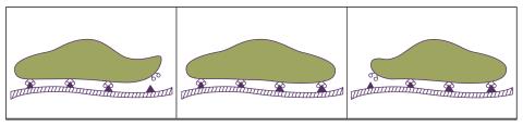
1292: Smooth ER
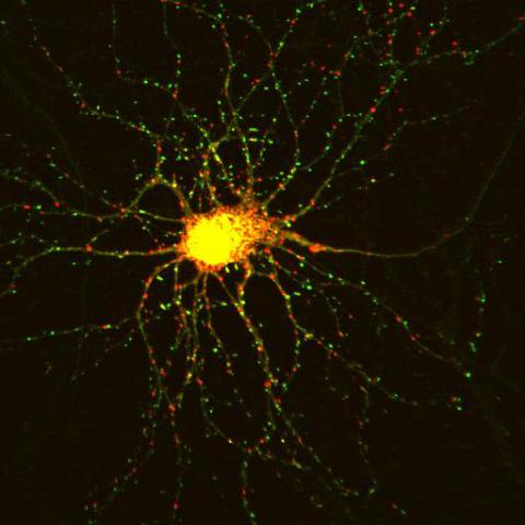
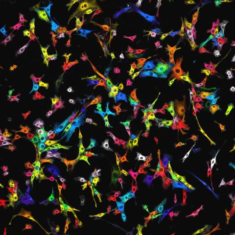
3509: Neuron with labeled synapses
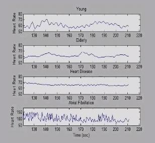
3596: Heart rates time series image
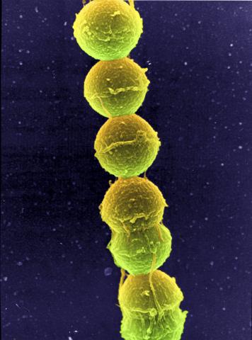
1157: Streptococcus bacteria
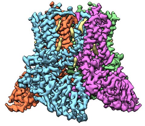
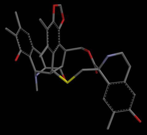
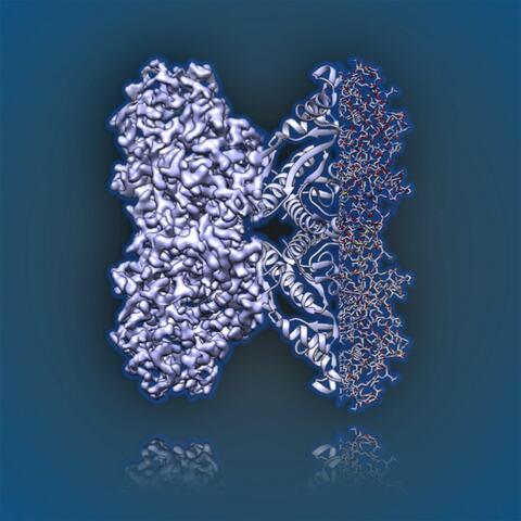
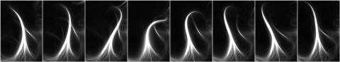
6577: Transient receptor potential channel TRPV5
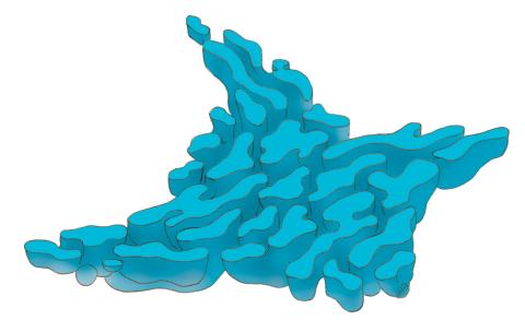
6573: Nuclear Lamina – Three Views
See 6572 for a 3D view of this structure.
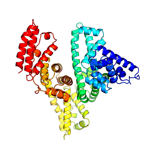
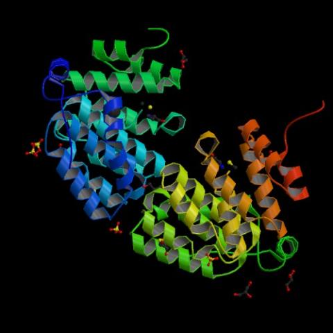
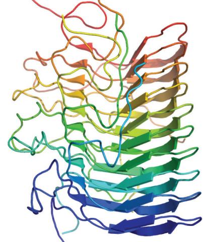
3745: Serum albumin structure 2
Related to entries 3744 and 3746
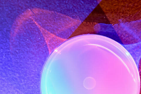
6752: Petri dish
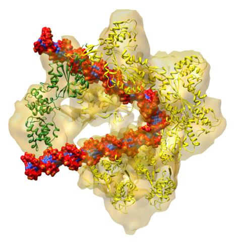
3597: DNA replication origin recognition complex (ORC)
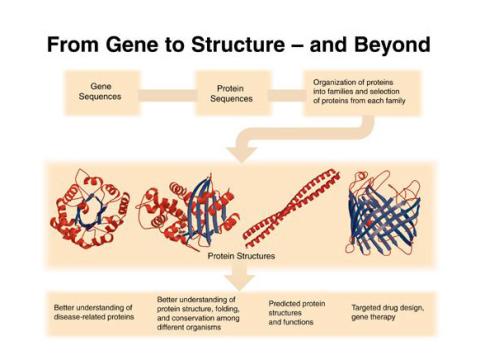
2363: PSI: from genes to structures
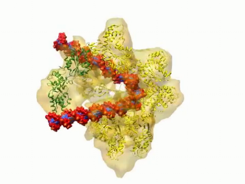
3307: DNA replication origin recognition complex (ORC)
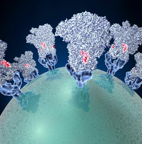
3753: Coronavirus spike protein structure
6350: Aldolase
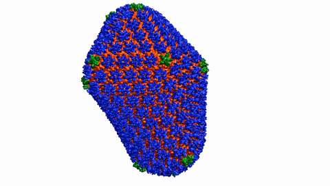
6601: Atomic-level structure of the HIV capsid
3628: Skin cancer cells (squamous cell carcinoma)
This image was part of the Life: Magnified exhibit that ran from June 3, 2014, to January 21, 2015, at Dulles International Airport.
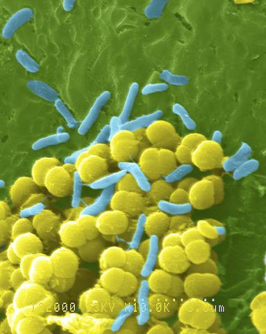
1158: Bacteria shapes
2502: Focal adhesions
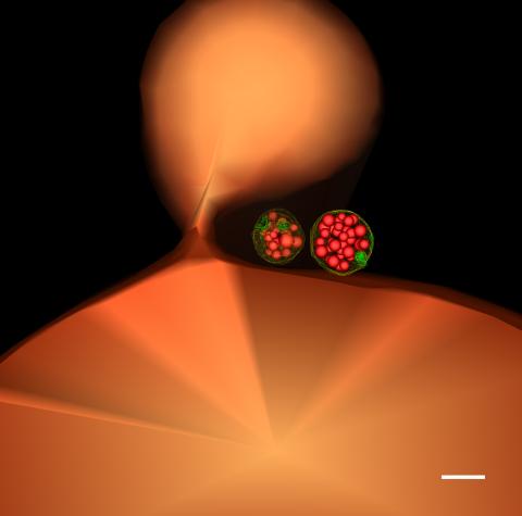
7021: Single-cell “radios” image
Related to video 7022.
2342: Protein from E. faecalis
6591: Cell-like compartments from frog eggs 4
For more photos of cell-like compartments from frog eggs view: 6584, 6585, 6586, 6592, and 6593.
For videos of cell-like compartments from frog eggs view: 6587, 6588, 6589, and 6590.
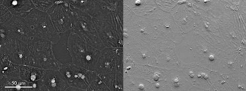
6899: Epithelial cell migration
More information about the research that produced this video can be found in the Journal of Microscopy paper “An Orientation-Independent DIC Microscope Allows High Resolution Imaging of Epithelial Cell Migration and Wound Healing in a Cnidarian Model” by Malamy and Shribak.
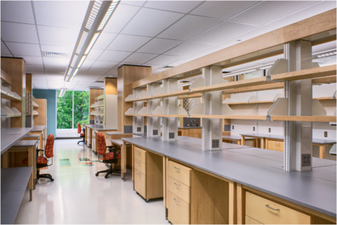
2723: iPS cell facility at the Coriell Institute for Medical Research
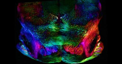
6901: Mouse brain slice showing nerve cells
The image was obtained with a polychromatic polarizing microscope that shows the polychromatic birefringent image with hue corresponding to the slow axis orientation. More information about the microscopy that produced this image can be found in the Scientific Reports paper “Polychromatic Polarization Microscope: Bringing Colors to a Colorless World” by Shribak.
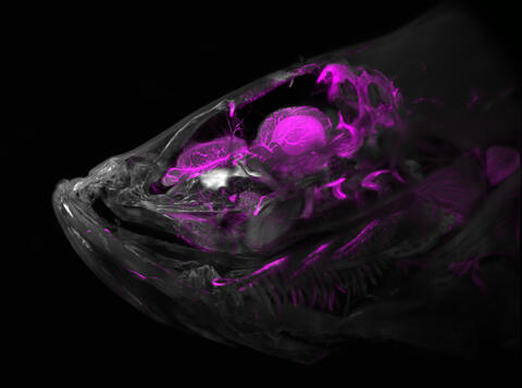
6934: Zebrafish head vasculature
This image was captured using a light sheet microscope.
Related to video 6933.
3344: Artificial cilia exhibit spontaneous beating
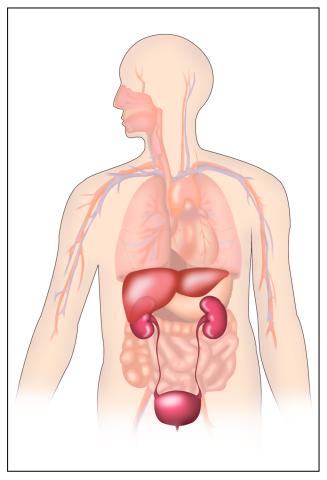
2496: Body toxins

2702: Thermotoga maritima and its metabolic network
5769: Multivesicular bodies containing intralumenal vesicles assemble at the vacuole 1
Scientists working with baker's yeast (Saccharomyces cerevisiae) study the budding inward of the limiting membrane (green lines on top of the yellow lines) into the intralumenal vesicles. This tomogram was shot with a Tecnai F-20 high-energy electron microscope, at 29,000x magnification, with a 0.7-nm pixel, ~4-nm resolution.
To learn more about endosomes, see the Biomedical Beat blog post The Cell’s Mailroom. Related to a microscopy photograph 5768 that was used to generate this illustration and a zoomed-in version 5767 of this illustration.

2709: Retroviruses as fossils
7018: Bacterial cells aggregating above the light organ of the Hawaiian bobtail squid
Related to images 7016, 7017, 7019, and 7020.
6968: Regenerating lizard tail
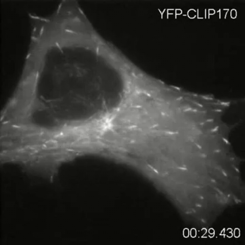
2784: Microtubule dynamics in real time
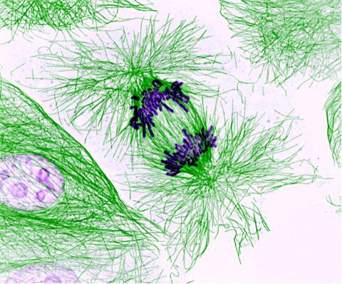
3631: Dividing cells showing chromosomes and cell skeleton
This image was part of the Life: Magnified exhibit that ran from June 3, 2014, to January 21, 2015, at Dulles International Airport.
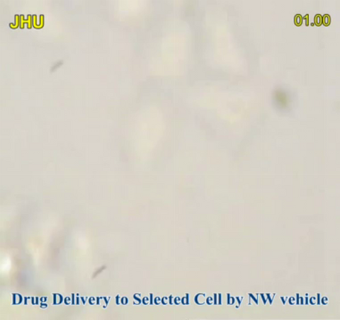
3779: Precisely Delivering Chemical Cargo to Cells
This movie shows the manipulation of the nanowires for drug delivery to a single cell. To learn more about this technique, see this post in the Computing Life series.
3542: Structure of amyloid-forming prion protein
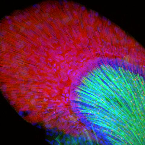
6930: Mouse brain 2
This image was captured using a light sheet microscope.
Related to image 6929 and video 6931.
2308: Cellular metropolis
2322: Modeling disease spread
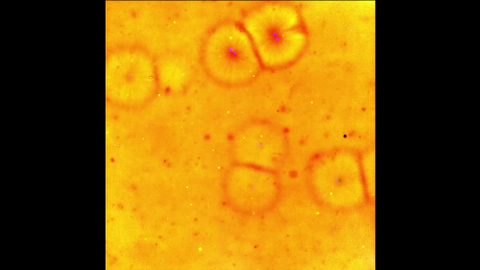
6584: Cell-like compartments from frog eggs
For more photos of cell-like compartments from frog eggs view: 6585, 6586, 6591, 6592, and 6593.
For videos of cell-like compartments from frog eggs view: 6587, 6588, 6589, and 6590.
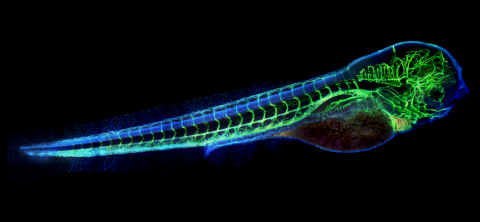
6661: Zebrafish embryo showing vasculature
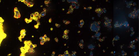
6588: Cell-like compartments emerging from scrambled frog eggs 2
For more photos of cell-like compartments from frog eggs view: 6584, 6585, 6586, 6591, 6592, and 6593.
For videos of cell-like compartments from frog eggs view: 6587, 6589, and 6590.
3580: V. Cholerae Biofilm
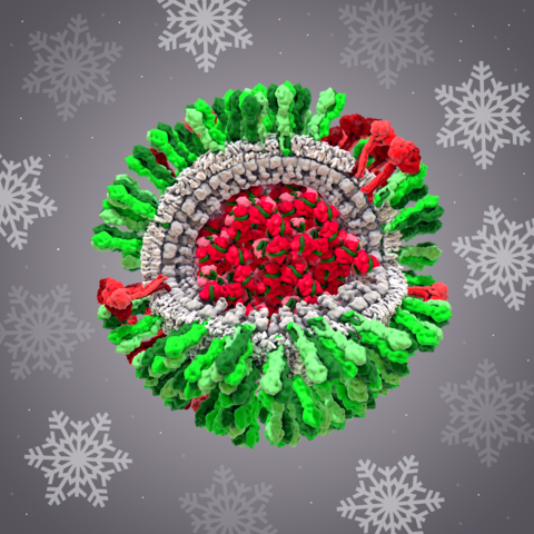
6355: H1N1 Influenza Virus
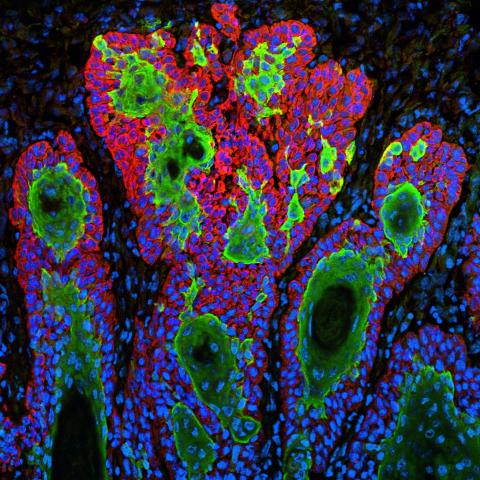
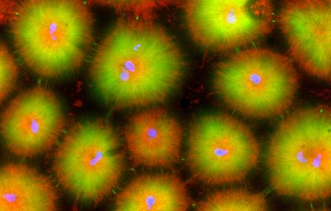
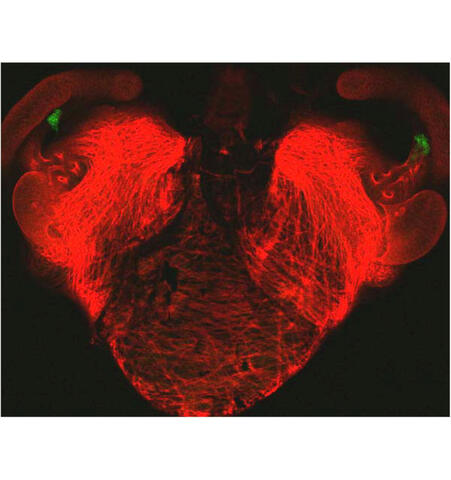
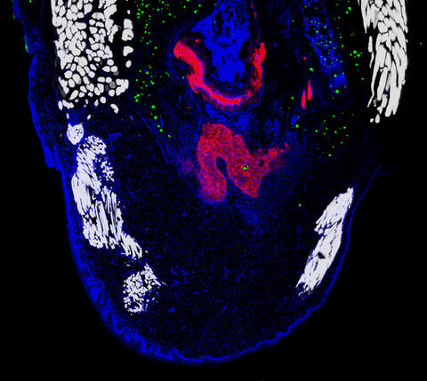
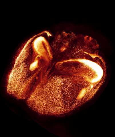
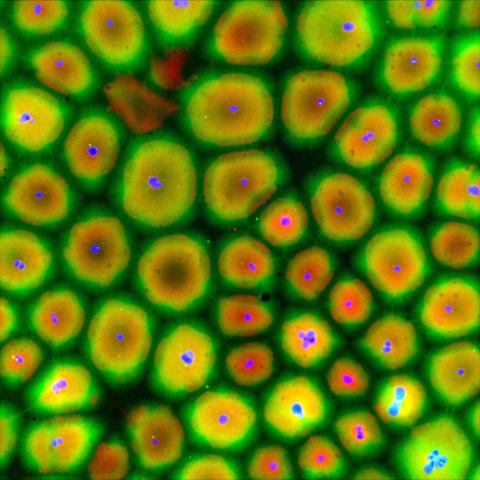
3598: Developing zebrafish fin
In this image, green fluorescent protein (GFP) is expressed where the gene sox9b is expressed. Collagen (red) marks the fin rays, and DNA, stained with a dye called DAPI, is in blue. sox9b plays many important roles during development, including the building of the heart and brain, and is also necessary for skeletal development. At the University of Wisconsin, researchers have found that exposure to contaminants that bind the aryl-hydrocarbon receptor results in the downregulation of sox9b. Loss of sox9b severely disrupts development in zebrafish and causes a life-threatening disorder called campomelic dysplasia (CD) in humans. CD is characterized by cardiovascular, neural, and skeletal defects. By studying the roles of genes such as sox9b in zebrafish, scientists hope to better understand normal development in humans as well as how to treat developmental disorders and diseases.
This image was part of the Life: Magnified exhibit that ran from June 3, 2014, to January 21, 2015, at Dulles International Airport.
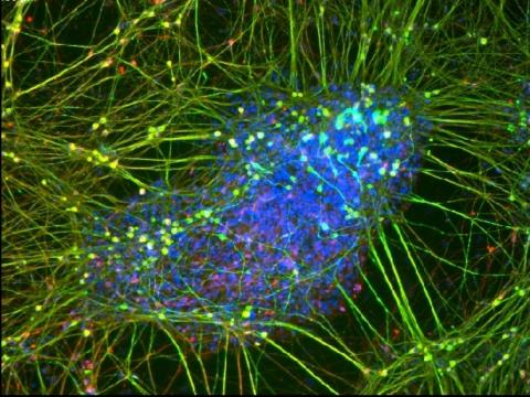
3264: Peripheral nerve cell derived from ES cells
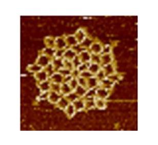
3724: Snowflake DNA origami
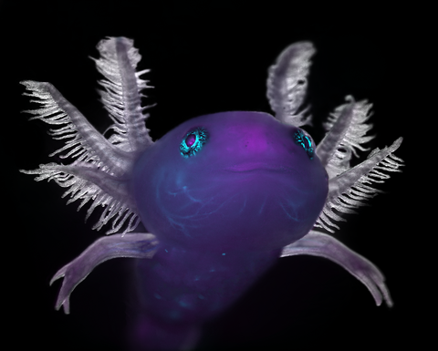
6932: Axolotl
This image was captured using a stereo microscope.
Related to images 6927 and 6928.
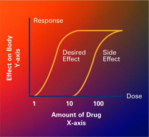
2533: Dose response curves
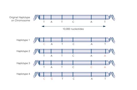
2567: Haplotypes (with labels)