
Switch to List View
Image and Video Gallery
This is a searchable collection of scientific photos, illustrations, and videos. The images and videos in this gallery are licensed under Creative Commons Attribution Non-Commercial ShareAlike 3.0. This license lets you remix, tweak, and build upon this work non-commercially, as long as you credit and license your new creations under identical terms.
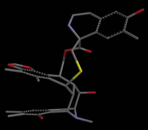
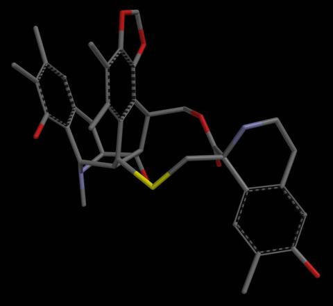
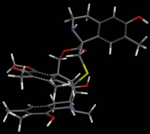
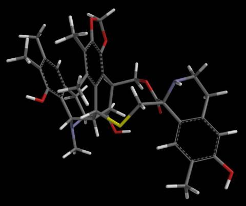
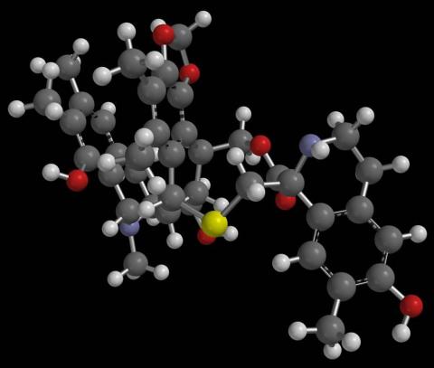
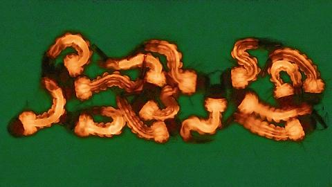
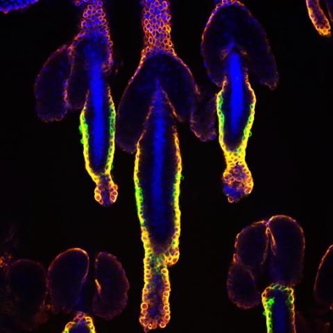
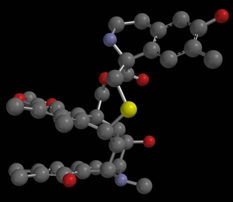
2795: Anti-tumor drug ecteinascidin 743 (ET-743), structure without hydrogens 02
2795: Anti-tumor drug ecteinascidin 743 (ET-743), structure without hydrogens 02
Ecteinascidin 743 (ET-743, brand name Yondelis), was discovered and isolated from a sea squirt, Ecteinascidia turbinata, by NIGMS grantee Kenneth Rinehart at the University of Illinois. It was synthesized by NIGMS grantees E.J. Corey and later by Samuel Danishefsky. Multiple versions of this structure are available as entries 2790-2797.
Timothy Jamison, Massachusetts Institute of Technology
View Media
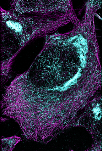
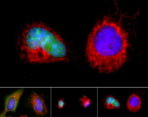
6892: Microtubules and tau aggregates
6892: Microtubules and tau aggregates
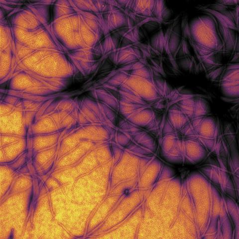
Microtubules (magenta) and tau protein (light blue) in a cell model of tauopathy. Researchers believe that tauopathy—the aggregation of tau protein—plays a role in Alzheimer’s disease and other neurodegenerative diseases. This image was captured using Stochastic Optical Reconstruction Microscopy (STORM).
Related to images 6889, 6890, and 6891.
Related to images 6889, 6890, and 6891.
Melike Lakadamyali, Perelman School of Medicine at the University of Pennsylvania.
View Media
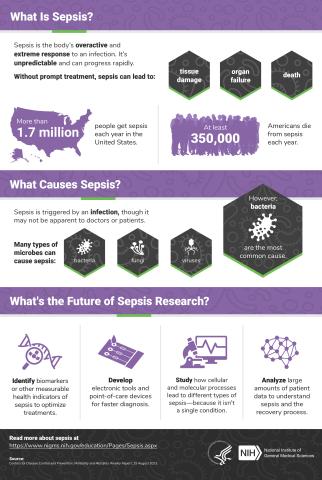
6536: Sepsis Infographic
6536: Sepsis Infographic
Sepsis is the body’s overactive and extreme response to an infection. More than 1.7 million people get sepsis each year in the United States. Without prompt treatment, sepsis can lead to tissue damage, organ failure, and death. Many NIGMS-supported researchers are working to improve sepsis diagnosis and treatment. Learn more with our sepsis featured topic page.
See 6551 for the Spanish version of this infographic.
See 6551 for the Spanish version of this infographic.
National Institute of General Medical Sciences
View Media
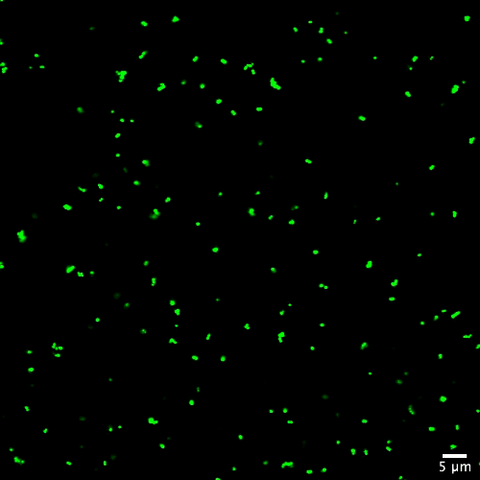
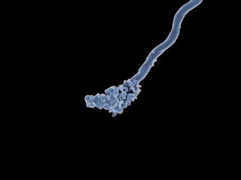
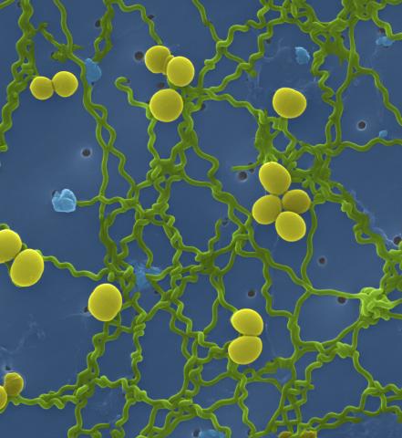
6805: Staphylococcus aureus aggregating upon contact with synovial fluid
6805: Staphylococcus aureus aggregating upon contact with synovial fluid
Staphylococcus aureus bacteria (green) grouping together upon contact with synovial fluid—a viscous substance found in joints. The formation of groups can help protect the bacteria from immune system defenses and from antibiotics, increasing the likelihood of an infection. This video is a 1-hour time lapse and was captured using a confocal laser scanning microscope.
More information about the research that produced this video can be found in the Journal of Bacteriology paper "In Vitro Staphylococcal Aggregate Morphology and Protection from Antibiotics Are Dependent on Distinct Mechanisms Arising from Postsurgical Joint Components and Fluid Motion" by Staats et al.
Related to images 6803 and 6804.
Paul Stoodley, The Ohio State University.
View Media

2335: Virtual snow world
2335: Virtual snow world
Glide across an icy canyon, where you see smiling snowmen and waddling penguins. Toss a snowball, hear it smash against an igloo, and then watch it explode in bright colors. Psychologists David Patterson and Hunter Hoffman of the University of Washington in Seattle developed this virtual "Snow World" to test whether immersing someone in a pretend reality could ease pain during burn treatment and other medical procedures. They found that people fully engaged in the virtual reality experience reported 60 percent less pain. The technology offers a promising way to manage pain.
David Patterson and Hunter Hoffmann, University of Washington
View Media
1157: Streptococcus bacteria
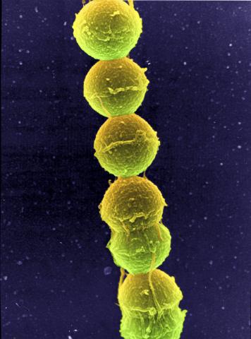
1157: Streptococcus bacteria
Image of Streptococcus, a type (genus) of spherical bacteria that can colonize the throat and back of the mouth. Stroptococci often occur in pairs or in chains, as shown here.
Tina Weatherby Carvalho, University of Hawaii at Manoa
View Media
7002: Plant resistosome
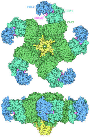
7002: Plant resistosome
The research organism Arabidopsis thaliana forms a large molecular machine called a resistosome to fight off infections. This illustration shows the top and side views of the fully-formed resistosome assembly (PDB entry 6J5T), composed of different proteins including one the plant uses as a decoy, PBL2 (dark blue), that gets uridylylated to begin the process of building the resistosome (uridylyl groups in magenta). Other proteins include RSK1 (turquoise) and ZAR1 (green) subunits. The ends of the ZAR1 subunits (yellow) form a funnel-like protrusion on one side of the assembly (seen in the side view). The funnel can carry out the critical protective function of the resistosome by inserting itself into the cell membrane to form a pore, which leads to a localized programmed cell death. The death of the infected cell helps protect the rest of the plant.
Amy Wu and Christine Zardecki, RCSB Protein Data Bank.
View Media
6998: Zika virus
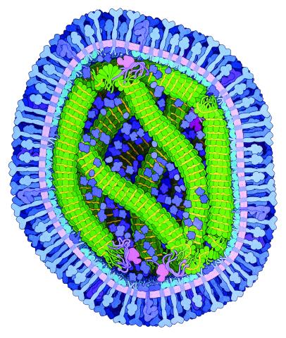
6998: Zika virus
Zika virus is shown in cross section at center left. On the outside, it includes envelope protein (red) and membrane protein (magenta) embedded in a lipid membrane (light purple). Inside, the RNA genome (yellow) is associated with capsid proteins (orange). The viruses are shown interacting with receptors on the cell surface (green) and are surrounded by blood plasma molecules at the top.
Amy Wu and Christine Zardecki, RCSB Protein Data Bank.
View Media
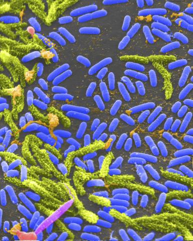
6804: Staphylococcus aureus in the porous coating of a femoral hip stem
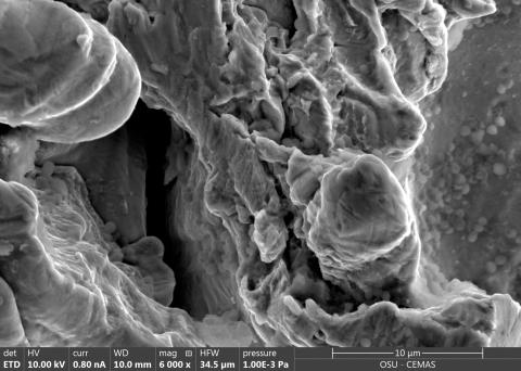
6804: Staphylococcus aureus in the porous coating of a femoral hip stem
Staphylococcus aureus bacteria (blue) on the porous coating of a femoral hip stem used in hip replacement surgery. The relatively rough surface of an implant is a favorable environment for bacteria to attach and grow. This can lead to the development of biofilms, which can cause infections. The researchers who took this image are working to understand where biofilms are likely to develop. This knowledge could support the prevention and treatment of infections. A scanning electron microscope was used to capture this image.
More information on the research that produced this image can be found in the Antibiotics paper "Free-floating aggregate and single-cell-initiated biofilms of Staphylococcus aureus" by Gupta et al.
Related to image 6803 and video 6805.
More information on the research that produced this image can be found in the Antibiotics paper "Free-floating aggregate and single-cell-initiated biofilms of Staphylococcus aureus" by Gupta et al.
Related to image 6803 and video 6805.
Paul Stoodley, The Ohio State University.
View Media
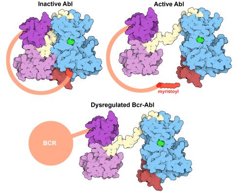
7004: Protein kinases as cancer chemotherapy targets
7004: Protein kinases as cancer chemotherapy targets
Protein kinases—enzymes that add phosphate groups to molecules—are cancer chemotherapy targets because they play significant roles in almost all aspects of cell function, are tightly regulated, and contribute to the development of cancer and other diseases if any alterations to their regulation occur. Genetic abnormalities affecting the c-Abl tyrosine kinase are linked to chronic myelogenous leukemia, a cancer of immature cells in the bone marrow. In the noncancerous form of the protein, binding of a myristoyl group to the kinase domain inhibits the activity of the protein until it is needed (top left shows the inactive form, top right shows the open and active form). The cancerous variant of the protein, called Bcr-Abl, lacks this autoinhibitory myristoyl group and is continually active (bottom). ATP is shown in green bound in the active site of the kinase.
Find these in the RCSB Protein Data Bank: c-Abl tyrosine kinase and regulatory domains (PDB entry 1OPL) and F-actin binding domain (PDB entry 1ZZP).
Find these in the RCSB Protein Data Bank: c-Abl tyrosine kinase and regulatory domains (PDB entry 1OPL) and F-actin binding domain (PDB entry 1ZZP).
Amy Wu and Christine Zardecki, RCSB Protein Data Bank.
View Media
1160: Vibrio bacteria
1160: Vibrio bacteria
Vibrio, a type (genus) of rod-shaped bacteria. Some Vibrio species cause cholera in humans.
Tina Weatherby Carvalho, University of Hawaii at Manoa
View Media

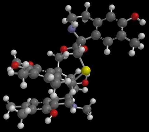
2796: Anti-tumor drug ecteinascidin 743 (ET-743), structure without hydrogens 03
2796: Anti-tumor drug ecteinascidin 743 (ET-743), structure without hydrogens 03
Ecteinascidin 743 (ET-743, brand name Yondelis), was discovered and isolated from a sea squirt, Ecteinascidia turbinata, by NIGMS grantee Kenneth Rinehart at the University of Illinois. It was synthesized by NIGMS grantees E.J. Corey and later by Samuel Danishefsky. Multiple versions of this structure are available as entries 2790-2797.
Timothy Jamison, Massachusetts Institute of Technology
View Media
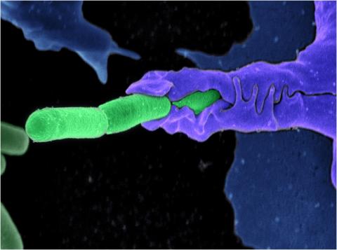
3612: Anthrax bacteria (green) being swallowed by an immune system cell
3612: Anthrax bacteria (green) being swallowed by an immune system cell
Multiple anthrax bacteria (green) being enveloped by an immune system cell (purple). Anthrax bacteria live in soil and form dormant spores that can survive for decades. When animals eat or inhale these spores, the bacteria activate and rapidly increase in number. Today, a highly effective and widely used vaccine has made the disease uncommon in domesticated animals and rare in humans.
This image was part of the Life: Magnified exhibit that ran from June 3, 2014, to January 21, 2015, at Dulles International Airport.
This image was part of the Life: Magnified exhibit that ran from June 3, 2014, to January 21, 2015, at Dulles International Airport.
Camenzind G. Robinson, Sarah Guilman, and Arthur Friedlander, United States Army Medical Research Institute of Infectious Diseases
View Media
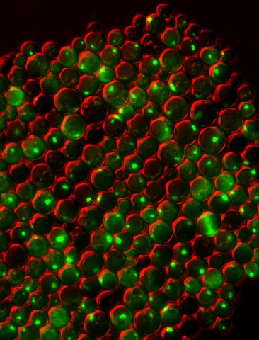
3550: Protein clumping in zinc-deficient yeast cells
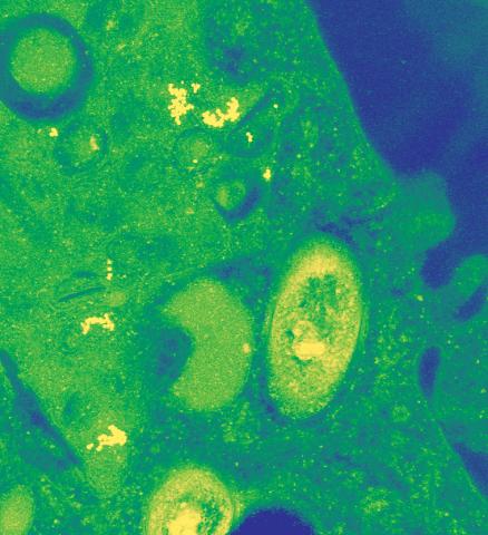
3550: Protein clumping in zinc-deficient yeast cells
The green spots in this image are clumps of protein inside yeast cells that are deficient in both zinc and a protein called Tsa1 that prevents clumping. Protein clumping plays a role in many diseases, including Parkinson's and Alzheimer's, where proteins clump together in the brain. Zinc deficiency within a cell can cause proteins to mis-fold and eventually clump together. Normally, in yeast, Tsa1 codes for so-called "chaperone proteins" which help proteins in stressed cells, such as those with a zinc deficiency, fold correctly. The research behind this image was published in 2013 in the Journal of Biological Chemistry.
Colin MacDiarmid and David Eide, University of Wisconsin--Madison
View Media
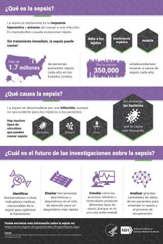
6551: ¿Qué es la sepsis? (Sepsis Infographic)
6551: ¿Qué es la sepsis? (Sepsis Infographic)
La sepsis o septicemia es la respuesta fulminante y extrema del cuerpo a una infección. En los Estados Unidos, más de 1.7 millones de personas contraen sepsis cada año. Sin un tratamiento rápido, la sepsis puede provocar daño de los tejidos, insuficiencia orgánica y muerte. El NIGMS apoya a muchos investigadores en su trabajo para mejorar el diagnóstico y el tratamiento de la sepsis.
Vea 6536 para la versión en inglés de esta infografía.
Vea 6536 para la versión en inglés de esta infografía.
Instituto Nacional de Ciencias Médicas Generales
View Media
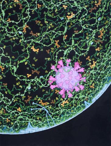
6994: Respiratory droplet
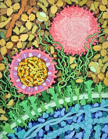
6994: Respiratory droplet
This painting shows a cross section of a small respiratory droplet, like the ones that are thought to transmit SARS-CoV-2, the virus that causes COVID-19. The virus is shown in pink, and the droplet is also filled with molecules that are present in the respiratory tract, including mucins (green), pulmonary surfactant proteins and lipids (blue), and antibodies (tan).
Amy Wu and Christine Zardecki, RCSB Protein Data Bank.
View Media
2797: Anti-tumor drug ecteinascidin 743 (ET-743), structure without hydrogens 04
2797: Anti-tumor drug ecteinascidin 743 (ET-743), structure without hydrogens 04
Ecteinascidin 743 (ET-743, brand name Yondelis), was discovered and isolated from a sea squirt, Ecteinascidia turbinata, by NIGMS grantee Kenneth Rinehart at the University of Illinois. It was synthesized by NIGMS grantees E.J. Corey and later by Samuel Danishefsky. Multiple versions of this structure are available as entries 2790-2797.
Timothy Jamison, Massachusetts Institute of Technology
View Media
3486: Apoptosis reversed
3486: Apoptosis reversed
Two healthy cells (bottom, left) enter into apoptosis (bottom, center) but spring back to life after a fatal toxin is removed (bottom, right; top).
Hogan Tang of the Denise Montell Lab, Johns Hopkins University School of Medicine
View Media
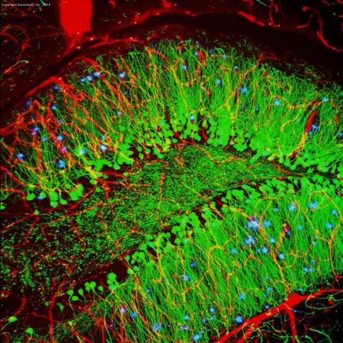
3604: Brain showing hallmarks of Alzheimer's disease
3604: Brain showing hallmarks of Alzheimer's disease
Along with blood vessels (red) and nerve cells (green), this mouse brain shows abnormal protein clumps known as plaques (blue). These plaques multiply in the brains of people with Alzheimer's disease and are associated with the memory impairment characteristic of the disease. Because mice have genomes nearly identical to our own, they are used to study both the genetic and environmental factors that trigger Alzheimer's disease. Experimental treatments are also tested in mice to identify the best potential therapies for human patients.
This image was part of the Life: Magnified exhibit that ran from June 3, 2014, to January 21, 2015, at Dulles International Airport.
This image was part of the Life: Magnified exhibit that ran from June 3, 2014, to January 21, 2015, at Dulles International Airport.
Alvin Gogineni, Genentech
View Media
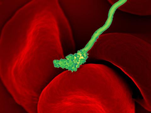
3586: Human blood cells with Borrelia hermsii, a bacterium that causes relapsing fever
3586: Human blood cells with Borrelia hermsii, a bacterium that causes relapsing fever
Relapsing fever is caused by a bacterium and transmitted by certain soft-bodied ticks or body lice. The disease is seldom fatal in humans, but it can be very serious and prolonged. This scanning electron micrograph shows Borrelia hermsii (green), one of the bacterial species that causes the disease, interacting with red blood cells. Micrograph by Robert Fischer, NIAID.
For more information on this see, relapsing fever.
This image was part of the Life: Magnified exhibit that ran from June 3, 2014, to January 21, 2015, at Dulles International Airport.
For more information on this see, relapsing fever.
This image was part of the Life: Magnified exhibit that ran from June 3, 2014, to January 21, 2015, at Dulles International Airport.
NIAID
View Media
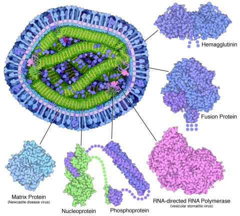
6996: Measles virus proteins
6996: Measles virus proteins
A cross section of the measles virus in which six proteins (enlarged on the outside of the virus) work together to infect cells. The measles virus is extremely infectious; 9 out of 10 people exposed will contract the disease. Fortunately, an effective vaccine protects against infection. Portions of the proteins that have not been determined are shown with dots.
Learn more about the six proteins on PDB 101’s Molecule of the Month: Measles Virus Proteins. Structures are available for the ordered regions of nucleoprotein and phosphoprotein (PDB entries 5E4V, 3ZDO, 1T6O), but the remaining regions are thought to form a flexible, random tangle. For a larger look at the measles virus, see 6995.
Learn more about the six proteins on PDB 101’s Molecule of the Month: Measles Virus Proteins. Structures are available for the ordered regions of nucleoprotein and phosphoprotein (PDB entries 5E4V, 3ZDO, 1T6O), but the remaining regions are thought to form a flexible, random tangle. For a larger look at the measles virus, see 6995.
Amy Wu and Christine Zardecki, RCSB Protein Data Bank.
View Media
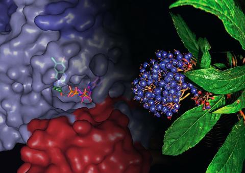
3483: Chang Shan
3483: Chang Shan
For thousands of years, Chinese herbalists have treated malaria using Chang Shan, a root extract from a type of hydrangea that grows in Tibet and Nepal. Recent studies have suggested Chang Shan can also reduce scar formation, treat multiple sclerosis and even slow cancer progression.
Paul Schimmel Lab, Scripps Research Institute
View Media
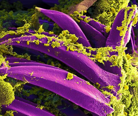
3576: Bubonic plague bacteria on part of the digestive system in a rat flea
3576: Bubonic plague bacteria on part of the digestive system in a rat flea
Here, bubonic plague bacteria (yellow) are shown in the digestive system of a rat flea (purple). The bubonic plague killed a third of Europeans in the mid-14th century. Today, it is still active in Africa, Asia, and the Americas, with as many as 2,000 people infected worldwide each year. If caught early, bubonic plague can be treated with antibiotics.
This image was part of the Life: Magnified exhibit that ran from June 3, 2014, to January 21, 2015, at Dulles International Airport.
This image was part of the Life: Magnified exhibit that ran from June 3, 2014, to January 21, 2015, at Dulles International Airport.
NIAID
View Media
2791: Anti-tumor drug ecteinascidin 743 (ET-743) with hydrogens 02
2791: Anti-tumor drug ecteinascidin 743 (ET-743) with hydrogens 02
Ecteinascidin 743 (ET-743, brand name Yondelis), was discovered and isolated from a sea squirt, Ecteinascidia turbinata, by NIGMS grantee Kenneth Rinehart at the University of Illinois. It was synthesized by NIGMS grantees E.J. Corey and later by Samuel Danishefsky. Multiple versions of this structure are available as entries 2790-2797.
Timothy Jamison, Massachusetts Institute of Technology
View Media
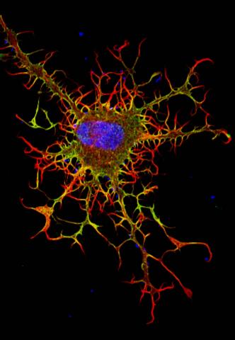
3613: Abnormal, spiky fibroblast
3613: Abnormal, spiky fibroblast
This is a fibroblast, a connective tissue cell that plays an important role in wound healing. Normal fibroblasts have smooth edges. In contrast, this spiky cell is missing a protein that is necessary for proper construction of the cell's skeleton. Its jagged shape makes it impossible for the cell to move normally. In addition to compromising wound healing, abnormal cell movement can lead to birth defects, faulty immune function, and other health problems.
This image was part of the Life: Magnified exhibit that ran from June 3, 2014, to January 21, 2015, at Dulles International Airport.
This image was part of the Life: Magnified exhibit that ran from June 3, 2014, to January 21, 2015, at Dulles International Airport.
Praveen Suraneni, Stowers Institute for Medical Research, Kansas City, Mo.
View Media
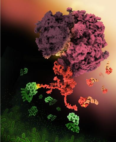
6997: Shiga toxin
6997: Shiga toxin
E. coli bacteria normally live harmlessly in our intestines, but some cause disease by making toxins. One of these toxins, called Shiga toxin (green), inactivates host ribosomes (purple) by mimicking their normal binding partners, the EF-Tu elongation factor (red) complexed with Phe-tRNAPhe (orange).
Find these in the RCSB Protein Data Bank: Shiga toxin 2 (PDB entry 7U6V) and Phe-tRNA (PDB entry 1TTT).
More information about this work can be found in the J. Biol. Chem. paper "Cryo-EM structure of Shiga toxin 2 in complex with the native ribosomal P-stalk reveals residues involved in the binding interaction" by Kulczyk et. al.
Amy Wu and Christine Zardecki, RCSB Protein Data Bank.
View Media
6769: Culex quinquefasciatus mosquito larva
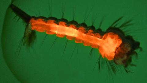
6769: Culex quinquefasciatus mosquito larva
A mosquito larva with genes edited by CRISPR. The red-orange glow is a fluorescent protein used to track the edits. This species of mosquito, Culex quinquefasciatus, can transmit West Nile virus, Japanese encephalitis virus, and avian malaria, among other diseases. The researchers who took this image developed a gene-editing toolkit for Culex quinquefasciatus that could ultimately help stop the mosquitoes from spreading pathogens. The work is described in the Nature Communications paper "Optimized CRISPR tools and site-directed transgenesis towards gene drive development in Culex quinquefasciatus mosquitoes" by Feng et al. Related to image 6770 and video 6771.
Valentino Gantz, University of California, San Diego.
View Media
2793: Anti-tumor drug ecteinascidin 743 (ET-743) with hydrogens 04
2793: Anti-tumor drug ecteinascidin 743 (ET-743) with hydrogens 04
Ecteinascidin 743 (ET-743, brand name Yondelis), was discovered and isolated from a sea squirt, Ecteinascidia turbinata, by NIGMS grantee Kenneth Rinehart at the University of Illinois. It was synthesized by NIGMS grantees E.J. Corey and later by Samuel Danishefsky. Multiple versions of this structure are available as entries 2790-2797.
Timothy Jamison, Massachusetts Institute of Technology
View Media
6999: HIV enzyme
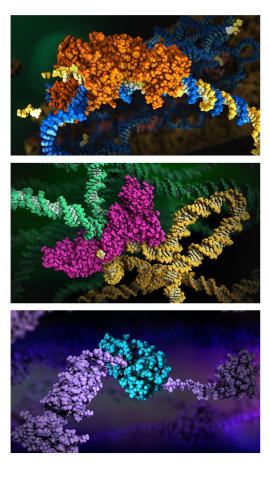
6999: HIV enzyme
These images model the molecular structures of three enzymes with critical roles in the life cycle of the human immunodeficiency virus (HIV). At the top, reverse transcriptase (orange) creates a DNA copy (yellow) of the virus's RNA genome (blue). In the middle image, integrase (magenta) inserts this DNA copy in the DNA genome (green) of the infected cell. At the bottom, much later in the viral life cycle, protease (turquoise) chops up a chain of HIV structural protein (purple) to generate the building blocks for making new viruses. See these enzymes in action on PDB 101’s video A Molecular View of HIV Therapy.
Amy Wu and Christine Zardecki, RCSB Protein Data Bank.
View Media
3497: Wound healing in process
3497: Wound healing in process
Wound healing requires the action of stem cells. In mice that lack the Sept2/ARTS gene, stem cells involved in wound healing live longer and wounds heal faster and more thoroughly than in normal mice. This confocal microscopy image from a mouse lacking the Sept2/ARTS gene shows a tail wound in the process of healing. See more information in the article in Science.
Related to images 3498 and 3500.
Related to images 3498 and 3500.
Hermann Steller, Rockefeller University
View Media
6771: Culex quinquefasciatus mosquito larvae
6771: Culex quinquefasciatus mosquito larvae
Mosquito larvae with genes edited by CRISPR swimming in water. This species of mosquito, Culex quinquefasciatus, can transmit West Nile virus, Japanese encephalitis virus, and avian malaria, among other diseases. The researchers who took this video optimized the gene-editing tool CRISPR for Culex quinquefasciatus that could ultimately help stop the mosquitoes from spreading pathogens. The work is described in the Nature Communications paper "Optimized CRISPR tools and site-directed transgenesis towards gene drive development in Culex quinquefasciatus mosquitoes" by Feng et al. Related to images 6769 and 6770.
Valentino Gantz, University of California, San Diego.
View Media
2792: Anti-tumor drug ecteinascidin 743 (ET-743) with hydrogens 03
2792: Anti-tumor drug ecteinascidin 743 (ET-743) with hydrogens 03
Ecteinascidin 743 (ET-743, brand name Yondelis), was discovered and isolated from a sea squirt, Ecteinascidia turbinata, by NIGMS grantee Kenneth Rinehart at the University of Illinois. It was synthesized by NIGMS grantees E.J. Corey and later by Samuel Danishefsky. Multiple versions of this structure are available as entries 2790-2797.
Timothy Jamison, Massachusetts Institute of Technology
View Media
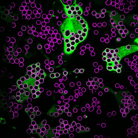
7009: Hungry, hungry macrophages
7009: Hungry, hungry macrophages
Macrophages (green) are the professional eaters of our immune system. They are constantly surveilling our tissues for targets—such as bacteria, dead cells, or even cancer—and clearing them before they can cause harm. In this image, researchers were testing how macrophages responded to different molecules that were attached to silica beads (magenta) coated with a lipid bilayer to mimic a cell membrane.
Find more information on this image in the NIH Director’s Blog post "How to Feed a Macrophage."
Find more information on this image in the NIH Director’s Blog post "How to Feed a Macrophage."
Meghan Morrissey, University of California, Santa Barbara.
View Media
2790: Anti-tumor drug ecteinascidin 743 (ET-743) with hydrogens 01
2790: Anti-tumor drug ecteinascidin 743 (ET-743) with hydrogens 01
Ecteinascidin 743 (ET-743, brand name Yondelis), was discovered and isolated from a sea squirt, Ecteinascidia turbinata, by NIGMS grantee Kenneth Rinehart at the University of Illinois. It was synthesized by NIGMS grantees E.J. Corey and later by Samuel Danishefsky. Multiple versions of this structure are available as entries 2790-2797.
Timothy Jamison, Massachusetts Institute of Technology
View Media
6770: Group of Culex quinquefasciatus mosquito larvae
6770: Group of Culex quinquefasciatus mosquito larvae
Mosquito larvae with genes edited by CRISPR. This species of mosquito, Culex quinquefasciatus, can transmit West Nile virus, Japanese encephalitis virus, and avian malaria, among other diseases. The researchers who took this image developed a gene-editing toolkit for Culex quinquefasciatus that could ultimately help stop the mosquitoes from spreading pathogens. The work is described in the Nature Communications paper "Optimized CRISPR tools and site-directed transgenesis towards gene drive development in Culex quinquefasciatus mosquitoes" by Feng et al. Related to image 6769 and video 6771.
Valentino Gantz, University of California, San Diego.
View Media
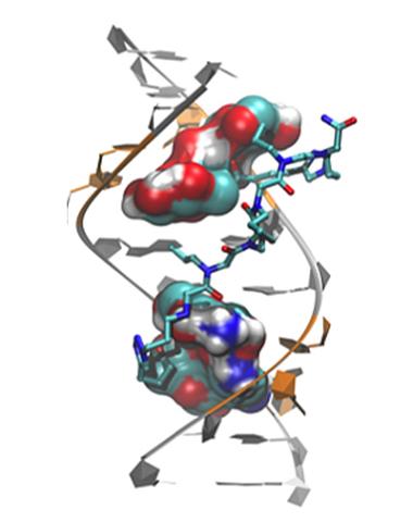
3573: Myotonic dystrophy type 2 genetic defect
3573: Myotonic dystrophy type 2 genetic defect
Scientists revealed a detailed image of the genetic defect that causes myotonic dystrophy type 2 and used that information to design drug candidates to counteract the disease.
Matthew Disney, Scripps Research Institute and Ilyas Yildirim, Northwestern University
View Media
3585: Relapsing fever bacterium (gray) and red blood cells
3585: Relapsing fever bacterium (gray) and red blood cells
Relapsing fever is caused by a bacterium and transmitted by certain soft-bodied ticks or body lice. The disease is seldom fatal in humans, but it can be very serious and prolonged. This scanning electron micrograph shows Borrelia hermsii (green), one of the bacterial species that causes the disease, interacting with red blood cells. Micrograph by Robert Fischer, NIAID. Related to image 3586.
For more information about relapsing fever, see https://www.cdc.gov/relapsing-fever/index.html.
This image is part of the Life: Magnified collection, which was displayed in the Gateway Gallery at Washington Dulles International Airport June 3, 2014, to January 21, 2015.
For more information about relapsing fever, see https://www.cdc.gov/relapsing-fever/index.html.
This image is part of the Life: Magnified collection, which was displayed in the Gateway Gallery at Washington Dulles International Airport June 3, 2014, to January 21, 2015.
NIAID
View Media
3498: Wound healing in process
3498: Wound healing in process
Wound healing requires the action of stem cells. In mice that lack the Sept2/ARTS gene, stem cells involved in wound healing live longer and wounds heal faster and more thoroughly than in normal mice. This confocal microscopy image from a mouse lacking the Sept2/ARTS gene shows a tail wound in the process of healing. See more information in the article in Science.
Related to images 3497 and 3500.
Related to images 3497 and 3500.
Hermann Steller, Rockefeller University
View Media
1166: Leptospira bacteria
1166: Leptospira bacteria
Leptospira, shown here in green, is a type (genus) of elongated, spiral-shaped bacteria. Infection can cause Weil's disease, a kind of jaundice, in humans.
Tina Weatherby Carvalho, University of Hawaii at Manoa
View Media
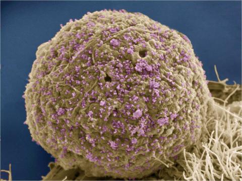
3386: HIV Infected Cell
3386: HIV Infected Cell
The human immunodeficiency virus (HIV), shown here as tiny purple spheres, causes the disease known as AIDS (for acquired immunodeficiency syndrome). HIV can infect multiple cells in your body, including brain cells, but its main target is a cell in the immune system called the CD4 lymphocyte (also called a T-cell or CD4 cell).
Tom Deerinck, National Center for Microscopy and Imaging Research (NCMIR)
View Media
3500: Wound healing in process
3500: Wound healing in process
Wound healing requires the action of stem cells. In mice that lack the Sept2/ARTS gene, stem cells involved in wound healing live longer and wounds heal faster and more thoroughly than in normal mice. This confocal microscopy image from a mouse lacking the Sept2/ARTS gene shows a tail wound in the process of healing. See more information in the article in Science.
Related to images 3497 and 3498.
Related to images 3497 and 3498.
Hermann Steller, Rockefeller University
View Media
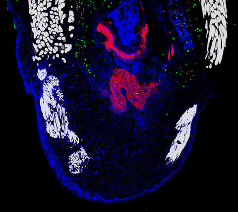
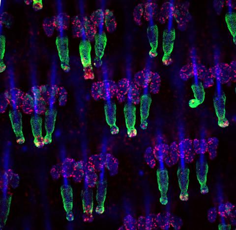
6968: Regenerating lizard tail
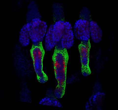
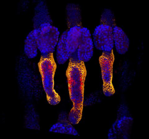
6968: Regenerating lizard tail
The interior of a regenerating lizard tail 14 days after the original tail was amputated. Cell nuclei (blue), proliferating cells (green), cartilage (red), and muscle (white) have been visualized with immunofluorescence staining.
Thomas Lozito, University of Southern California.
View Media
2716: Mycobacterium tuberculosis
2716: Mycobacterium tuberculosis
Mycobacterium tuberculosis, the bacterium that causes tuberculosis, has infected one-quarter of the world's population and causes more than one million deaths each year, according to the World Health Organization.
Reuben Peters, Iowa State University
View Media
3499: Growing hair follicle stem cells
3499: Growing hair follicle stem cells
Wound healing requires the action of stem cells. In mice that lack the Sept2/ARTS gene, stem cells involved in wound healing live longer and wounds heal faster and more thoroughly than in normal mice. This confocal microscopy image from a mouse lacking the Sept2/ARTS gene shows a tail wound in the process of healing. Cell nuclei are in blue. Red and orange mark hair follicle stem cells (hair follicle stem cells activate to cause hair regrowth, which indicates healing). See more information in the article in Science.
Hermann Steller, Rockefeller University
View Media
2573: Simulation of controlled avian flu outbreak
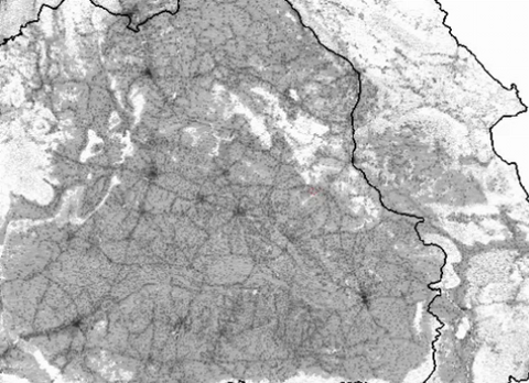
2573: Simulation of controlled avian flu outbreak
This video shows a controlled outbreak of transmissible avian flu among people living in Thailand. Red indicates areas of infection while blue indicates areas where a combination of control measures were implemented. The video shows how control measures contained the infection in 90 days, before it spread elsewhere.
Neil M. Ferguson, Imperial College London
View Media
2574: Simulation of uncontrolled avian flu outbreak
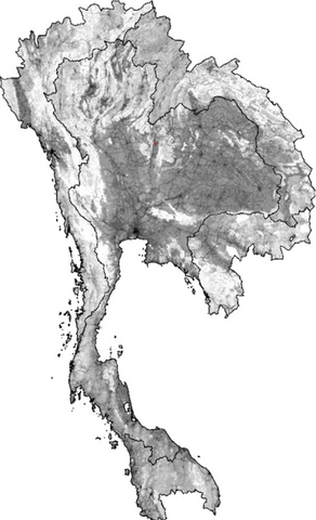
2574: Simulation of uncontrolled avian flu outbreak
This video simulation shows what an uncontrolled outbreak of transmissible avian flu among people living in Thailand might look like. Red indicates new cases while green indicates areas where the epidemic has finished. The video shows the spread of infection and recovery over 300 days in Thailand and neighboring countries.
Neil M. Ferguson, Imperial College London
View Media
6995: Measles virus
6995: Measles virus
A cross section of the measles virus in which six proteins work together to infect cells. The measles virus is extremely infectious; 9 out of 10 people exposed will contract the disease. Fortunately, an effective vaccine protects against infection.
For a zoomed-in look at the six important proteins, see Measles Virus Proteins.
For a zoomed-in look at the six important proteins, see Measles Virus Proteins.
Amy Wu and Christine Zardecki, RCSB Protein Data Bank.
View Media
2794: Anti-tumor drug ecteinascidin 743 (ET-743), structure without hydrogens 01
2794: Anti-tumor drug ecteinascidin 743 (ET-743), structure without hydrogens 01
Ecteinascidin 743 (ET-743, brand name Yondelis), was discovered and isolated from a sea squirt, Ecteinascidia turbinata, by NIGMS grantee Kenneth Rinehart at the University of Illinois. It was synthesized by NIGMS grantees E.J. Corey and later by Samuel Danishefsky. Multiple versions of this structure are available as entries 2790-2797.
Timothy Jamison, Massachusetts Institute of Technology
View Media
3460: Prion protein fibrils 1
3460: Prion protein fibrils 1
Recombinant proteins such as the prion protein shown here are often used to model how proteins misfold and sometimes polymerize in neurodegenerative disorders. This prion protein was expressed in E. coli, purified and fibrillized at pH 7. Image taken in 2004 for a research project by Roger Moore, Ph.D., at Rocky Mountain Laboratories that was published in 2007 in Biochemistry. This image was not used in the publication.
Ken Pekoc (public affairs officer) and Julie Marquardt, NIAID/ Rocky Mountain Laboratories
View Media
6991: SARS-CoV-2 nucleocapsid dimer
6991: SARS-CoV-2 nucleocapsid dimer
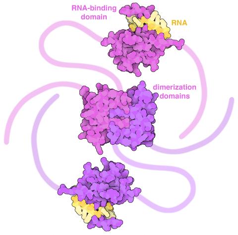
In SARS-CoV-2, the virus that causes COVID-19, nucleocapsid is a complex molecule with many functional parts. One section folds into an RNA-binding domain, with a groove that grips a short segment of the viral genomic RNA. Another section folds into a dimerization domain that brings two nucleocapsid molecules together. The rest of the protein is intrinsically disordered, forming tails at each end of the protein chain and a flexible linker that connects the two structured domains. These disordered regions assist with RNA binding and orchestrate association of nucleocapsid dimers into larger assemblies that package the RNA in the small space inside virions. Nucleocapsid is in magenta and purple, and short RNA strands are in yellow.
Find these in the RCSB Protein Data Bank: RNA-binding domain (PDB entry 7ACT) and Dimerization domain (PDB entry 6WJI).
Find these in the RCSB Protein Data Bank: RNA-binding domain (PDB entry 7ACT) and Dimerization domain (PDB entry 6WJI).
Amy Wu and Christine Zardecki, RCSB Protein Data Bank.
View Media