
Switch to List View
Image and Video Gallery
This is a searchable collection of scientific photos, illustrations, and videos. The images and videos in this gallery are licensed under Creative Commons Attribution Non-Commercial ShareAlike 3.0. This license lets you remix, tweak, and build upon this work non-commercially, as long as you credit and license your new creations under identical terms.
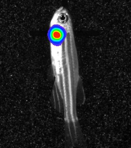
3558: Bioluminescent imaging in adult zebrafish - lateral view
3558: Bioluminescent imaging in adult zebrafish - lateral view
Luciferase-based imaging enables visualization and quantification of internal organs and transplanted cells in live adult zebrafish. In this image, a cardiac muscle-restricted promoter drives firefly luciferase expression (lateral view).
For imagery of both the lateral and overhead view go to 3556.
For imagery of the overhead view go to 3557.
For more information about the illumated area go to 3559.
For imagery of both the lateral and overhead view go to 3556.
For imagery of the overhead view go to 3557.
For more information about the illumated area go to 3559.
Kenneth Poss, Duke University
View Media
2431: Fruit fly embryo
2431: Fruit fly embryo
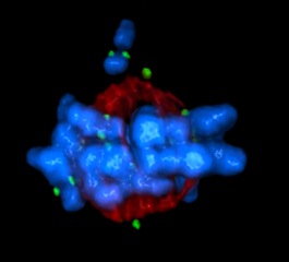
Cells in an early-stage fruit fly embryo, showing the DIAP1 protein (pink), an inhibitor of apoptosis.
Hermann Steller, Rockefeller University
View Media
3277: Human ES cells turn into insulin-producing cells
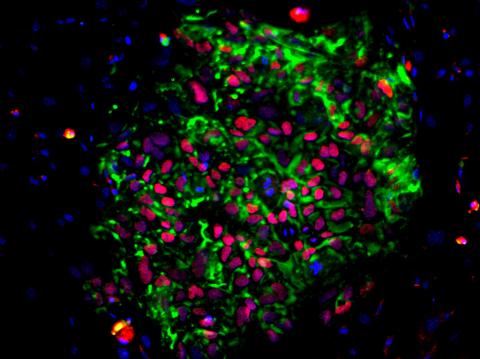
3277: Human ES cells turn into insulin-producing cells
Human embryonic stem cells were differentiated into cells like those found in the pancreas (blue), which give rise to insulin-producing cells (red). When implanted in mice, the stem cell-derived pancreatic cells can replace the insulin that isn't produced in type 1 diabetes. Image and caption information courtesy of the California Institute for Regenerative Medicine.
Eugene Brandon, ViaCyte, via CIRM
View Media
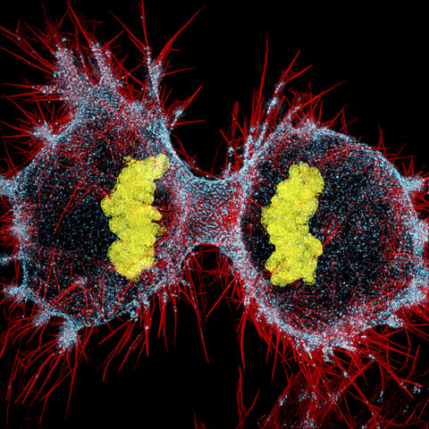
6520: HeLa cell undergoing division into two daughter cells
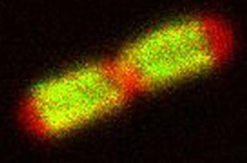
6520: HeLa cell undergoing division into two daughter cells
Here, a human HeLa cell (a type of immortal cell line used in laboratory experiments) is undergoing cell division. They come from cervical cancer cells that were obtained in 1951 from Henrietta Lacks, a patient at the Johns Hopkins Hospital. The final stage of division, called cytokinesis, occurs after the genomes—shown in yellow—have split into two new daughter cells. The myosin II is a motor protein shown in blue, and the actin filaments, which are types of protein that support cell structure, are shown in red.
Dylan T. Burnette, Ph.D., Vanderbilt University School of Medicine.
View Media
6503: Arabidopsis Thaliana: Flowers Spring to Life
6503: Arabidopsis Thaliana: Flowers Spring to Life
This image capture shows how a single gene, STM, plays a starring role in plant development. This gene acts like a molecular fountain of youth, keeping cells ever-young until it’s time to grow up and commit to making flowers and other plant parts. Because of its ease of use and low cost, Arabidopsis is a favorite model for scientists to learn the basic principles driving tissue growth and regrowth for humans as well as the beautiful plants outside your window. Image captured from video Watch Flowers Spring to Life, featured in the NIH Director's Blog: Watch Flowers Spring to Life.
Nathanaёl Prunet NIH Support: National Institute of General Medical Sciences
View Media
3363: Dopamine D3 receptor
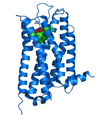
3363: Dopamine D3 receptor
The receptor is shown bound to an antagonist, eticlopride
Raymond Stevens, The Scripps Research Institute
View Media
2494: VDAC-1 (3)
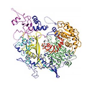
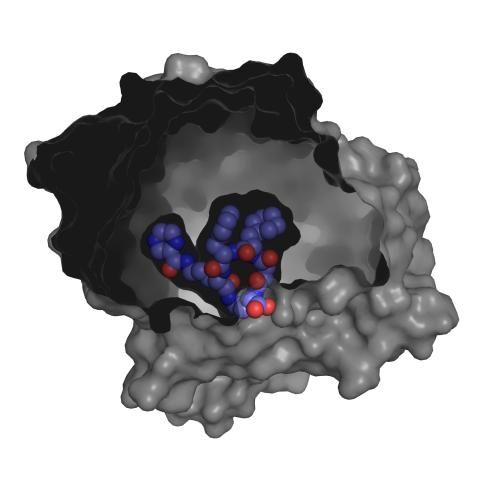
2494: VDAC-1 (3)
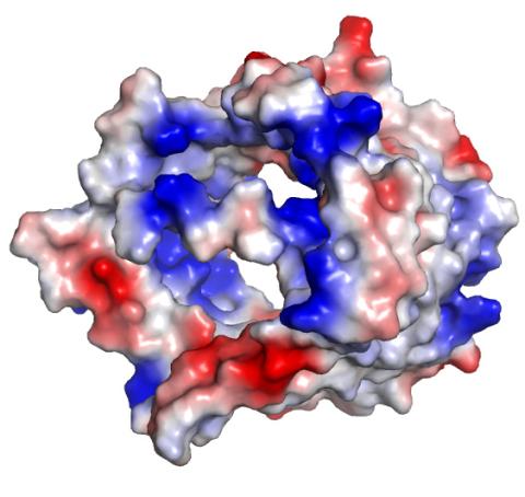
The structure of the pore-forming protein VDAC-1 from humans. This molecule mediates the flow of products needed for metabolism--in particular the export of ATP--across the outer membrane of mitochondria, the power plants for eukaryotic cells. VDAC-1 is involved in metabolism and the self-destruction of cells--two biological processes central to health.
Related to images 2491, 2495, and 2488.
Related to images 2491, 2495, and 2488.
Gerhard Wagner, Harvard Medical School
View Media
3567: RSV-Infected Cell
3567: RSV-Infected Cell
Viral RNA (red) in an RSV-infected cell. More information about the research behind this image can be found in a Biomedical Beat Blog posting from January 2014.
Eric Alonas and Philip Santangelo, Georgia Institute of Technology and Emory University
View Media
6750: C. elegans with blue and yellow lights in the background
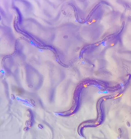
6750: C. elegans with blue and yellow lights in the background
These microscopic roundworms, called Caenorhabditis elegans, lack eyes and the opsin proteins used by visual systems to detect colors. However, researchers found that the worms can still sense the color of light in a way that enables them to avoid pigmented toxins made by bacteria. This image was captured using a stereo microscope.
H. Robert Horvitz and Dipon Ghosh, Massachusetts Institute of Technology.
View Media

6521: Yeast art depicting the New York City skyline
6521: Yeast art depicting the New York City skyline
This skyline of New York City was created by “printing” nanodroplets containing yeast (Saccharomyces cerevisiae) onto a large plate. Each dot is a separate yeast colony. As the colonies grew, a picture emerged, creating art. To make the different colors shown here, yeast strains were genetically engineered to produce pigments naturally made by bacteria, fungi, and sea creatures such as coral and sea anemones. Using genes from other organisms to make biological compounds paves the way toward harnessing yeast in the production of other useful molecules, from food to fuels and drugs.
Michael Shen, Ph.D., Jasmine Temple, Leslie Mitchell, Ph.D., and Jef Boeke, Ph.D., New York University School of Medicine; and Nick Phillips, James Chuang, Ph.D., and Jiarui Wang, Johns Hopkins University.
View Media
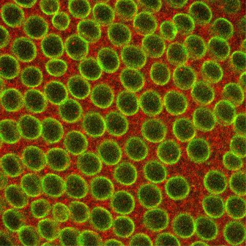
3793: Nucleolus subcompartments spontaneously self-assemble 4
3793: Nucleolus subcompartments spontaneously self-assemble 4
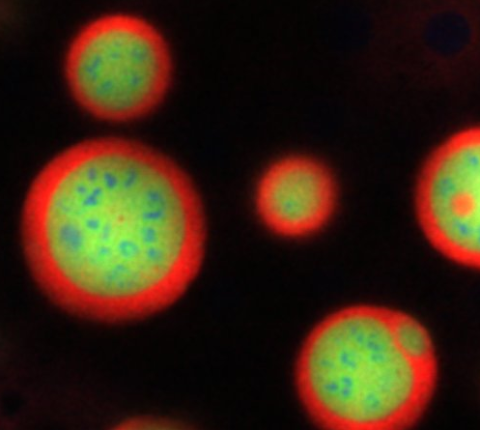
What looks a little like distant planets with some mysterious surface features are actually assemblies of proteins normally found in the cell's nucleolus, a small but very important protein complex located in the cell's nucleus. It forms on the chromosomes at the location where the genes for the RNAs are that make up the structure of the ribosome, the indispensable cellular machine that makes proteins from messenger RNAs.
However, how the nucleolus grows and maintains its structure has puzzled scientists for some time. It turns out that even though it looks like a simple liquid blob, it's rather well-organized, consisting of three distinct layers: the fibrillar center, where the RNA polymerase is active; the dense fibrillar component, which is enriched in the protein fibrillarin; and the granular component, which contains a protein called nucleophosmin. Researchers have now discovered that this multilayer structure of the nucleolus arises from differences in how the proteins in each compartment mix with water and with each other. These differences let the proteins readily separate from each other into the three nucleolus compartments.
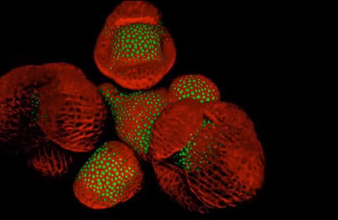
This photo of nucleolus proteins in the eggs of a commonly used lab animal, the frog Xenopus laevis, shows each of the nucleolus compartments (the granular component is shown in red, the fibrillarin in yellow-green, and the fibrillar center in blue). The researchers have found that these compartments spontaneously fuse with each other on encounter without mixing with the other compartments.
For more details on this research, see this press release from Princeton. Related to video 3789, video 3791 and image 3792.
However, how the nucleolus grows and maintains its structure has puzzled scientists for some time. It turns out that even though it looks like a simple liquid blob, it's rather well-organized, consisting of three distinct layers: the fibrillar center, where the RNA polymerase is active; the dense fibrillar component, which is enriched in the protein fibrillarin; and the granular component, which contains a protein called nucleophosmin. Researchers have now discovered that this multilayer structure of the nucleolus arises from differences in how the proteins in each compartment mix with water and with each other. These differences let the proteins readily separate from each other into the three nucleolus compartments.
This photo of nucleolus proteins in the eggs of a commonly used lab animal, the frog Xenopus laevis, shows each of the nucleolus compartments (the granular component is shown in red, the fibrillarin in yellow-green, and the fibrillar center in blue). The researchers have found that these compartments spontaneously fuse with each other on encounter without mixing with the other compartments.
For more details on this research, see this press release from Princeton. Related to video 3789, video 3791 and image 3792.
Nilesh Vaidya, Princeton University
View Media
6932: Axolotl
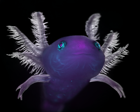
6932: Axolotl
An axolotl—a type of salamander—that has been genetically modified so that its developing nervous system glows purple and its Schwann cell nuclei appear light blue. Schwann cells insulate and provide nutrients to peripheral nerve cells. Researchers often study axolotls for their extensive regenerative abilities. They can regrow tails, limbs, spinal cords, brains, and more. The researcher who took this image focuses on the role of the peripheral nervous system during limb regeneration.
This image was captured using a stereo microscope.
Related to images 6927 and 6928.
This image was captured using a stereo microscope.
Related to images 6927 and 6928.
Prayag Murawala, MDI Biological Laboratory and Hannover Medical School.
View Media
5882: Beta-galactosidase montage showing cryo-EM improvement--transparent background
5882: Beta-galactosidase montage showing cryo-EM improvement--transparent background
Composite image of beta-galactosidase showing how cryo-EM’s resolution has improved dramatically in recent years. Older images to the left, more recent to the right. Related to image 5883. NIH Director Francis Collins featured this on his blog on January 14, 2016.
Veronica Falconieri, Sriram Subramaniam Lab, National Cancer Institute
View Media
3295: Cluster analysis of mysterious protein
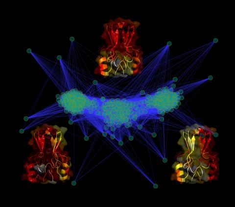
3295: Cluster analysis of mysterious protein
Researchers use cluster analysis to study protein shape and function. Each green circle represents one potential shape of the protein mitoNEET. The longer the blue line between two circles, the greater the differences between the shapes. Most shapes are similar; they fall into three clusters that are represented by the three images of the protein. From a Rice University news release. Graduate student Elizabeth Baxter and Patricia Jennings, professor of chemistry and biochemistry at UCSD, collaborated with José Onuchic, a physicist at Rice University, on this work.
Patricia Jennings and Elizabeth Baxter, University of California, San Diego
View Media
2752: Bacterial spore
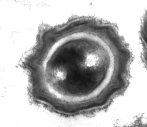
2752: Bacterial spore
A spore from the bacterium Bacillus subtilis shows four outer layers that protect the cell from harsh environmental conditions.
Patrick Eichenberger, New York University
View Media
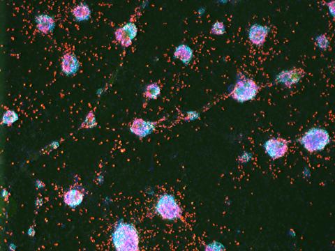
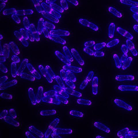
6984: Fruit fly starvation leads to adipokine accumulation
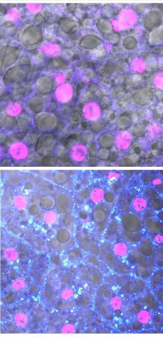
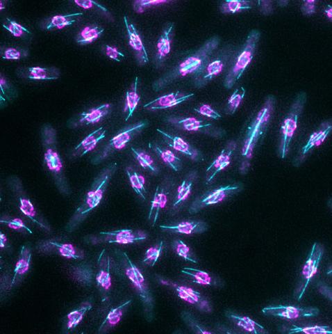
6984: Fruit fly starvation leads to adipokine accumulation
Adult Drosophila abdominal fat tissue showing cell nuclei labelled in magenta. The upper panel is from well-fed flies, and the lower panel is from flies that have been deprived of food for 4 hours. Starvation results in the accumulation of a key adipokine—a fat hormone (blue-green dots).
Related to images 6982, 6983, and 6985.
Related to images 6982, 6983, and 6985.
Akhila Rajan, Fred Hutchinson Cancer Center
View Media
2442: Hydra 06
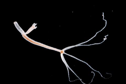
2442: Hydra 06
Hydra magnipapillata is an invertebrate animal used as a model organism to study developmental questions, for example the formation of the body axis.
Hiroshi Shimizu, National Institute of Genetics in Mishima, Japan
View Media
3483: Chang Shan
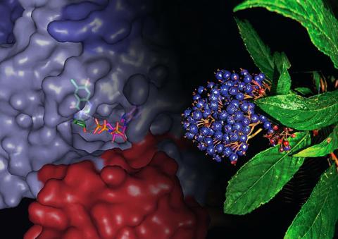
3483: Chang Shan
For thousands of years, Chinese herbalists have treated malaria using Chang Shan, a root extract from a type of hydrangea that grows in Tibet and Nepal. Recent studies have suggested Chang Shan can also reduce scar formation, treat multiple sclerosis and even slow cancer progression.
Paul Schimmel Lab, Scripps Research Institute
View Media

3475: Automated Worm Sorter - 4
3475: Automated Worm Sorter - 4
Georgia Tech associate professor Hang Lu holds a microfluidic chip that is part of a system that uses artificial intelligence and cutting-edge image processing to automatically examine large number of nematodes used for genetic research.
Georgia Tech/Gary Meek
View Media
3432: Mouse mammary cells lacking anti-cancer protein
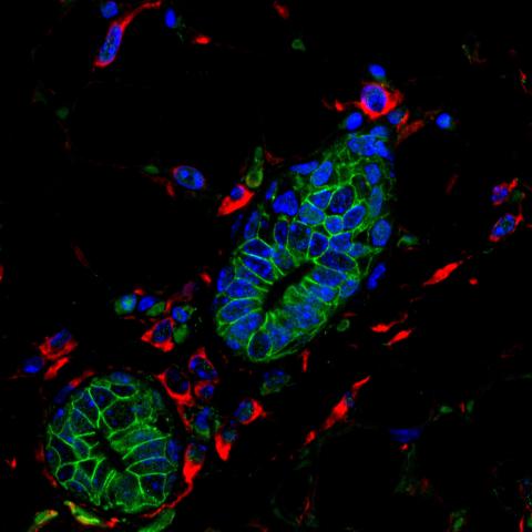
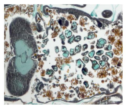
3432: Mouse mammary cells lacking anti-cancer protein
Shortly after a pregnant woman gives birth, her breasts start to secrete milk. This process is triggered by hormonal and genetic cues, including the protein Elf5. Scientists discovered that Elf5 also has another job--it staves off cancer. Early in the development of breast cancer, human breast cells often lose Elf5 proteins. Cells without Elf5 change shape and spread readily--properties associated with metastasis. This image shows cells in the mouse mammary gland that are lacking Elf5, leading to the overproduction of other proteins (red) that increase the likelihood of metastasis.
Nature Cell Biology, November 2012, Volume 14 No 11 pp1113-1231
View Media
6966: Dying melanoma cells
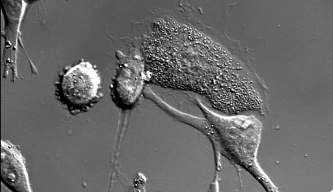
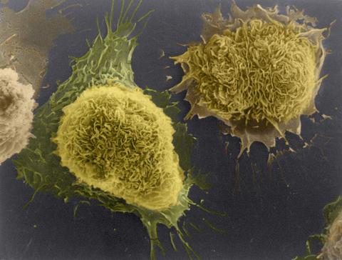
6966: Dying melanoma cells
Melanoma (skin cancer) cells undergoing programmed cell death, also called apoptosis. This process was triggered by raising the pH of the medium that the cells were growing in. Melanoma in people cannot be treated by raising pH because that would also kill healthy cells. This video was taken using a differential interference contrast (DIC) microscope.
Dylan T. Burnette, Vanderbilt University School of Medicine.
View Media
3278: Induced pluripotent stem cells from skin
3278: Induced pluripotent stem cells from skin
These induced pluripotent stem cells (iPS cells) were derived from a woman's skin. Green and red indicate proteins found in reprogrammed cells but not in skin cells (TRA1-62 and NANOG). These cells can then develop into different cell types. Image and caption information courtesy of the California Institute for Regenerative Medicine. Related to image 3279.
Kathrin Plath lab, University of California, Los Angeles, via CIRM
View Media
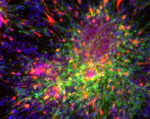
3270: Dopaminergic neurons from ES cells
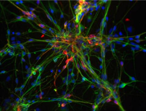
3270: Dopaminergic neurons from ES cells
Human embryonic stem cells differentiated into dopaminergic neurons, the type that degenerate in Parkinson's disease. Image courtesy of the California Institute for Regenerative Medicine. Related to images 3271 and 3285.
Jeannie Liu, Lab of Jan Nolta, University of California, Davis, via CIRM
View Media
3408: Kluyveromyces polysporus Argonaute bound to guide RNA
3408: Kluyveromyces polysporus Argonaute bound to guide RNA
A segment of siRNA, shown in red, guides a "slicer" protein called Argonaute (multi-colored twists and corkscrews) to the target RNA molecules.
Kotaro Nakanishi and David Weinberg, Massachusetts Institute of Technology
View Media
3639: Cerebellum: the brain's locomotion control center
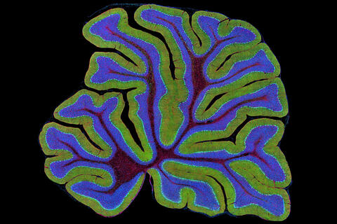
3639: Cerebellum: the brain's locomotion control center
The cerebellum of a mouse is shown here in cross-section. The cerebellum is the brain's locomotion control center. Every time you shoot a basketball, tie your shoe or chop an onion, your cerebellum fires into action. Found at the base of your brain, the cerebellum is a single layer of tissue with deep folds like an accordion. People with damage to this region of the brain often have difficulty with balance, coordination and fine motor skills. For a higher magnification, see image 3371.
This image was part of the Life: Magnified exhibit that ran from June 3, 2014, to January 21, 2015, at Dulles International Airport.
This image was part of the Life: Magnified exhibit that ran from June 3, 2014, to January 21, 2015, at Dulles International Airport.
Thomas Deerinck, National Center for Microscopy and Imaging Research, University of California, San Diego
View Media
2310: Cellular traffic
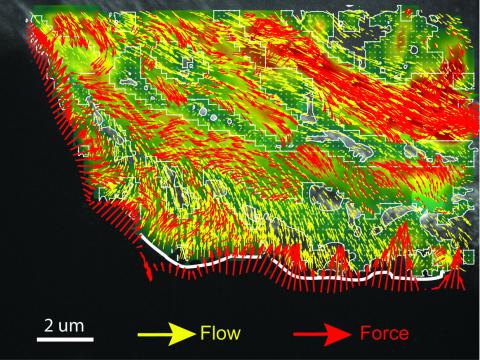
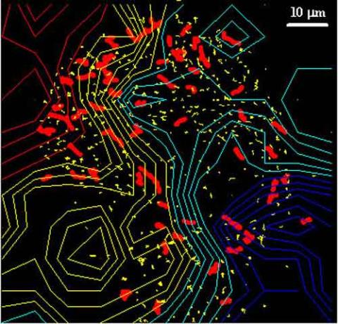
2310: Cellular traffic
Like tractor-trailers on a highway, small sacs called vesicles transport substances within cells. This image tracks the motion of vesicles in a living cell. The short red and yellow marks offer information on vesicle movement. The lines spanning the image show overall traffic trends. Typically, the sacs flow from the lower right (blue) to the upper left (red) corner of the picture. Such maps help researchers follow different kinds of cellular processes as they unfold.
Alexey Sharonov and Robin Hochstrasser, University of Pennsylvania
View Media
3415: X-ray co-crystal structure of Src kinase bound to a DNA-templated macrocycle inhibitor 3
3415: X-ray co-crystal structure of Src kinase bound to a DNA-templated macrocycle inhibitor 3
X-ray co-crystal structure of Src kinase bound to a DNA-templated macrocycle inhibitor. Related to 3413, 3414, 3416, 3417, 3418, and 3419.
Markus A. Seeliger, Stony Brook University Medical School and David R. Liu, Harvard University
View Media

2759: Cross section of a Drosophila melanogaster pupa lacking Draper
2759: Cross section of a Drosophila melanogaster pupa lacking Draper
In the absence of the engulfment receptor Draper, salivary gland cells (light blue) persist in the thorax of a developing Drosophila melanogaster pupa. See image 2758 for a cross section of a normal pupa that does express Draper.
Christina McPhee and Eric Baehrecke, University of Massachusetts Medical School
View Media
1178: Cultured cells
1178: Cultured cells
This image of laboratory-grown cells was taken with the help of a scanning electron microscope, which yields detailed images of cell surfaces.
Tina Weatherby Carvalho, University of Hawaii at Manoa
View Media
2386: Sortase b from B. anthracis
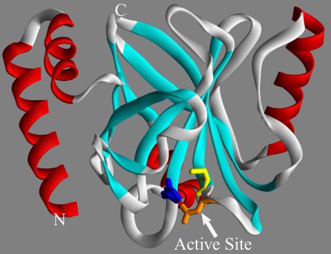
2386: Sortase b from B. anthracis
Structure of sortase b from the bacterium B. anthracis, which causes anthrax. Sortase b is an enzyme used to rob red blood cells of iron, which the bacteria need to survive.
Midwest Center for Structural Genomics, PSI
View Media
3613: Abnormal, spiky fibroblast
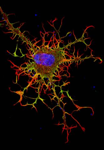
3613: Abnormal, spiky fibroblast
This is a fibroblast, a connective tissue cell that plays an important role in wound healing. Normal fibroblasts have smooth edges. In contrast, this spiky cell is missing a protein that is necessary for proper construction of the cell's skeleton. Its jagged shape makes it impossible for the cell to move normally. In addition to compromising wound healing, abnormal cell movement can lead to birth defects, faulty immune function, and other health problems.
This image was part of the Life: Magnified exhibit that ran from June 3, 2014, to January 21, 2015, at Dulles International Airport.
This image was part of the Life: Magnified exhibit that ran from June 3, 2014, to January 21, 2015, at Dulles International Airport.
Praveen Suraneni, Stowers Institute for Medical Research, Kansas City, Mo.
View Media
6994: Respiratory droplet
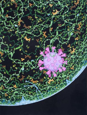
6994: Respiratory droplet
This painting shows a cross section of a small respiratory droplet, like the ones that are thought to transmit SARS-CoV-2, the virus that causes COVID-19. The virus is shown in pink, and the droplet is also filled with molecules that are present in the respiratory tract, including mucins (green), pulmonary surfactant proteins and lipids (blue), and antibodies (tan).
Amy Wu and Christine Zardecki, RCSB Protein Data Bank.
View Media
3549: TonB protein in gram-negative bacteria
3549: TonB protein in gram-negative bacteria
The green in this image highlights a protein called TonB, which is produced by many gram-negative bacteria, including those that cause typhoid fever, meningitis and dysentery. TonB lets bacteria take up iron from the host's body, which they need to survive. More information about the research behind this image can be found in a Biomedical Beat Blog posting from August 2013.
Phillip Klebba, Kansas State University
View Media
6961: C. elegans showing internal structures
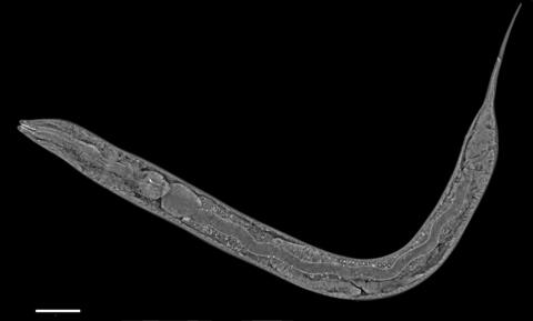
6961: C. elegans showing internal structures
An image of Caenorhabditis elegans, a tiny roundworm, showing internal structures including the intestine, pharynx, and body wall muscle. C. elegans is one of the simplest organisms with a nervous system. Scientists use it to study nervous system development, among other things. This image was captured with a quantitative orientation-independent differential interference contrast (OI-DIC) microscope. The scale bar is 100 µm.
More information about the microscopy that produced this image can be found in the Journal of Microscopy paper by Malamy and Shribak.
More information about the microscopy that produced this image can be found in the Journal of Microscopy paper by Malamy and Shribak.
Michael Shribak, Marine Biological Laboratory/University of Chicago.
View Media
5896: Stetten Lecture 2017poster image
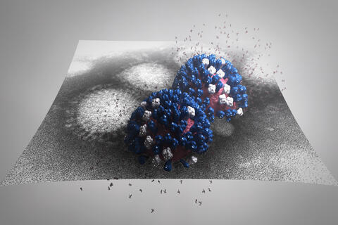
5896: Stetten Lecture 2017poster image
This image is featured on the poster for Dr. Rommie Amaro's 2017 Stetten Lecture. It depicts a detailed physical model of an influenza virus, incorporating information from several structural data sources. The small molecules around the virus are sialic acid molecules. The virus binds to and cleaves sialic acid as it enters and exits host cells. Researchers are building these highly detailed molecular scale models of different biomedical systems and then “bringing them to life” with physics-based methods, either molecular or Brownian dynamics simulations, to understand the structural dynamics of the systems and their complex interactions with drug or substrate molecules.
Dr. Rommie Amaro, University of California, San Diego
View Media
3641: A mammalian eye has approximately 70 different cell types
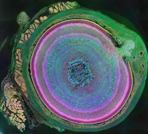
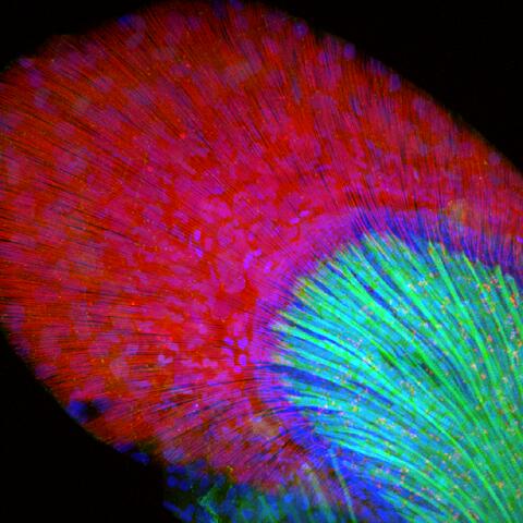
3641: A mammalian eye has approximately 70 different cell types
The incredible complexity of a mammalian eye (in this case from a mouse) is captured here. Each color represents a different type of cell. In total, there are nearly 70 different cell types, including the retina's many rings and the peach-colored muscle cells clustered on the left.
This image was part of the Life: Magnified exhibit that ran from June 3, 2014, to January 21, 2015, at Dulles International Airport.
This image was part of the Life: Magnified exhibit that ran from June 3, 2014, to January 21, 2015, at Dulles International Airport.
Bryan William Jones and Robert E. Marc, University of Utah
View Media
3677: Human skeletal muscle
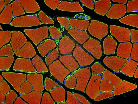
3677: Human skeletal muscle
Cross section of human skeletal muscle. Image taken with a confocal fluorescent light microscope.
Tom Deerinck, National Center for Microscopy and Imaging Research (NCMIR)
View Media
5765: Mitotic cell awaits chromosome alignment
5765: Mitotic cell awaits chromosome alignment
During mitosis, spindle microtubules (red) attach to chromosome pairs (blue), directing them to the spindle equator. This midline alignment is critical for equal distribution of chromosomes in the dividing cell. Scientists are interested in how the protein kinase Plk1 (green) regulates this activity in human cells. Image is a volume projection of multiple deconvolved z-planes acquired with a Nikon widefield fluorescence microscope. This image was chosen as a winner of the 2016 NIH-funded research image call. Related to image 5766.
The research that led to this image was funded by NIGMS.
View Media
The research that led to this image was funded by NIGMS.
3546: Insulin and protein interact in pancreatic beta cells
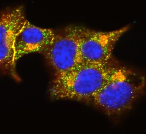
3546: Insulin and protein interact in pancreatic beta cells
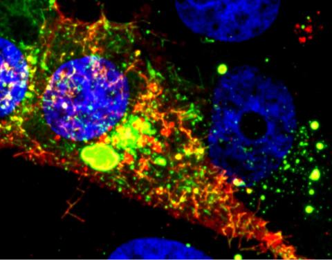
A large number of proteins interact with the hormone insulin as it is produced in and secreted from the beta cells of the pancreas. In this image, the interactions of TMEM24 protein (green) and insulin (red) in pancreatic beta cells are shown in yellow. More information about the research behind this image can be found in a Biomedical Beat Blog posting from November 2013.
William E. Balch, The Scripps Research Institute
View Media
6551: ¿Qué es la sepsis? (Sepsis Infographic)
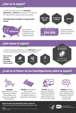
6551: ¿Qué es la sepsis? (Sepsis Infographic)
La sepsis o septicemia es la respuesta fulminante y extrema del cuerpo a una infección. En los Estados Unidos, más de 1.7 millones de personas contraen sepsis cada año. Sin un tratamiento rápido, la sepsis puede provocar daño de los tejidos, insuficiencia orgánica y muerte. El NIGMS apoya a muchos investigadores en su trabajo para mejorar el diagnóstico y el tratamiento de la sepsis.
Vea 6536 para la versión en inglés de esta infografía.
Vea 6536 para la versión en inglés de esta infografía.
Instituto Nacional de Ciencias Médicas Generales
View Media
3443: Interphase in Xenopus frog cells
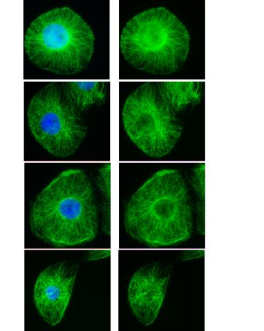
3443: Interphase in Xenopus frog cells
These images show frog cells in interphase. The cells are Xenopus XL177 cells, which are derived from tadpole epithelial cells. The microtubules are green and the chromosomes are blue. Related to 3442.
Claire Walczak, who took them while working as a postdoc in the laboratory of Timothy Mitchison.
View Media
2542: Nucleotides make up DNA (with labels)
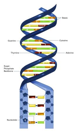
2542: Nucleotides make up DNA (with labels)
DNA consists of two long, twisted chains made up of nucleotides. Each nucleotide contains one base, one phosphate molecule, and the sugar molecule deoxyribose. The bases in DNA nucleotides are adenine, thymine, cytosine, and guanine. See image 2541 for an unlabeled version of this illustration. Featured in The New Genetics.
Crabtree + Company
View Media
3598: Developing zebrafish fin
3598: Developing zebrafish fin
Originally from the waters of India, Nepal, and neighboring countries, zebrafish can now be found swimming in science labs (and home aquariums) throughout the world. This fish is a favorite study subject for scientists interested in how genes guide the early stages of prenatal development (including the developing fin shown here) and in the effects of environmental contamination on embryos.
In this image, green fluorescent protein (GFP) is expressed where the gene sox9b is expressed. Collagen (red) marks the fin rays, and DNA, stained with a dye called DAPI, is in blue. sox9b plays many important roles during development, including the building of the heart and brain, and is also necessary for skeletal development. At the University of Wisconsin, researchers have found that exposure to contaminants that bind the aryl-hydrocarbon receptor results in the downregulation of sox9b. Loss of sox9b severely disrupts development in zebrafish and causes a life-threatening disorder called campomelic dysplasia (CD) in humans. CD is characterized by cardiovascular, neural, and skeletal defects. By studying the roles of genes such as sox9b in zebrafish, scientists hope to better understand normal development in humans as well as how to treat developmental disorders and diseases.
This image was part of the Life: Magnified exhibit that ran from June 3, 2014, to January 21, 2015, at Dulles International Airport.
Jessica Plavicki
View Media
2750: Antibodies in silica honeycomb
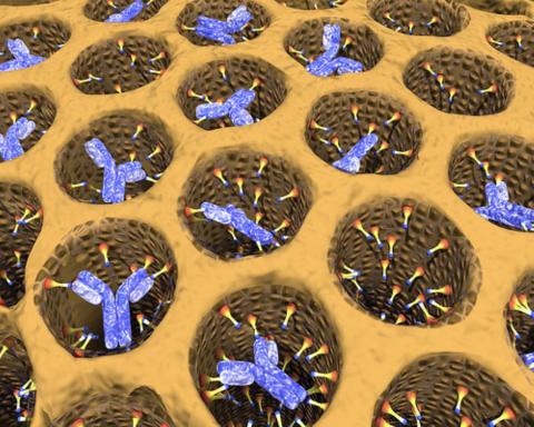
2750: Antibodies in silica honeycomb
Antibodies are among the most promising therapies for certain forms of cancer, but patients must take them intravenously, exposing healthy tissues to the drug and increasing the risk of side effects. A team of biochemists packed the anticancer antibodies into porous silica particles to deliver a heavy dose directly to tumors in mice.
Chenghong Lei, Pacific Northwest National Laboratory & Karl Erik Hellstrom, University of Washington
View Media
6798: Yeast cells with nuclear envelopes and tubulin
6798: Yeast cells with nuclear envelopes and tubulin
Yeast cells with nuclear envelopes shown in magenta and tubulin shown in light blue. The nuclear envelope defines the borders of the nucleus, which houses DNA. Tubulin is a protein that makes up microtubules—strong, hollow fibers that provide structure to cells and help direct chromosomes during cell division. This image was captured using wide-field microscopy with deconvolution.
Related to images 6791, 6792, 6793, 6794, 6797, and videos 6795 and 6796.
Related to images 6791, 6792, 6793, 6794, 6797, and videos 6795 and 6796.
Alaina Willet, Kathy Gould’s lab, Vanderbilt University.
View Media
6799: Phagosome in macrophage cell
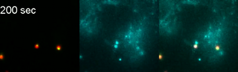
6799: Phagosome in macrophage cell
A sensor particle being engulfed by a macrophage—an immune cell—and encapsuled in a compartment called a phagosome. The phagosome then fuses with lysosomes—another type of compartment. The left video shows snowman-shaped sensor particles with fluorescent green nanoparticle “heads” and “bodies” colored red by Förster Resonance Energy Transfer (FRET)-donor fluorophores. The middle video visualizes light blue FRET signals that are only generated when the “snowman” sensor—the FRET-donor—fuses with the lysosomes, which are loaded with FRET-acceptors. The right video combines the other two. The videos were captured using epi-fluorescence microscopy.
More details can be found in the paper “Transport motility of phagosomes on actin and microtubules regulates timing and kinetics of their maturation” by Yu et al.
More details can be found in the paper “Transport motility of phagosomes on actin and microtubules regulates timing and kinetics of their maturation” by Yu et al.
Yan Yu, Indiana University, Bloomington.
View Media
3271: Dopaminergic neurons derived from mouse embryonic stem cells
3271: Dopaminergic neurons derived from mouse embryonic stem cells
These neurons are derived from mouse embryonic stem cells. Red shows cells making a protein called TH that is characteristic of the neurons that degenerate in Parkinson's disease. Green indicates a protein that's found in all neurons. Blue indicates the nuclei of all cells. Studying dopaminergic neurons can help researchers understand the origins of Parkinson's disease and could be used to screen potential new drugs. Image and caption information courtesy of the California Institute for Regenerative Medicine. Related to images 3270 and 3285.
Yaping Sun, lab of Su Guo, University of California, San Francisco, via CIRM
View Media
6794: Yeast cells with Fimbrin Fim1
6794: Yeast cells with Fimbrin Fim1
Yeast cells with the protein Fimbrin Fim1 shown in magenta. This protein plays a role in cell division. This image was captured using wide-field microscopy with deconvolution.
Related to images 6791, 6792, 6793, 6797, 6798, and videos 6795 and 6796.
Related to images 6791, 6792, 6793, 6797, 6798, and videos 6795 and 6796.
Alaina Willet, Kathy Gould’s lab, Vanderbilt University.
View Media