
Switch to List View
Image and Video Gallery
This is a searchable collection of scientific photos, illustrations, and videos. The images and videos in this gallery are licensed under Creative Commons Attribution Non-Commercial ShareAlike 3.0. This license lets you remix, tweak, and build upon this work non-commercially, as long as you credit and license your new creations under identical terms.
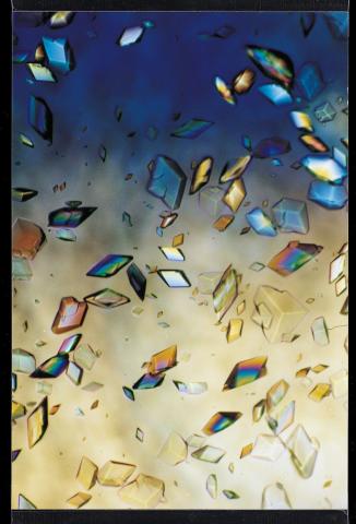
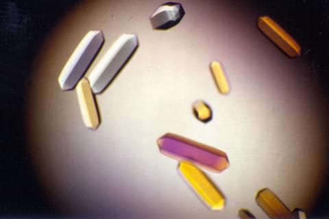
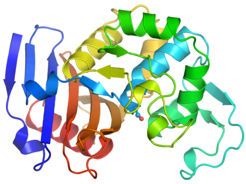
1060: Protein crystals
1060: Protein crystals
Structural biologists create crystals of proteins, shown here, as a first step in a process called X-ray crystallography, which can reveal detailed, three-dimensional protein structures.
Alex McPherson, University of California, Irvine
View Media

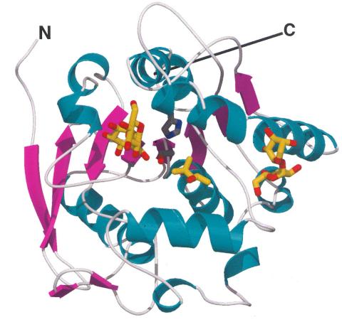
2387: Thymidylate synthase complementing protein from Thermotoga maritime
2387: Thymidylate synthase complementing protein from Thermotoga maritime
A model of thymidylate synthase complementing protein from Thermotoga maritime.
Joint Center for Structural Genomics, PSI
View Media
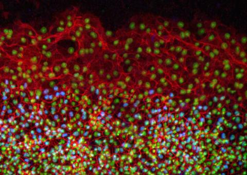
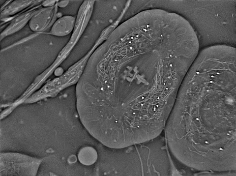
2808: Cell proliferation in a quail embryo
2808: Cell proliferation in a quail embryo
Image showing that the edge zone (top of image) of the quail embryo shows no proliferating cells (cyan), unlike the interior zone (bottom of image). Non-proliferating cell nuclei are labeled green. This image was obtained as part of a study to understand cell migration in embryos. More specifically, cell proliferation at the edge of the embryo was studied by examining the cellular uptake of a chemical compound called BrDU, which incorporates into the DNA during the S-phase of the cell cycle. Here, the cells that are positive for BrDU uptake are labeled in cyan, while other non-proliferating cell nuclei are labeled green. Notice that the vast majority of BrDU+ cells are located far away from the edge, indicating that edge cells are mostly non-proliferating. An NIGMS grant to Professor Garcia was used to purchase the confocal microscope that collected this image. Related to image 2807 and video 2809.
Andrés Garcia, Georgia Tech
View Media
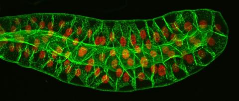
3603: Salivary gland in the developing fruit fly
3603: Salivary gland in the developing fruit fly
For fruit flies, the salivary gland is used to secrete materials for making the pupal case, the protective enclosure in which a larva transforms into an adult fly. For scientists, this gland provided one of the earliest glimpses into the genetic differences between individuals within a species. Chromosomes in the cells of these salivary glands replicate thousands of times without dividing, becoming so huge that scientists can easily view them under a microscope and see differences in genetic content between individuals.
This image was part of the Life: Magnified exhibit that ran from June 3, 2014, to January 21, 2015, at Dulles International Airport.
This image was part of the Life: Magnified exhibit that ran from June 3, 2014, to January 21, 2015, at Dulles International Airport.
Richard Fehon, University of Chicago
View Media
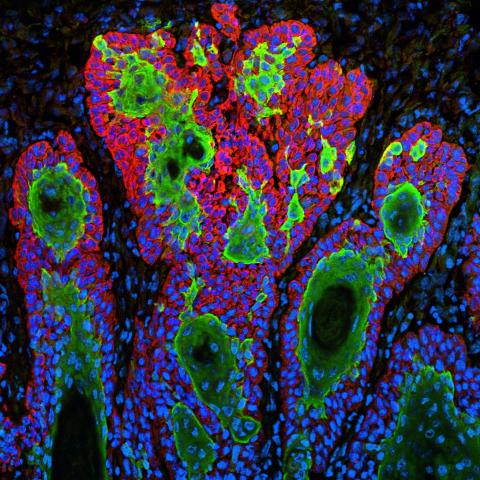
3628: Skin cancer cells (squamous cell carcinoma)
3628: Skin cancer cells (squamous cell carcinoma)
This image shows the uncontrolled growth of cells in squamous cell carcinoma, the second most common form of skin cancer. If caught early, squamous cell carcinoma is usually not life-threatening.
This image was part of the Life: Magnified exhibit that ran from June 3, 2014, to January 21, 2015, at Dulles International Airport.
This image was part of the Life: Magnified exhibit that ran from June 3, 2014, to January 21, 2015, at Dulles International Airport.
Markus Schober and Elaine Fuchs, The Rockefeller University
View Media
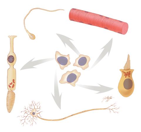
1294: Stem cell differentiation
1294: Stem cell differentiation
Undifferentiated embryonic stem cells cease to exist a few days after conception. In this image, ES cells are shown to differentiate into sperm, muscle fiber, hair cells, nerve cells, and cone cells.
Judith Stoffer
View Media
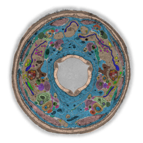
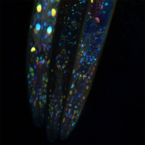
5759: TEM cross-section of C. elegans (roundworm)
5759: TEM cross-section of C. elegans (roundworm)
The worm Caenorhabditis elegans is a popular laboratory animal because its small size and fairly simple body make it easy to study. Scientists use this small worm to answer many research questions in developmental biology, neurobiology, and genetics. This image, which was taken with transmission electron microscopy (TEM), shows a cross-section through C. elegans, revealing various internal structures.
The image is from a figure in an article published in the journal eLife. There is an annotated version of this graphic at 5760.
The image is from a figure in an article published in the journal eLife. There is an annotated version of this graphic at 5760.
Piali Sengupta, Brandeis University
View Media
3481: Bacillus anthracis being killed
3481: Bacillus anthracis being killed
Bacillus anthracis (anthrax) cells being killed by a fluorescent trans-translation inhibitor, which disrupts bacterial protein synthesis. The inhibitor is naturally fluorescent and looks blue when it is excited by ultraviolet light in the microscope. This is a black-and-white version of Image 3525.
John Alumasa, Keiler Laboratory, Pennsylvania State University
View Media
2397: Bovine milk alpha-lactalbumin (1)
2397: Bovine milk alpha-lactalbumin (1)
A crystal of bovine milk alpha-lactalbumin protein created for X-ray crystallography, which can reveal detailed, three-dimensional protein structures.
Alex McPherson, University of California, Irvine
View Media
2378: Most abundant protein in M. tuberculosis
2378: Most abundant protein in M. tuberculosis
Model of a protein, antigen 85B, that is the most abundant protein exported by Mycobacterium tuberculosis, which causes most cases of tuberculosis. Antigen 85B is involved in building the bacterial cell wall and is an attractive drug target. Based on its structure, scientists have suggested a new class of antituberculous drugs.
Mycobacterium Tuberculosis Center, PSI
View Media
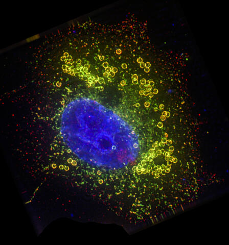
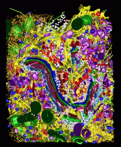
6547: Cell Nucleus and Lipid Droplets
6547: Cell Nucleus and Lipid Droplets
A cell nucleus (blue) surrounded by lipid droplets (yellow). Exogenously expressed, S-tagged UBXD8 (green) recruits endogenous p97/VCP (red) to the surface of lipid droplets in oleate-treated HeLa cells. Nucleus stained with DAPI.
James Olzmann, University of California, Berkeley
View Media
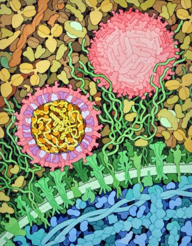
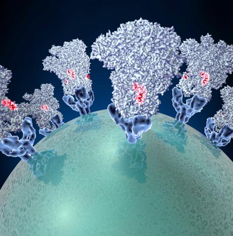
6998: Zika virus
6998: Zika virus
Zika virus is shown in cross section at center left. On the outside, it includes envelope protein (red) and membrane protein (magenta) embedded in a lipid membrane (light purple). Inside, the RNA genome (yellow) is associated with capsid proteins (orange). The viruses are shown interacting with receptors on the cell surface (green) and are surrounded by blood plasma molecules at the top.
Amy Wu and Christine Zardecki, RCSB Protein Data Bank.
View Media
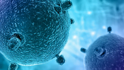
6597: Pathways – Bacteria vs. Viruses: What's the Difference?
6597: Pathways – Bacteria vs. Viruses: What's the Difference?
Learn about how bacteria and viruses differ, how they each can make you sick, and how they can or cannot be treated. Discover more resources from NIGMS’ Pathways collaboration with Scholastic. View the video on YouTube for closed captioning.
National Institute of General Medical Sciences
View Media
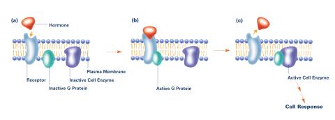
2538: G switch (with labels and stages)
2538: G switch (with labels and stages)
The G switch allows our bodies to respond rapidly to hormones. G proteins act like relay batons to pass messages from circulating hormones into cells. A hormone (red) encounters a receptor (blue) in the membrane of a cell. Next, a G protein (green) becomes activated and makes contact with the receptor to which the hormone is attached. Finally, the G protein passes the hormone's message to the cell by switching on a cell enzyme (purple) that triggers a response. See image 2536 and 2537 for other versions of this image. Featured in Medicines By Design.
Crabtree + Company
View Media
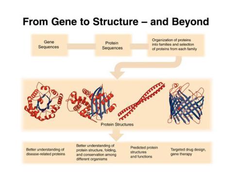
2363: PSI: from genes to structures
2363: PSI: from genes to structures
The goal of the Protein Structure Initiative (PSI) is to determine the three-dimensional shapes of a wide range of proteins by solving the structures of representative members of each protein family found in nature. The collection of structures should serve as a valuable resource for biomedical research scientists.
National Institute of General Medical Sciences
View Media
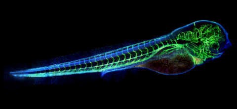
6661: Zebrafish embryo showing vasculature
6661: Zebrafish embryo showing vasculature
A zebrafish embryo. The blue areas are cell bodies, the green lines are blood vessels, and the red glow is blood. This image was created by stitching together five individual images captured with a hyperspectral multipoint confocal fluorescence microscope that was developed at the Eliceiri Lab.
Kevin Eliceiri, University of Wisconsin-Madison.
View Media
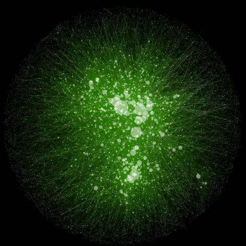
2749: Cytoscape network wiring diagram 2
2749: Cytoscape network wiring diagram 2
This image integrates the thousands of known molecular and genetic interactions happening inside our bodies using a computer program called Cytoscape. Images like this are known as network wiring diagrams, but Cytoscape creator Trey Ideker somewhat jokingly calls them "hairballs" because they can be so complicated, intricate and hard to tease apart. Cytoscape comes with tools to help scientists study specific interactions, such as differences between species or between sick and diseased cells. Related to 2737.
Trey Ideker, University of California, San Diego
View Media
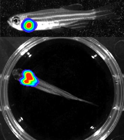
3556: Bioluminescent imaging in adult zebrafish - lateral and overhead view
3556: Bioluminescent imaging in adult zebrafish - lateral and overhead view
Luciferase-based imaging enables visualization and quantification of internal organs and transplanted cells in live adult zebrafish. In this image, a cardiac muscle-restricted promoter drives firefly luciferase expression. This is the lateral and overhead (Bottom) view.
For imagery of the overhead view go to 3557.
For imagery of the lateral view go to 3558.
For more information about the illumated area go to 3559.
For imagery of the overhead view go to 3557.
For imagery of the lateral view go to 3558.
For more information about the illumated area go to 3559.
Kenneth Poss, Duke University
View Media
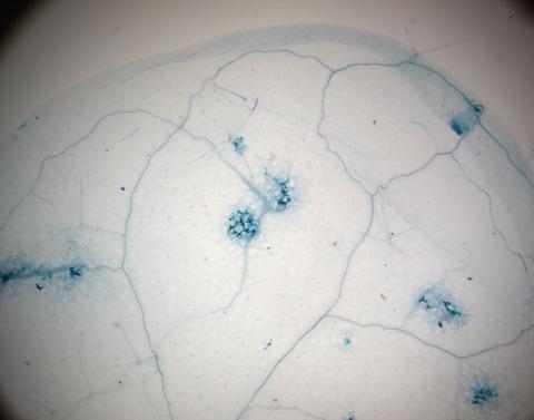
2781: Disease-resistant Arabidopsis leaf
2781: Disease-resistant Arabidopsis leaf
This is a magnified view of an Arabidopsis thaliana leaf a few days after being exposed to the pathogen Hyaloperonospora arabidopsidis. The plant from which this leaf was taken is genetically resistant to the pathogen. The spots in blue show areas of localized cell death where infection occurred, but it did not spread. Compare this response to that shown in Image 2782. Jeff Dangl has been funded by NIGMS to study the interactions between pathogens and hosts that allow or suppress infection.
Jeff Dangl, University of North Carolina, Chapel Hill
View Media
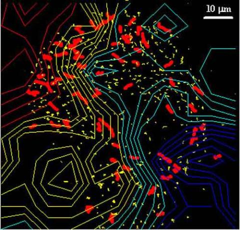
2310: Cellular traffic
2310: Cellular traffic
Like tractor-trailers on a highway, small sacs called vesicles transport substances within cells. This image tracks the motion of vesicles in a living cell. The short red and yellow marks offer information on vesicle movement. The lines spanning the image show overall traffic trends. Typically, the sacs flow from the lower right (blue) to the upper left (red) corner of the picture. Such maps help researchers follow different kinds of cellular processes as they unfold.
Alexey Sharonov and Robin Hochstrasser, University of Pennsylvania
View Media
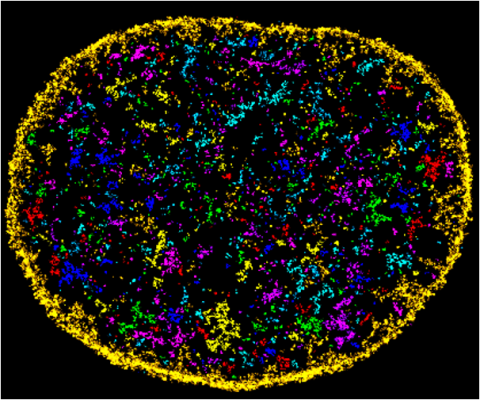
6888: Chromatin in human fibroblast
6888: Chromatin in human fibroblast
The nucleus of a human fibroblast cell with chromatin—a substance made up of DNA and proteins—shown in various colors. Fibroblasts are one of the most common types of cells in mammalian connective tissue, and they play a key role in wound healing and tissue repair. This image was captured using Stochastic Optical Reconstruction Microscopy (STORM).
Related to images 6887 and 6893.
Related to images 6887 and 6893.
Melike Lakadamyali, Perelman School of Medicine at the University of Pennsylvania.
View Media

1335: Telomerase illustration
1335: Telomerase illustration
Reactivating telomerase in our cells does not appear to be a good way to extend the human lifespan. Cancer cells reactivate telomerase.
Judith Stoffer
View Media
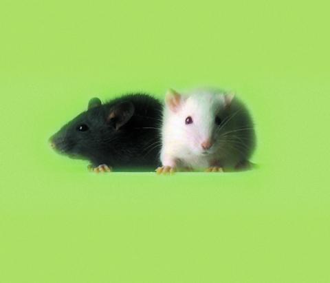
1069: Lab mice
1069: Lab mice
Many researchers use the mouse (Mus musculus) as a model organism to study mammalian biology. Mice carry out practically all the same life processes as humans and, because of their small size and short generation times, are easily raised in labs. Scientists studying a certain cellular activity or disease can choose from tens of thousands of specially bred strains of mice to select those prone to developing certain tumors, neurological diseases, metabolic disorders, premature aging, or other conditions.
Bill Branson, National Institutes of Health
View Media
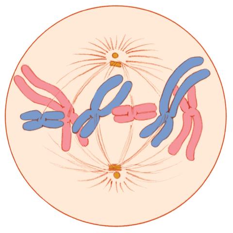
1329: Mitosis - metaphase
1329: Mitosis - metaphase
A cell in metaphase during mitosis: The copied chromosomes align in the middle of the spindle. Mitosis is responsible for growth and development, as well as for replacing injured or worn out cells throughout the body. For simplicity, mitosis is illustrated here with only six chromosomes.
Judith Stoffer
View Media
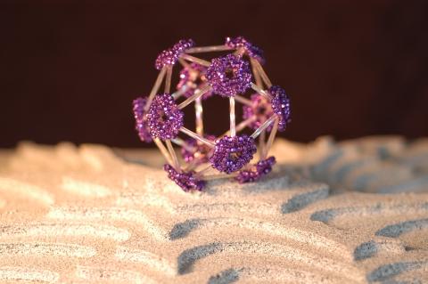
2305: Beaded bacteriophage
2305: Beaded bacteriophage
This sculpture made of purple and clear glass beads depicts bacteriophage Phi174, a virus that infects bacteria. It rests on a surface that portrays an adaptive landscape, a conceptual visualization. The ridges represent the gene combinations associated with the greatest fitness levels of the virus, as measured by how quickly the virus can reproduce itself. Phi174 is an important model system for studies of viral evolution because its genome can readily be sequenced as it evolves under defined laboratory conditions.
Holly Wichman, University of Idaho. (Surface by A. Johnston; photo by J. Palmersheim)
View Media
2324: Movements of myosin
2324: Movements of myosin
Inside the fertilized egg cell of a fruit fly, we see a type of myosin (related to the protein that helps muscles contract) made to glow by attaching a fluorescent protein. After fertilization, the myosin proteins are distributed relatively evenly near the surface of the embryo. The proteins temporarily vanish each time the cells' nuclei--initially buried deep in the cytoplasm--divide. When the multiplying nuclei move to the surface, they shift the myosin, producing darkened holes. The glowing myosin proteins then gather, contract, and start separating the nuclei into their own compartments.
Victoria Foe, University of Washington
View Media
6580: Bacterial nanowire model
6580: Bacterial nanowire model
A model of a Geobacter sulfurreducens nanowire created from cryo-electron microscopy images. The bacterium conducts electricity through these nanowires, which are made up of protein and iron-containing molecules.
Edward Egelman, University of Virginia.
View Media
3753: Coronavirus spike protein structure
3753: Coronavirus spike protein structure
Coronaviruses are enveloped viruses responsible for 30 percent of mild respiratory infections and atypical deadly pneumonia in humans worldwide. These deadly pneumonia include those caused by infections with severe acute respiratory syndrome coronavirus (SARS-CoV) and Middle East respiratory syndrome coronavirus (MERS-CoV). The coronavirus spike glycoprotein mediates virus entry into cells and represents an important therapeutic target. The illustration shows a viral membrane decorated with spike glycoproteins; highlighted in red is a potential neutralization site, which is a protein sequence that might be used as a target for vaccines to combat viruses such as MERS-CoV and other coronaviruses.
Melody Campbell, UCSF
View Media
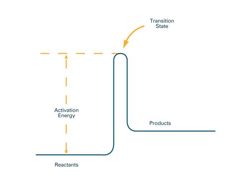
2526: Activation energy (with labels)
2526: Activation energy (with labels)
To become products, reactants must overcome an energy hill. See image 2525 for an unlabeled version of this illustration. Featured in The Chemistry of Health.
Crabtree + Company
View Media
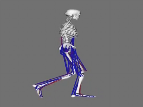
6598: Simulation of leg muscles moving
6598: Simulation of leg muscles moving
When we walk, muscles and nerves interact in intricate ways. This simulation, which is based on data from a six-foot-tall man, shows these interactions.
Chand John and Eran Guendelman, Stanford University
View Media
6532: Mosaicism in C. elegans (Black Background)
6532: Mosaicism in C. elegans (Black Background)
In the worm C. elegans, double-stranded RNA made in neurons can silence matching genes in a variety of cell types through the transport of RNA between cells. The head region of three worms that were genetically modified to express a fluorescent protein were imaged and the images were color-coded based on depth. The worm on the left lacks neuronal double-stranded RNA and thus every cell is fluorescent. In the middle worm, the expression of the fluorescent protein is silenced by neuronal double-stranded RNA and thus most cells are not fluorescent. The worm on the right lacks an enzyme that amplifies RNA for silencing. Surprisingly, the identities of the cells that depend on this enzyme for gene silencing are unpredictable. As a result, worms of identical genotype are nevertheless random mosaics for how the function of gene silencing is carried out. For more, see journal article and press release. Related to image 6534.
Snusha Ravikumar, Ph.D., University of Maryland, College Park, and Antony M. Jose, Ph.D., University of Maryland, College Park
View Media
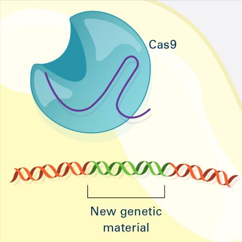
6488: CRISPR Illustration Frame 4
6488: CRISPR Illustration Frame 4
This illustration shows, in simplified terms, how the CRISPR-Cas9 system can be used as a gene-editing tool. The CRISPR system has two components joined together: a finely tuned targeting device (a small strand of RNA programmed to look for a specific DNA sequence) and a strong cutting device (an enzyme called Cas9 that can cut through a double strand of DNA). This frame (4 out of 4) shows a repaired DNA strand with new genetic material that researchers can introduce, which the cell automatically incorporates into the gap when it repairs the broken DNA.
For an explanation and overview of the CRISPR-Cas9 system, see the iBiology video, and find the full CRIPSR illustration here.
For an explanation and overview of the CRISPR-Cas9 system, see the iBiology video, and find the full CRIPSR illustration here.
National Institute of General Medical Sciences.
View Media
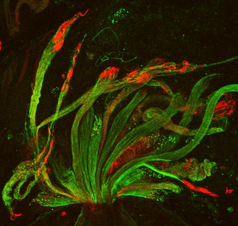
3590: Fruit fly spermatids
3590: Fruit fly spermatids
Developing spermatids (precursors of mature sperm cells) begin as small, round cells and mature into long-tailed, tadpole-shaped ones. In the sperm cell's head is the cell nucleus; in its tail is the power to outswim thousands of competitors to fertilize an egg. As seen in this microscopy image, fruit fly spermatids start out as groups of interconnected cells. A small lipid molecule called PIP2 helps spermatids tell their heads from their tails. Here, PIP2 (red) marks the nuclei and a cell skeleton-building protein called tubulin (green) marks the tails. When PIP2 levels are too low, some spermatids get mixed up and grow with their heads at the wrong end. Because sperm development is similar across species, studies in fruit flies could help researchers understand male infertility in humans.
Lacramioara Fabian, The Hospital for Sick Children, Toronto, Canada
View Media
2315: Fly cells live
2315: Fly cells live
If a picture is worth a thousand words, what's a movie worth? For researchers studying cell migration, a "documentary" of fruit fly cells (bright green) traversing an egg chamber could answer longstanding questions about cell movement. Historically, researchers have been unable to watch this cell migration unfold in living ovarian tissue in real time. But by developing a culture medium that allows fly eggs to survive outside their ovarian homes, scientists can observe the nuances of cell migration as it happens. Such details may shed light on how immune cells move to a wound and why cancer cells spread to other sites. See 3594 for still image.
Denise Montell, Johns Hopkins University School of Medicine
View Media
2308: Cellular metropolis
2308: Cellular metropolis
Like a major city, a cell teems with specialized workers that carry out its daily operations--making energy, moving proteins, or helping with other tasks. Researchers took microscopic pictures of thin layers of a cell and then combined them to make this 3-D image featuring color-coded organelles--the cell's "workers." Using this image, scientists can understand how these specialized components fit together in the cell's packed inner world.
Kathryn Howell, University of Colorado Health Sciences Center
View Media
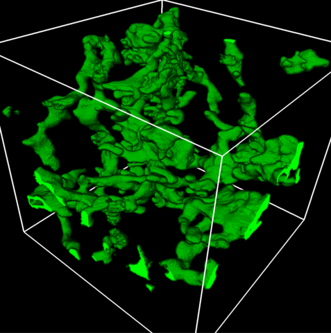
5857: 3D reconstruction of a tubular matrix in peripheral endoplasmic reticulum
5857: 3D reconstruction of a tubular matrix in peripheral endoplasmic reticulum
Detailed three-dimensional reconstruction of a tubular matrix in a thin section of the peripheral endoplasmic reticulum between the plasma membranes of the cell. The endoplasmic reticulum (ER) is a continuous membrane that extends like a net from the envelope of the nucleus outward to the cell membrane. The ER plays several roles within the cell, such as in protein and lipid synthesis and transport of materials between organelles. Shown here is a three-dimensional representation of the peripheral ER microtubules. Related to images 5855 and 5856
Jennifer Lippincott-Schwartz, Howard Hughes Medical Institute Janelia Research Campus, Virginia
View Media
2395: Fungal lipase (1)
2395: Fungal lipase (1)
Crystals of fungal lipase protein created for X-ray crystallography, which can reveal detailed, three-dimensional protein structures.
Alex McPherson, University of California, Irvine
View Media
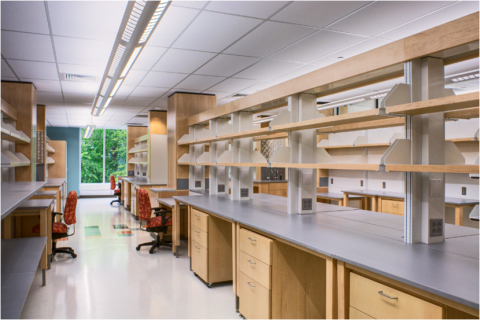
2723: iPS cell facility at the Coriell Institute for Medical Research
2723: iPS cell facility at the Coriell Institute for Medical Research
This lab space was designed for work on the induced pluripotent stem (iPS) cell collection, part of the NIGMS Human Genetic Cell Repository at the Coriell Institute for Medical Research.
Courtney Sill, Coriell Institute for Medical Research
View Media
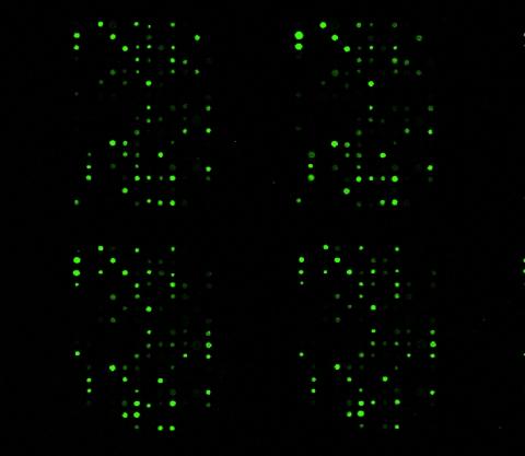
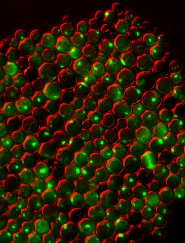
1265: Glycan arrays
1265: Glycan arrays
The signal is obtained by allowing proteins in human serum to interact with glycan (polysaccharide) arrays. The arrays are shown in replicate so the pattern is clear. Each spot contains a specific type of glycan. Proteins have bound to the spots highlighted in green.
Ola Blixt, Scripps Research Institute
View Media
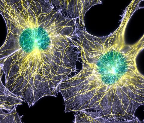
2428: Colorful cells
2428: Colorful cells
Actin (purple), microtubules (yellow), and nuclei (green) are labeled in these cells by immunofluorescence. This image won first place in the Nikon 2003 Small World photo competition.
Torsten Wittmann, Scripps Research Institute
View Media
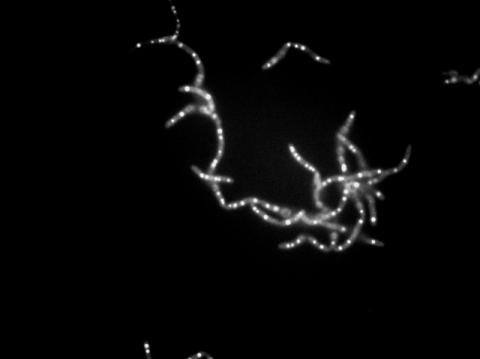
6771: Culex quinquefasciatus mosquito larvae
6771: Culex quinquefasciatus mosquito larvae
Mosquito larvae with genes edited by CRISPR swimming in water. This species of mosquito, Culex quinquefasciatus, can transmit West Nile virus, Japanese encephalitis virus, and avian malaria, among other diseases. The researchers who took this video optimized the gene-editing tool CRISPR for Culex quinquefasciatus that could ultimately help stop the mosquitoes from spreading pathogens. The work is described in the Nature Communications paper "Optimized CRISPR tools and site-directed transgenesis towards gene drive development in Culex quinquefasciatus mosquitoes" by Feng et al. Related to images 6769 and 6770.
Valentino Gantz, University of California, San Diego.
View Media
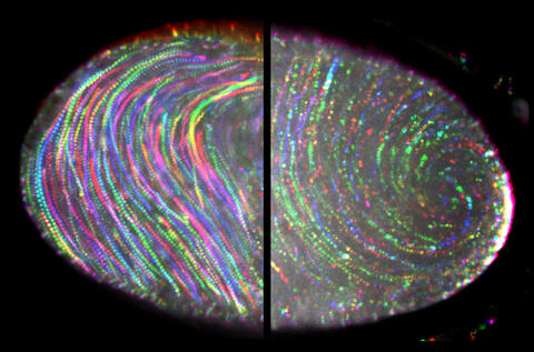
6809: Fruit fly egg ooplasmic streaming
6809: Fruit fly egg ooplasmic streaming
Two fruit fly (Drosophila melanogaster) egg cells, one on each side of the central black line. The colorful swirls show the circular movement of cytoplasm—called ooplasmic streaming—that occurs in late egg cell development in wild-type (right) and mutant (left) oocytes. This image was captured using confocal microscopy.
More information on the research that produced this image can be found in the Journal of Cell Biology paper “Ooplasmic flow cooperates with transport and anchorage in Drosophila oocyte posterior determination” by Lu et al.
More information on the research that produced this image can be found in the Journal of Cell Biology paper “Ooplasmic flow cooperates with transport and anchorage in Drosophila oocyte posterior determination” by Lu et al.
Vladimir I. Gelfand, Feinberg School of Medicine, Northwestern University.
View Media
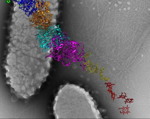
6766: Ribbon diagram of a cefotaxime-CCD-1 complex
6766: Ribbon diagram of a cefotaxime-CCD-1 complex
CCD-1 is an enzyme produced by the bacterium Clostridioides difficile that helps it resist antibiotics. Using X-ray crystallography, researchers determined the structure of a CCD-1 molecule and a molecule of the antibiotic cefotaxime bound together. The structure revealed that CCD-1 provides extensive hydrogen bonding and stabilization of the antibiotic in the active site, leading to efficient degradation of the antibiotic.
Related to images 6764, 6765, and 6767.
Related to images 6764, 6765, and 6767.
Keith Hodgson, Stanford University.
View Media
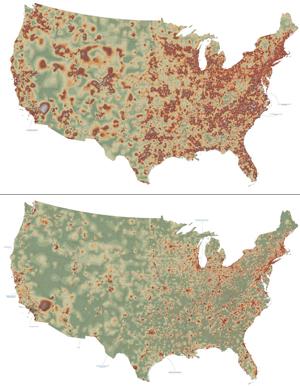
2320: Mapping disease spread
2320: Mapping disease spread
How far and fast an infectious disease spreads across a community depends on many factors, including transportation. These U.S. maps, developed as part of an international study to simulate and analyze disease spread, chart daily commuting patterns. They show where commuters live (top) and where they travel for work (bottom). Green represents the fewest number of people whereas orange, brown, and white depict the most. Such information enables researchers and policymakers to visualize how an outbreak in one area can spread quickly across a geographic region.
David Chrest, RTI International
View Media
3550: Protein clumping in zinc-deficient yeast cells
3550: Protein clumping in zinc-deficient yeast cells
The green spots in this image are clumps of protein inside yeast cells that are deficient in both zinc and a protein called Tsa1 that prevents clumping. Protein clumping plays a role in many diseases, including Parkinson's and Alzheimer's, where proteins clump together in the brain. Zinc deficiency within a cell can cause proteins to mis-fold and eventually clump together. Normally, in yeast, Tsa1 codes for so-called "chaperone proteins" which help proteins in stressed cells, such as those with a zinc deficiency, fold correctly. The research behind this image was published in 2013 in the Journal of Biological Chemistry.
Colin MacDiarmid and David Eide, University of Wisconsin--Madison
View Media
5825: A Growing Bacterial Biofilm
5825: A Growing Bacterial Biofilm
A growing Vibrio cholerae (cholera) biofilm. Cholera bacteria form colonies called biofilms that enable them to resist antibiotic therapy within the body and other challenges to their growth.
Each slightly curved comma shape represents an individual bacterium from assembled confocal microscopy images. Different colors show each bacterium’s position in the biofilm in relation to the surface on which the film is growing.
Each slightly curved comma shape represents an individual bacterium from assembled confocal microscopy images. Different colors show each bacterium’s position in the biofilm in relation to the surface on which the film is growing.
Jing Yan, Ph.D., and Bonnie Bassler, Ph.D., Department of Molecular Biology, Princeton University, Princeton, NJ.
View Media
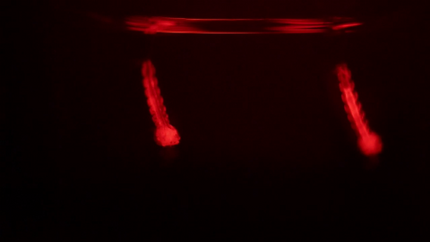
6898: Crane fly spermatocyte undergoing meiosis
6898: Crane fly spermatocyte undergoing meiosis
A crane fly spermatocyte during metaphase of meiosis-I, a step in the production of sperm. A meiotic spindle pulls apart three pairs of autosomal chromosomes, along with a sex chromosome on the right. Tubular mitochondria surround the spindle and chromosomes. This video was captured with quantitative orientation-independent differential interference contrast and is a time lapse showing a 1-second image taken every 30 seconds over the course of 30 minutes.
More information about the research that produced this video can be found in the J. Biomed Opt. paper “Orientation-Independent Differential Interference Contrast (DIC) Microscopy and Its Combination with Orientation-Independent Polarization System” by Shribak et. al.
More information about the research that produced this video can be found in the J. Biomed Opt. paper “Orientation-Independent Differential Interference Contrast (DIC) Microscopy and Its Combination with Orientation-Independent Polarization System” by Shribak et. al.
Michael Shribak, Marine Biological Laboratory/University of Chicago.
View Media
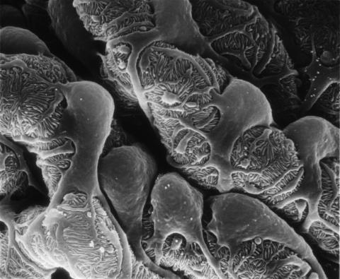
3565: Podocytes from a chronically diseased kidney
3565: Podocytes from a chronically diseased kidney
This scanning electron microscope (SEM) image shows podocytes--cells in the kidney that play a vital role in filtering waste from the bloodstream--from a patient with chronic kidney disease. This image first appeared in Princeton Journal Watch on October 4, 2013.
Olga Troyanskaya, Princeton University and Matthias Kretzler, University of Michigan
View Media
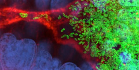
7015: Bacterial cells migrating through the tissues of the squid light organ
7015: Bacterial cells migrating through the tissues of the squid light organ
Vibrio fischeri cells (~ 2 mm), labeled with green fluorescent protein (GFP), passing through a very narrow bottleneck in the tissues (red) of the Hawaiian bobtail squid, Euprymna scolopes, on the way to the crypts where the symbiont population resides. This image was taken using a confocal fluorescence microscope.
Margaret J. McFall-Ngai, Carnegie Institution for Science/California Institute of Technology, and Edward G. Ruby, California Institute of Technology.
View Media
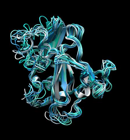
5866: Structure of a key antigen protein involved with Hepatitis C Virus infection
5866: Structure of a key antigen protein involved with Hepatitis C Virus infection
A three-dimensional representation of the structure of E2, a key antigen protein involved with hepatitis C virus infection.
Mansun Law Associate Professor Department of Immunolgy and Microbial Science The Scripps Research Institute
View Media