
Switch to List View
Image and Video Gallery
This is a searchable collection of scientific photos, illustrations, and videos. The images and videos in this gallery are licensed under Creative Commons Attribution Non-Commercial ShareAlike 3.0. This license lets you remix, tweak, and build upon this work non-commercially, as long as you credit and license your new creations under identical terms.
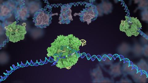
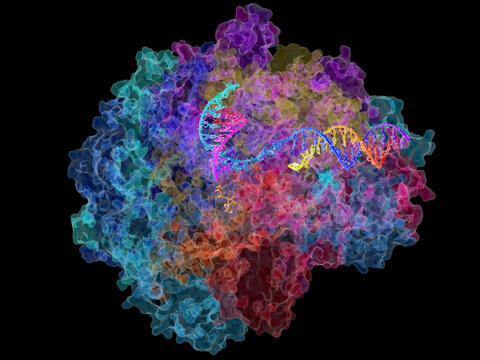
5816: Cas9 protein involved in the CRISPR gene-editing technology
5816: Cas9 protein involved in the CRISPR gene-editing technology
In the gene-editing tool CRISPR, a small strand of RNA identifies a specific chunk of DNA. Then the enzyme Cas9 (green) swoops in and cuts the double-stranded DNA (blue/purple) in two places, removing the specific chunk.
Janet Iwasa
View Media
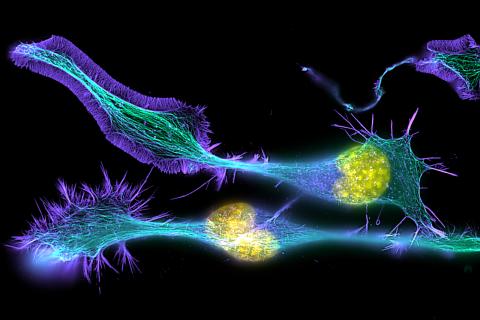
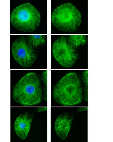
1272: Cytoskeleton
1272: Cytoskeleton
The three fibers of the cytoskeleton--microtubules in blue, intermediate filaments in red, and actin in green--play countless roles in the cell.
Judith Stoffer
View Media
2312: Color-coded chromosomes
2312: Color-coded chromosomes
By mixing fluorescent dyes like an artist mixes paints, scientists are able to color code individual chromosomes. The technique, abbreviated multicolor-FISH, allows researchers to visualize genetic abnormalities often linked to disease. In this image, "painted" chromosomes from a person with a hereditary disease called Werner Syndrome show where a piece of one chromosome has fused to another (see the gold-tipped maroon chromosome in the center). As reported by molecular biologist Jan Karlseder of the Salk Institute for Biological Studies, such damage is typical among people with this rare syndrome.
Anna Jauch, Institute of Human Genetics, Heidelberg, Germany
View Media
5885: 3-D Architecture of a Synapse
5885: 3-D Architecture of a Synapse
This image shows the structure of a synapse, or junction between two nerve cells in three dimensions. From the brain of a mouse.
Anton Maximov, The Scripps Research Institute, La Jolla, CA
View Media
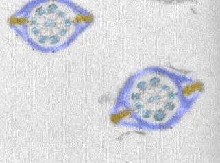
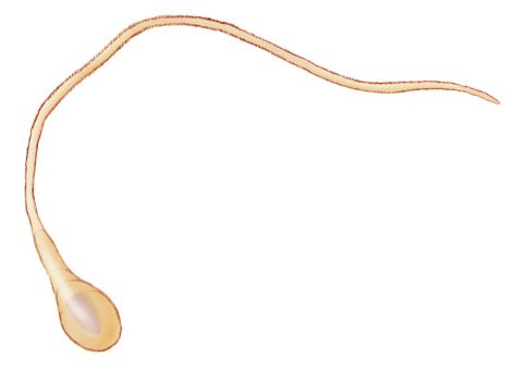
1191: Mouse sperm sections
1191: Mouse sperm sections
This transmission electron micrograph shows sections of mouse sperm tails, or flagella.
Tina Weatherby Carvalho, University of Hawaii at Manoa
View Media
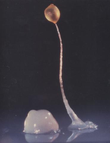
2684: Dicty fruit
2684: Dicty fruit
Dictyostelium discoideum is a microscopic amoeba. A group of 100,000 form a mound as big as a grain of sand. Featured in The New Genetics.
View Media
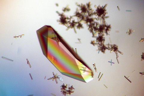
2396: Hen egg lysozyme (1)
2396: Hen egg lysozyme (1)
Crystals of hen egg lysozyme protein created for X-ray crystallography, which can reveal detailed, three-dimensional protein structures.
Alex McPherson, University of California, Irvine
View Media
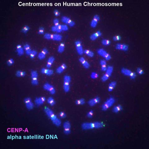
3255: Centromeres on human chromosomes
3255: Centromeres on human chromosomes
Human metaphase chromosomes are visible with fluorescence in vitro hybridization (FISH). Centromeric alpha satellite DNA (green) are found in the heterochromatin at each centromere. Immunofluorescence with CENP-A (red) shows the centromere-specific histone H3 variant that specifies the kinetochore.
Peter Warburton, Mount Sinai School of Medicine
View Media
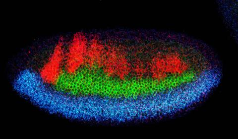
2327: Neural development
2327: Neural development
Using techniques that took 4 years to design, a team of developmental biologists showed that certain proteins can direct the subdivision of fruit fly and chicken nervous system tissue into the regions depicted here in blue, green, and red. Molecules called bone morphogenetic proteins (BMPs) helped form this fruit fly embryo. While scientists knew that BMPs play a major role earlier in embryonic development, they didn't know how the proteins help organize nervous tissue. The findings suggest that BMPs are part of an evolutionarily conserved mechanism for organizing the nervous system. The National Institute of Neurological Disorders and Stroke also supported this work.
Mieko Mizutani and Ethan Bier, University of California, San Diego, and Henk Roelink, University of Washington
View Media
3573: Myotonic dystrophy type 2 genetic defect
3573: Myotonic dystrophy type 2 genetic defect
Scientists revealed a detailed image of the genetic defect that causes myotonic dystrophy type 2 and used that information to design drug candidates to counteract the disease.
Matthew Disney, Scripps Research Institute and Ilyas Yildirim, Northwestern University
View Media
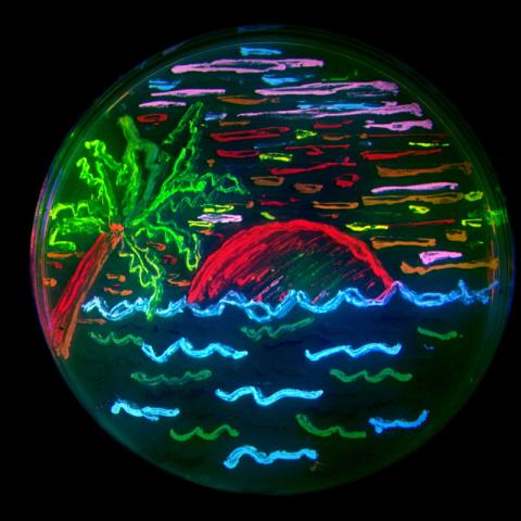
3492: Glowing bacteria make a pretty postcard
3492: Glowing bacteria make a pretty postcard
This tropical scene, reminiscent of a postcard from Key West, is actually a petri dish containing an artistic arrangement of genetically engineered bacteria. The image showcases eight of the fluorescent proteins created in the laboratory of the late Roger Y. Tsien, a cell biologist at the University of California, San Diego. Tsien, along with Osamu Shimomura of the Marine Biology Laboratory and Martin Chalfie of Columbia University, share the 2008 Nobel Prize in chemistry for their work on green fluorescent protein-a naturally glowing molecule from jellyfish that has become a powerful tool for studying molecules inside living cells.
Nathan C. Shaner, The Scintillon Institute
View Media
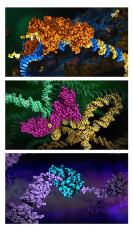
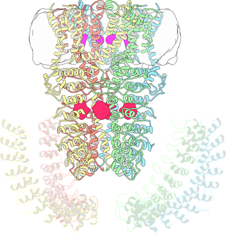
6999: HIV enzyme
6999: HIV enzyme
These images model the molecular structures of three enzymes with critical roles in the life cycle of the human immunodeficiency virus (HIV). At the top, reverse transcriptase (orange) creates a DNA copy (yellow) of the virus's RNA genome (blue). In the middle image, integrase (magenta) inserts this DNA copy in the DNA genome (green) of the infected cell. At the bottom, much later in the viral life cycle, protease (turquoise) chops up a chain of HIV structural protein (purple) to generate the building blocks for making new viruses. See these enzymes in action on PDB 101’s video A Molecular View of HIV Therapy.
Amy Wu and Christine Zardecki, RCSB Protein Data Bank.
View Media

2398: RNase A (1)
2398: RNase A (1)
A crystal of RNase A protein created for X-ray crystallography, which can reveal detailed, three-dimensional protein structures.
Alex McPherson, University of California, Irvine
View Media
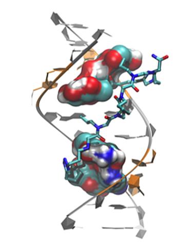
3414: X-ray co-crystal structure of Src kinase bound to a DNA-templated macrocycle inhibitor 2
3414: X-ray co-crystal structure of Src kinase bound to a DNA-templated macrocycle inhibitor 2
X-ray co-crystal structure of Src kinase bound to a DNA-templated macrocycle inhibitor. Related to 3413, 3415, 3416, 3417, 3418, and 3419.
Markus A. Seeliger, Stony Brook University Medical School and David R. Liu, Harvard University
View Media
2600: Molecules blocking Huntington's protein production
2600: Molecules blocking Huntington's protein production
The molecules that glow blue in these cultured cells prevent the expression of the mutant proteins that cause Huntington's disease. Biochemist David Corey and others at UT Southwestern Medical Center designed the molecules to specifically target the genetic repeats that code for harmful proteins in people with Huntington's disese. People with Huntington's disease and similar neurodegenerative disorders often have extra copies of a gene segment. Moving from cell cultures to animals will help researchers further explore the potential of their specially crafted molecule to treat brain disorders. In addition to NIGMS, NIH's National Institute of Neurological Disorders and Stroke and National Institute of Biomedical Imaging and Bioengineering also funded this work.
Jiaxin Hu, David W. Dodd and Robert H. E. Hudson, UT Southwestern Medical Center
View Media
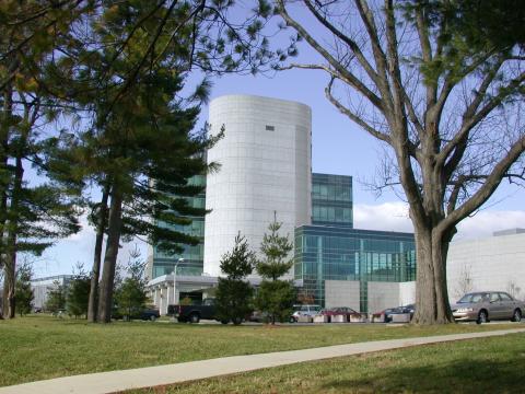
1083: Natcher Building 03
1083: Natcher Building 03
NIGMS staff are located in the Natcher Building on the NIH campus.
Alisa Machalek, National Institute of General Medical Sciences
View Media
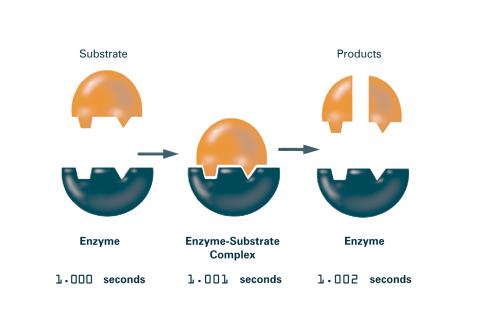
2522: Enzymes convert subtrates into products (with labels)
2522: Enzymes convert subtrates into products (with labels)
Enzymes convert substrates into products very quickly. See image 2521 for an unlabeled version of this illustration. Featured in The Chemistry of Health.
Crabtree + Company
View Media
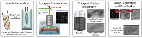
6568: Correlative imaging by annotation with single molecules (CIASM) process
6568: Correlative imaging by annotation with single molecules (CIASM) process
These images illustrate a technique combining cryo-electron tomography and super-resolution fluorescence microscopy called correlative imaging by annotation with single molecules (CIASM). CIASM enables researchers to identify small structures and individual molecules in cells that they couldn’t using older techniques.
Peter Dahlberg, Stanford University.
View Media
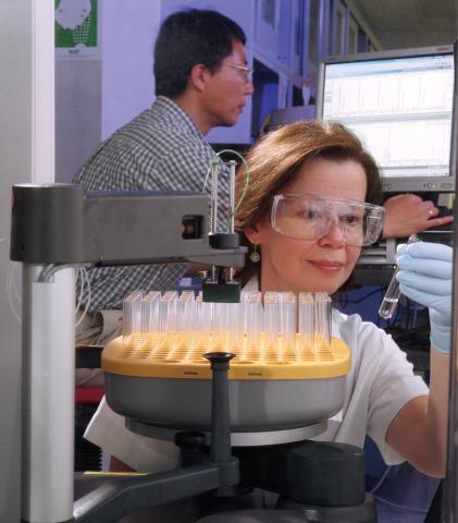
2375: Protein purification robot
2375: Protein purification robot
Irina Dementieva, a biochemist, and Youngchang Kim, a biophysicist and crystallographer, work with the first robot of its type in the U.S. to automate protein purification. The robot, which is housed in a refrigerator, is an integral part of the Midwest Structural Genomics Center's plan to automate the protein crystallography process.
Midwest Center for Structural Genomics
View Media
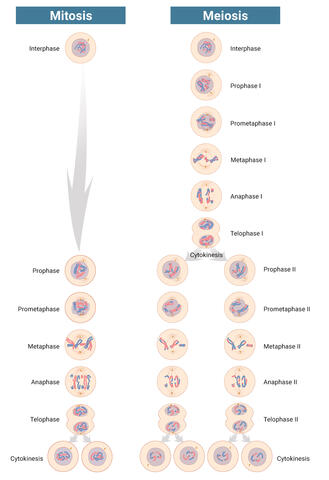
6788: Mitosis and meiosis compared-labeled
6788: Mitosis and meiosis compared-labeled
Meiosis is used to make sperm and egg cells. During meiosis, a cell's chromosomes are copied once, but the cell divides twice. During mitosis, the chromosomes are copied once, and the cell divides once. For simplicity, cells are illustrated with only three pairs of chromosomes.
See image 1333 for an unlabeled version of this illustration.
See image 1333 for an unlabeled version of this illustration.
Judith Stoffer
View Media

2371: NMR spectrometer
2371: NMR spectrometer
This photo shows a Varian Unity Inova 900 MHz, 21.1 T standard bore magnet Nuclear Magnetic Resonnance (NMR) spectrometer. NMR spectroscopy provides data used to determine the structures of proteins in solution, rather than in crystal form, as in X-ray crystallography. The technique is limited to smaller proteins or protein fragments in a high throughput approach.
Center for Eukaryotic Structural Genomics
View Media
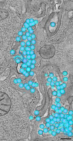
3571: HIV-1 virus in the colon
3571: HIV-1 virus in the colon
A tomographic reconstruction of the colon shows the location of large pools of HIV-1 virus particles (in blue) located in the spaces between adjacent cells. The purple objects within each sphere represent the conical cores that are one of the structural hallmarks of the HIV virus.
Mark Ladinsky, California Institute of Technology
View Media
3782: A multicolored fish scale 1
3782: A multicolored fish scale 1
Each of the colored specs in this image is a cell on the surface of a fish scale. To better understand how wounds heal, scientists have inserted genes that make cells brightly glow in different colors into the skin cells of zebrafish, a fish often used in laboratory research. The colors enable the researchers to track each individual cell, for example, as it moves to the location of a cut or scrape over the course of several days. These technicolor fish endowed with glowing skin cells dubbed "skinbow" provide important insight into how tissues recover and regenerate after an injury.
For more information on skinbow fish, see the Biomedical Beat blog post Visualizing Skin Regeneration in Real Time and a press release from Duke University highlighting this research. Related to image 3783.
For more information on skinbow fish, see the Biomedical Beat blog post Visualizing Skin Regeneration in Real Time and a press release from Duke University highlighting this research. Related to image 3783.
Chen-Hui Chen and Kenneth Poss, Duke University
View Media
5777: Microsporidia in roundworm 1
5777: Microsporidia in roundworm 1
Many disease-causing microbes manipulate their host’s metabolism and cells for their own ends. Microsporidia—which are parasites closely related to fungi—infect and multiply inside animal cells, and take the rearranging of cells’ interiors to a new level. They reprogram animal cells such that the cells start to fuse, causing them to form long, continuous tubes. As shown in this image of the roundworm Caenorhabditis elegans, microsporidia (shown in magenta) have invaded the worm’s gut cells (shown in yellow; the cells’ nuclei are shown in blue) and have instructed the cells to merge. The cell fusion enables the microsporidia to thrive and propagate in the expanded space. Scientists study microsporidia in worms to gain more insight into how these parasites manipulate their host cells. This knowledge might help researchers devise strategies to prevent or treat infections with microsporidia. For more on the research into microsporidia, see this news release from the University of California San Diego. Related to images 5778 and 5779.
Keir Balla and Emily Troemel, University of California San Diego
View Media
3747: Cryo-electron microscopy revealing the "wasabi receptor"
3747: Cryo-electron microscopy revealing the "wasabi receptor"
The TRPA1 protein is responsible for the burn you feel when you taste a bite of sushi topped with wasabi. Known therefore informally as the "wasabi receptor," this protein forms pores in the membranes of nerve cells that sense tastes or odors. Pungent chemicals like wasabi or mustard oil cause the pores to open, which then triggers a tingling or burn on our tongue. This receptor also produces feelings of pain in response to chemicals produced within our own bodies when our tissues are damaged or inflamed. Researchers used cryo-EM to reveal the structure of the wasabi receptor at a resolution of about 4 angstroms (a credit card is about 8 million angstroms thick). This detailed structure can help scientists understand both how we feel pain and how we can limit it by developing therapies to block the receptor. For more on cryo-EM see the blog post Cryo-Electron Microscopy Reveals Molecules in Ever Greater Detail.
Jean-Paul Armache, UCSF
View Media
6795: Dividing yeast cells with nuclear envelopes and spindle pole bodies
6795: Dividing yeast cells with nuclear envelopes and spindle pole bodies
Time-lapse video of yeast cells undergoing cell division. Nuclear envelopes are shown in green, and spindle pole bodies, which help pull apart copied genetic information, are shown in magenta. This video was captured using wide-field microscopy with deconvolution.
Related to images 6791, 6792, 6793, 6794, 6797, 6798, and video 6796.
Related to images 6791, 6792, 6793, 6794, 6797, 6798, and video 6796.
Alaina Willet, Kathy Gould’s lab, Vanderbilt University.
View Media
2484: RNA Polymerase II
2484: RNA Polymerase II
NIGMS-funded researchers led by Roger Kornberg solved the structure of RNA polymerase II. This is the enzyme in mammalian cells that catalyzes the transcription of DNA into messenger RNA, the molecule that in turn dictates the order of amino acids in proteins. For his work on the mechanisms of mammalian transcription, Kornberg received the Nobel Prize in Chemistry in 2006.
David Bushnell, Ken Westover and Roger Kornberg, Stanford University
View Media
3632: Developing nerve cells
3632: Developing nerve cells
These developing mouse nerve cells have a nucleus (yellow) surrounded by a cell body, with long extensions called axons and thin branching structures called dendrites. Electrical signals travel from the axon of one cell to the dendrites of another.
This image was part of the Life: Magnified exhibit that ran from June 3, 2014, to January 21, 2015, at Dulles International Airport.
This image was part of the Life: Magnified exhibit that ran from June 3, 2014, to January 21, 2015, at Dulles International Airport.
Torsten Wittmann, University of California, San Francisco
View Media
5855: Dense tubular matrices in the peripheral endoplasmic reticulum (ER) 1
5855: Dense tubular matrices in the peripheral endoplasmic reticulum (ER) 1
Superresolution microscopy work on endoplasmic reticulum (ER) in the peripheral areas of the cell showing details of the structure and arrangement in a complex web of tubes. The ER is a continuous membrane that extends like a net from the envelope of the nucleus outward to the cell membrane. The ER plays several roles within the cell, such as in protein and lipid synthesis and transport of materials between organelles. The ER has a flexible structure to allow it to accomplish these tasks by changing shape as conditions in the cell change. Shown here an image created by super-resolution microscopy of the ER in the peripheral areas of the cell showing details of the structure and the arrangements in a complex web of tubes. Related to images 5856 and 5857.
Jennifer Lippincott-Schwartz, Howard Hughes Medical Institute Janelia Research Campus, Virginia
View Media
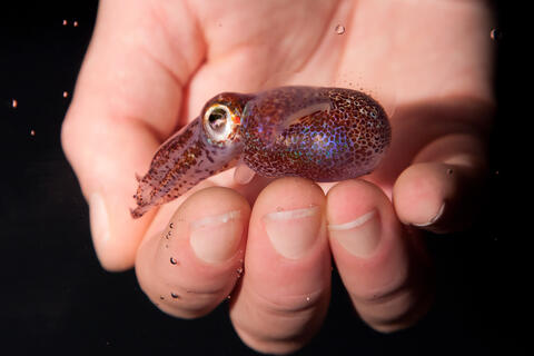
7011: Hawaiian bobtail squid
7011: Hawaiian bobtail squid
An adult Hawaiian bobtail squid, Euprymna scolopes, swimming next to a submerged hand.
Related to image 7010 and video 7012.
Related to image 7010 and video 7012.
Margaret J. McFall-Ngai, Carnegie Institution for Science/California Institute of Technology, and Edward G. Ruby, California Institute of Technology.
View Media
3443: Interphase in Xenopus frog cells
3443: Interphase in Xenopus frog cells
These images show frog cells in interphase. The cells are Xenopus XL177 cells, which are derived from tadpole epithelial cells. The microtubules are green and the chromosomes are blue. Related to 3442.
Claire Walczak, who took them while working as a postdoc in the laboratory of Timothy Mitchison.
View Media
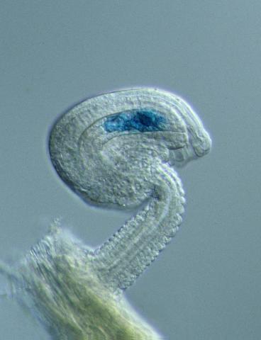
1293: Sperm cell
5881: Zebrafish larva
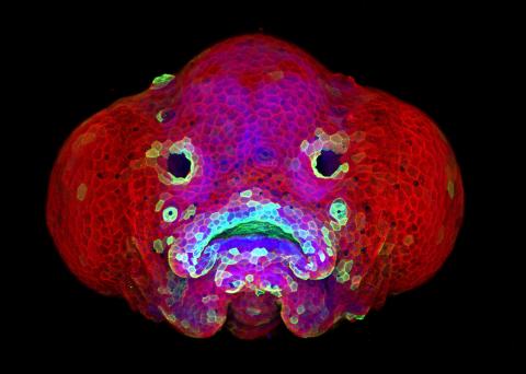
5881: Zebrafish larva
You are face to face with a 6-day-old zebrafish larva. What look like eyes will become nostrils, and the bulges on either side will become eyes. Scientists use fast-growing, transparent zebrafish to see body shapes form and organs develop over the course of just a few days. Images like this one help researchers understand how gene mutations can lead to facial abnormalities such as cleft lip and palate in people.
This image won a 2016 FASEB BioArt award. In addition, NIH Director Francis Collins featured this on his blog on January 26, 2017.
This image won a 2016 FASEB BioArt award. In addition, NIH Director Francis Collins featured this on his blog on January 26, 2017.
Oscar Ruiz and George Eisenhoffer, University of Texas MD Anderson Cancer Center, Houston
View Media
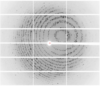
6765: X-ray diffraction pattern from a crystallized cefotaxime-CCD-1 complex
6765: X-ray diffraction pattern from a crystallized cefotaxime-CCD-1 complex
CCD-1 is an enzyme produced by the bacterium Clostridioides difficile that helps it resist antibiotics. Researchers crystallized complexes where a CCD-1 molecule and a molecule of the antibiotic cefotaxime were bound together. Then, they shot X-rays at the complexes to determine their structure—a process known as X-ray crystallography. This image shows the X-ray diffraction pattern of a complex.
Related to images 6764, 6766, and 6767.
Related to images 6764, 6766, and 6767.
Keith Hodgson, Stanford University.
View Media
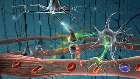
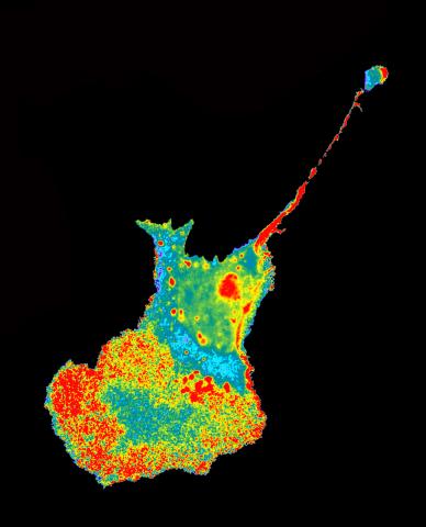
2323: Motion in the brain
2323: Motion in the brain
Amid a network of blood vessels and star-shaped support cells, neurons in the brain signal each other. The mists of color show the flow of important molecules like glucose and oxygen. This image is a snapshot from a 52-second simulation created by an animation artist. Such visualizations make biological processes more accessible and easier to understand.
Kim Hager and Neal Prakash, University of California, Los Angeles
View Media
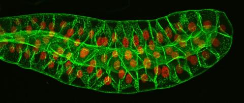
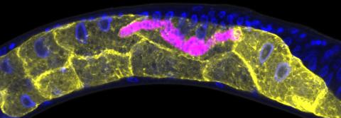
3603: Salivary gland in the developing fruit fly
3603: Salivary gland in the developing fruit fly
For fruit flies, the salivary gland is used to secrete materials for making the pupal case, the protective enclosure in which a larva transforms into an adult fly. For scientists, this gland provided one of the earliest glimpses into the genetic differences between individuals within a species. Chromosomes in the cells of these salivary glands replicate thousands of times without dividing, becoming so huge that scientists can easily view them under a microscope and see differences in genetic content between individuals.
This image was part of the Life: Magnified exhibit that ran from June 3, 2014, to January 21, 2015, at Dulles International Airport.
This image was part of the Life: Magnified exhibit that ran from June 3, 2014, to January 21, 2015, at Dulles International Airport.
Richard Fehon, University of Chicago
View Media
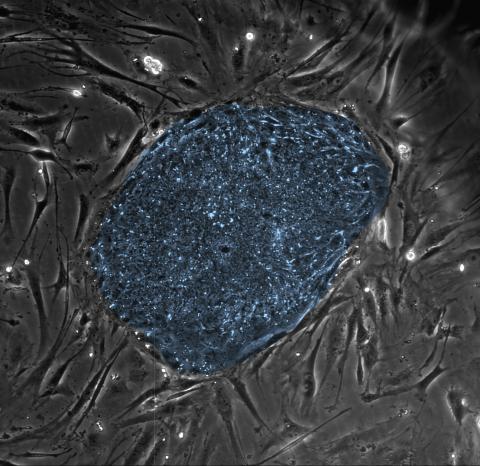
2608: Human embryonic stem cells
2608: Human embryonic stem cells
The center cluster of cells, colored blue, shows a colony of human embryonic stem cells. These cells, which arise at the earliest stages of development, are capable of differentiating into any of the 220 types of cells in the human body and can provide access to cells for basic research and potential therapies. This image is from the lab of the University of Wisconsin-Madison's James Thomson.
James Thomson, University of Wisconsin-Madison
View Media
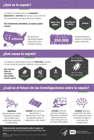
6551: ¿Qué es la sepsis? (Sepsis Infographic)
6551: ¿Qué es la sepsis? (Sepsis Infographic)
La sepsis o septicemia es la respuesta fulminante y extrema del cuerpo a una infección. En los Estados Unidos, más de 1.7 millones de personas contraen sepsis cada año. Sin un tratamiento rápido, la sepsis puede provocar daño de los tejidos, insuficiencia orgánica y muerte. El NIGMS apoya a muchos investigadores en su trabajo para mejorar el diagnóstico y el tratamiento de la sepsis.
Vea 6536 para la versión en inglés de esta infografía.
Vea 6536 para la versión en inglés de esta infografía.
Instituto Nacional de Ciencias Médicas Generales
View Media
2454: Seeing signaling protein activation in cells 04
2454: Seeing signaling protein activation in cells 04
Cdc42, a member of the Rho family of small guanosine triphosphatase (GTPase) proteins, regulates multiple cell functions, including motility, proliferation, apoptosis, and cell morphology. In order to fulfill these diverse roles, the timing and location of Cdc42 activation must be tightly controlled. Klaus Hahn and his research group use special dyes designed to report protein conformational changes and interactions, here in living neutrophil cells. Warmer colors in this image indicate higher levels of activation. Cdc42 looks to be activated at cell protrusions.
Related to images 2451, 2452, and 2453.
Related to images 2451, 2452, and 2453.
Klaus Hahn, University of North Carolina, Chapel Hill Medical School
View Media
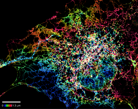
6572: Nuclear Lamina
6572: Nuclear Lamina
The 3D single-molecule super-resolution reconstruction of the entire nuclear lamina in a HeLa cell was acquired using the TILT3D platform. TILT3D combines a tilted light sheet with point-spread function (PSF) engineering to provide a flexible imaging platform for 3D single-molecule super-resolution imaging in mammalian cells.
See 6573 for 3 separate views of this structure.
See 6573 for 3 separate views of this structure.
Anna-Karin Gustavsson, Ph.D.
View Media
5779: Microsporidia in roundworm 3
5779: Microsporidia in roundworm 3
Many disease-causing microbes manipulate their host’s metabolism and cells for their own ends. Microsporidia—which are parasites closely related to fungi—infect and multiply inside animal cells, and take the rearranging of cells’ interiors to a new level. They reprogram animal cells such that the cells start to fuse, causing them to form long, continuous tubes. As shown in this image of the roundworm Caenorhabditis elegans, microsporidia (shown in red) have invaded the worm’s gut cells (the large blue dots are the cells' nuclei) and have instructed the cells to merge. The cell fusion enables the microsporidia to thrive and propagate in the expanded space. Scientists study microsporidia in worms to gain more insight into how these parasites manipulate their host cells. This knowledge might help researchers devise strategies to prevent or treat infections with microsporidia.
For more on the research into microsporidia, see this news release from the University of California San Diego. Related to images 5777 and 5778.
For more on the research into microsporidia, see this news release from the University of California San Diego. Related to images 5777 and 5778.
Keir Balla and Emily Troemel, University of California San Diego
View Media
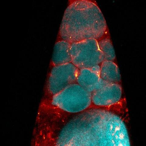
2418: Genetic imprinting in Arabidopsis
2418: Genetic imprinting in Arabidopsis
This delicate, birdlike projection is an immature seed of the Arabidopsis plant. The part in blue shows the cell that gives rise to the endosperm, the tissue that nourishes the embryo. The cell is expressing only the maternal copy of a gene called MEDEA. This phenomenon, in which the activity of a gene can depend on the parent that contributed it, is called genetic imprinting. In Arabidopsis, the maternal copy of MEDEA makes a protein that keeps the paternal copy silent and reduces the size of the endosperm. In flowering plants and mammals, this sort of genetic imprinting is thought to be a way for the mother to protect herself by limiting the resources she gives to any one embryo. Featured in the May 16, 2006, issue of Biomedical Beat.
Robert Fischer, University of California, Berkeley
View Media
2723: iPS cell facility at the Coriell Institute for Medical Research
2723: iPS cell facility at the Coriell Institute for Medical Research
This lab space was designed for work on the induced pluripotent stem (iPS) cell collection, part of the NIGMS Human Genetic Cell Repository at the Coriell Institute for Medical Research.
Courtney Sill, Coriell Institute for Medical Research
View Media
6770: Group of Culex quinquefasciatus mosquito larvae
6770: Group of Culex quinquefasciatus mosquito larvae
Mosquito larvae with genes edited by CRISPR. This species of mosquito, Culex quinquefasciatus, can transmit West Nile virus, Japanese encephalitis virus, and avian malaria, among other diseases. The researchers who took this image developed a gene-editing toolkit for Culex quinquefasciatus that could ultimately help stop the mosquitoes from spreading pathogens. The work is described in the Nature Communications paper "Optimized CRISPR tools and site-directed transgenesis towards gene drive development in Culex quinquefasciatus mosquitoes" by Feng et al. Related to image 6769 and video 6771.
Valentino Gantz, University of California, San Diego.
View Media
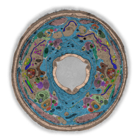
5759: TEM cross-section of C. elegans (roundworm)
5759: TEM cross-section of C. elegans (roundworm)
The worm Caenorhabditis elegans is a popular laboratory animal because its small size and fairly simple body make it easy to study. Scientists use this small worm to answer many research questions in developmental biology, neurobiology, and genetics. This image, which was taken with transmission electron microscopy (TEM), shows a cross-section through C. elegans, revealing various internal structures.
The image is from a figure in an article published in the journal eLife. There is an annotated version of this graphic at 5760.
The image is from a figure in an article published in the journal eLife. There is an annotated version of this graphic at 5760.
Piali Sengupta, Brandeis University
View Media
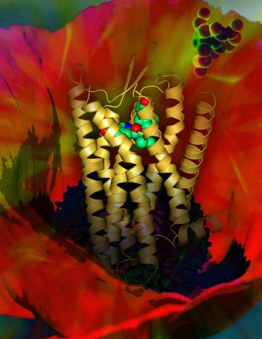
3314: Human opioid receptor structure superimposed on poppy
3314: Human opioid receptor structure superimposed on poppy
Opioid receptors on the surfaces of brain cells are involved in pleasure, pain, addiction, depression, psychosis, and other conditions. The receptors bind to both innate opioids and drugs ranging from hospital anesthetics to opium. Researchers at The Scripps Research Institute, supported by the NIGMS Protein Structure Initiative, determined the first three-dimensional structure of a human opioid receptor, a kappa-opioid receptor. In this illustration, the submicroscopic receptor structure is shown while bound to an agonist (or activator). The structure is superimposed on a poppy flower, the source of opium.
Raymond Stevens, The Scripps Research Institute
View Media
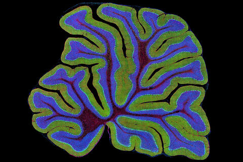
3639: Cerebellum: the brain's locomotion control center
3639: Cerebellum: the brain's locomotion control center
The cerebellum of a mouse is shown here in cross-section. The cerebellum is the brain's locomotion control center. Every time you shoot a basketball, tie your shoe or chop an onion, your cerebellum fires into action. Found at the base of your brain, the cerebellum is a single layer of tissue with deep folds like an accordion. People with damage to this region of the brain often have difficulty with balance, coordination and fine motor skills. For a higher magnification, see image 3371.
This image was part of the Life: Magnified exhibit that ran from June 3, 2014, to January 21, 2015, at Dulles International Airport.
This image was part of the Life: Magnified exhibit that ran from June 3, 2014, to January 21, 2015, at Dulles International Airport.
Thomas Deerinck, National Center for Microscopy and Imaging Research, University of California, San Diego
View Media
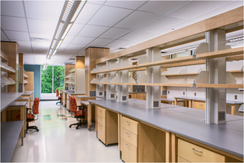
2376: Protein purification facility
2376: Protein purification facility
The Center for Eukaryotic Structural Genomics protein purification facility is responsible for purifying all recombinant proteins produced by the center. The facility performs several purification steps, monitors the quality of the processes, and stores information about the biochemical properties of the purified proteins in the facility database.
Center for Eukaryotic Structural Genomics
View Media
6808: Fruit fly larvae brains showing tubulin
6808: Fruit fly larvae brains showing tubulin
Two fruit fly (Drosophila melanogaster) larvae brains with neurons expressing fluorescently tagged tubulin protein. Tubulin makes up strong, hollow fibers called microtubules that play important roles in neuron growth and migration during brain development. This image was captured using confocal microscopy, and the color indicates the position of the neurons within the brain.
Vladimir I. Gelfand, Feinberg School of Medicine, Northwestern University.
View Media
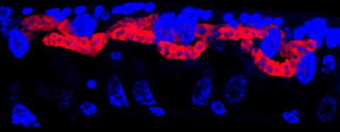
6753: Fruit fly nurse cells during egg development
6753: Fruit fly nurse cells during egg development
In many animals, the egg cell develops alongside sister cells. These sister cells are called nurse cells in the fruit fly (Drosophila melanogaster), and their job is to “nurse” an immature egg cell, or oocyte. Toward the end of oocyte development, the nurse cells transfer all their contents into the oocyte in a process called nurse cell dumping. This process involves significant shape changes on the part of the nurse cells (blue), which are powered by wavelike activity of the protein myosin (red). This image was captured using a confocal laser scanning microscope. Related to video 6754.
Adam C. Martin, Massachusetts Institute of Technology.
View Media